Dogs are mobile and active animals. Therefore, among the various types of injuries in pets, the owner encounters bone fractures quite often. Mechanical damage to bone elements leads to disruption of the integrity of the organ, which is accompanied by loss of the limb from motor function.
The owner must have an idea not only of the symptoms of bone fracture, but also be able to competently provide first aid to the animal in case of the corresponding pathology.
Causes of injury
Veterinary surgeons divide the reasons that led to a violation of the integrity of the bone of a limb in a dog into two groups:
- Injuries and damage. Blows, severe bruises, a pet falling from a height (important for miniature breeds), fights with relatives, unsuccessful jumps are common causes of damage to the integrity of the limbs.
Dogs often receive fractures of their hind and front legs when they are hit by vehicles, bicycles, or moving parts of machines and equipment. Bone fracture can be caused by pinching a limb and an attempt by the pet to escape from captivity.
An animal can become a victim of human cruelty or fall into a hunting trap. Gunshot wounds can also be accompanied by broken bones.
- Pathological conditions. Disturbances in mineral metabolism, the development of osteoporosis in animals, the presence of inflammatory and destructive processes in bone tissue, and the development of neoplasms lead to the fact that even a slight influence of an external factor leads to a fracture.
Most often, severe injury is observed in mobile and active individuals, in hunting and working dogs, as well as in dwarf and miniature pets.
We recommend reading about arthritis in dogs. You will learn about the causes and types of arthritis in dogs, symptoms of the disease, diagnosis, treatment and prevention of rheumatoid arthritis. And here is more information about spinal fractures in dogs.
Postoperative care
The period of adaptation and recovery is very important. Just as important as surgery is that it is performed in the right place by the right specialist. Be sure to follow all directions from your veterinarian. Don't be lazy and take care of your pet. He will express dissatisfaction when processing stitches or injections. Don't scold him for this. Show him that you do this with care and love, and not through force, and he will reciprocate your feelings. There's no need to shout. If you feel anger starting to arise inside you, stop. Think about the fact that his life is now in your hands.
Happy joyful days spent with your pet!
An accident, a fall from a height, an unsuccessful jump, excessive loads - it is unlikely that it will be possible to completely protect the dog from all risks. But even if the bone is severely crushed, today it is technically easy to cure almost any fracture in a dog. Of course, if you contact a veterinarian in time. But how can you help your pet before arriving at the clinic?
1. A crack is the safest of fractures; the bone is not completely broken, and the adjacent tissues are not damaged. Often confused with a bruise, because
The pet generally feels good, just moves with caution. If help is not provided in time, the situation may worsen
For example, an uncomplicated fracture of a dog’s paw without treatment becomes an open fracture after jumping over a high barrier (the partially damaged bone cannot withstand the usual loads).
It is impossible to visually determine a crack in the bone
Therefore, in all cases when, for no apparent reason, the dog does not allow touching any part of the body, moves cautiously, limps (paws, spine), chews slowly (jaws), etc., you need to take an x-ray.. 2
Impacted fracture - parts of the bone in the damaged area fit into each other. This happens when the blow is directed longitudinally to the bone, and not across. For example, when a puppy (elastic bones), a small or very large dog jumps from a height, leaning on its front paws. Symptoms of an impacted fracture of a dog’s paw are similar to those of a crack (lameness, pain), the danger is the same - parts of the bone can move if help is not provided in time. The tissues are not damaged or only slightly damaged; the broken bone partially fixes itself.
2. Impacted fracture
– parts of the bone in the damaged area fit into each other. This happens when the blow is directed longitudinally to the bone, and not across. For example, when a puppy (elastic bones), a small or very large dog jumps from a height, leaning on its front paws. Symptoms of an impacted fracture of a dog’s paw are similar to those of a crack (lameness, pain), the danger is the same - parts of the bone can move if help is not provided in time. The tissues are not damaged or only slightly damaged; the broken bone partially fixes itself.
3. Closed fracture without displacement
– the ends of the broken bone are in a natural position relative to each other. Severe pain, redness and/or swelling, the pet does not allow you to touch the site of injury and tries not to put stress on the painful area (does not lean on the paw, does not chew - the jaw, sleeps on one side - the ribs).
4. Closed fracture with displacement
– the ends of the broken bone diverge, damaging blood vessels and muscles. The pain is stronger, the swelling is greater, on light-colored dogs the hematoma is clearly visible, and a bulge may be noticeable (if the displaced edge of the broken bone rests on the skin from the inside).
Closed bone fractures in dogs are extremely dangerous - on the outside there seems to be nothing wrong, but the tissues under the skin can be seriously damaged: a large vessel - loss of blood, nerves - partial paralysis, the end of a broken rib can pierce an internal organ.
5. Open fracture
– the edge of the bone sticks out, tearing the muscles and skin. The bone itself may not be visible (especially in muscular or obese dogs), but the wound is always visible. The most dangerous fracture (severely damaged tissue, risk of blood loss), but there is also a plus - the signs are so obvious that the owner rushes to the clinic immediately.
In addition to fractures resulting from trauma, there are pathological fractures - the bone cannot withstand normal loads, and sometimes breaks even under the weight of the body while walking. Causes: calcium deficiency, bone cancer, cystic formations, any disease that causes bones to become brittle, old age. Such fractures in dogs require treatment in two directions - to restore the integrity of the bone and to eliminate (if possible) the root cause.
Types of Fractures
In veterinary practice, the following types of bone fractures in domestic animals are distinguished:
- Crack or incomplete linear fracture . The pathology is of the mildest form, passes for the owner with virtually no symptoms and is detected only by X-ray examination. If there is a crack in the bones of a limb, the dog may experience lameness and a cautious gait. The animal avoids active games, jumping, and is reluctant to go down the stairs.
- Impacted fracture. It is characterized by a blurred clinical picture, since the damaged bones fix each other. This type of injury is often observed in representatives of large breeds, as well as in puppies due to underdevelopment of the musculo-ligamentous apparatus.
- Closed fracture of a bone element without changing the anatomical position. The injury is characterized by the fact that the damaged bone structure remains in place. The pathology is characterized by severe pain and loss of limb from motor function.
- Closed bone fracture with displacement. Severe injury is associated not only with a violation of the integrity of the organ, but also with the fact that the bone leaves its anatomical location as a result of a fracture. The condition is dangerous because it can lead to damage to large vessels and injury to vital organs.
- Open fractures. The most complex injury, characterized by damage not only to bone, but also to internal and external tissues, rupture of blood and lymphatic vessels, damage to the ligamentous apparatus and joints.
Depending on the direction of the limb fracture in dogs, veterinary surgeons distinguish longitudinal, transverse, oblique, helical and marginal injuries. Taking into account the size of the bone fragments, a distinction is made between large-fragmented and finely fragmented fractures, also crushed and crushed. If the violation of integrity reaches the joint, then we are talking about a complex intra-articular pathology.
Daily regimen and food intake during a fracture
There should be at least 8 hours of sleep per day, since the body does not waste energy on other tasks and switches to recovery after a fracture. You need to drink at least 2 liters of liquid per day, and not only during meals. It is useful to eat 5 to 6 times a day in small warm portions. It is strictly forbidden to overeat; the diet for bone fractures includes freshly prepared food. You need to chew slowly, chewing foods thoroughly. The diet must be varied as much as possible; there should be vegetables and fruits on the table every day.
You can eat milk and products made from it every day, but not more than 200 grams. Cottage cheese, preferably made from natural raw materials, is useful at least 2 times a week.
The recovery period lasts on average from 30 to 80 days, provided that all specialist recommendations and diet are followed. Much depends on what part of the bone is broken and how advanced the immune system is. Along with this, a lot depends on nutrition, since it is this that ensures that the body receives the necessary substances.
Feedback from a veterinarian from the NIKA clinic. Vladivostok, Chkalova 6. tel. 316882 I am a veterinarian. Specialization: surgery. Work experience 15 years.
In my practice, I often encounter fractures in dogs and cats. Fractures vary in severity, and the speed of regeneration depends on this. Fractures without displacement, with displacement, diaphyseal, metaphyseal and others. they all heal at different times and give rise to different callus formations.
The rate of bone regeneration depends on many reasons, including the location of the fracture and the severity of the injury, and most importantly, drug therapy in the treatment regimen and understanding the health status of the animal, as well as the age and breed of the animal. Problems with gastrointestinal diseases also affect calcium metabolism.
The postoperative period for such injuries is rest, good nutrition and medications that affect bone metabolism and maintain a normal calcium-phosphorus ratio for the rapid growth of callus.
The effect of any drug is best seen in various pathologies, when you know the cause of the disease, the mechanism of its course, as well as the principle of action of a particular drug.
Naturally, any veterinarian is wary of new products because he does not know what to expect. We love to work with what we are confident in.
That's why I stopped at fractures and came up with Calcium Peptide. You can compare and actually see how the drug works using a type of diagnostic test such as x-ray. Our clinic was one of the first to work with calcium peptide; we participated in clinical trials.
The developer of the drug, A.B. Kolesnikov, also participated in the clinical trials. We already knew about positive trials on humans, our task was to select dosages and identify side effects in dogs.
It was interesting to compare and see the effect of the drug in real conditions, with pronounced pathologies in the body, in which the bone callus grew slowly, due to poor absorption of calcium. (This category includes dogs of dwarf breeds: toy terriers, chihauas, Yorkshire terriers, etc. It is known that these dogs have a genetic deviation in the absorption of calcium).
In clinical trials, we described everything in detail, compared it with different drugs and treatment regimens (this will be more interesting for veterinarians, but not dog owners), but the positive dynamics when using calcium peptide were obvious in all cases.
Callus was increased in case of any pathologies in the body, in a much shorter time, without additional correction of the body. For example, without prescribing vitamin D.
I can say the same about the chondrodog. We observed dogs with arthrosis. In the initial stage of the disease, the effect of chondrodog is visible already on the third day (pain relief), which is much safer for the body than non-steroidal drugs. (I do not want to say by this that non-steroidal drugs should not be given for injuries). Positive dynamics for age-related problems in large breeds (ligament weakness and sprains), without any additional treatment.
As a veterinarian, and even more so as a surgeon, I am interested in the results of my work not being useless, therefore, I recommend to animal owners those drugs that give a real positive effect, these drugs include “Calcium-peptide” and “Chondrodog”.
On my own behalf, I want to say that misunderstanding of this or that drug or supplement or medicine is not a problem for manufacturers, but for animal owners.
Often, certain drugs are used for other purposes than for their intended purpose, as in the example with Gamavit.
Its breeders use it in any situation, and when no dynamics of treatment with gamavit are observed during a viral disease, they conclude that this drug is bad.
Forgetting that gastroenteritis cannot be treated with gamavit alone, and there are more targeted means to stimulate the immune system and antiviral protection. This is how the reputation of a particular product is created.
From the subjective perceptions of a dog breeder or breeder, which have nothing to do with veterinary medicine.
The diagnosis must be made by a veterinarian, not the owner. Unfortunately, the scourge of modern dog breeding is self-medication.
And an incorrect diagnosis leads to incorrect conclusions, which can be found about any drug on the market.
Calcium peptide and Chondrodog work effectively in their field, but if lameness is caused by myositis, then calcium peptide will not help, because the cause of myositis is not an imbalance of calcium, but inflammation of the skeletal muscles.
If the dog has weak ligaments due to impaired bone mineralization, i.e., first you need to put the bone tissue in order, and not give chondroprotectors, i.e. this is the work of “Calcium-peptide”, not “Chondrodog”, if the dog is limping , you need to take an x-ray of the bone, it may have a crack, and not a problem with the ligaments or joints,
It is written above that the genetic position of the limbs also cannot be corrected by any calcium supplements or chondroprotectors.
I hope I explained clearly that diagnosis is important.
From practice, most breeders themselves give advice to the owners of their dogs and recommend not to consult a doctor for trifles.
This is where false conclusions and incorrect judgments about this or that drug appear. (I’m generally talking about any medicine).
Our clinic, as I said, has been working for a long time on the developments of Far Eastern scientists, we have positive experience and each supplement is effective in its own area, both for the prevention and recovery of animals.
Galina Shchetinina. Veterinarian. Clinic "Nika".
How to visually determine
The owner can quickly detect a serious injury in a four-legged friend if there is an open fracture, or if the damage is accompanied by displacement of bone elements. With an open injury, a bone fragment can be visually observed protruding through the damaged muscles and skin. Open damage is accompanied by bleeding.
In the case of pathology with displacement, the owner, as a rule, notices a change in the configuration of the damaged limb, its asymmetry due to the deviation of the bone from its anatomical position.
First aid for a broken paw
It is only necessary for open fractures. The main task is to stop the bleeding. Usually you don’t have a tourniquet at hand (but if you do, use it), so you’ll have to use improvised materials. To do this, you need to wrap the area above the wound with a piece of cloth, a scarf, or gauze pads, and then bandage it tightly with a bandage or a simple rope.
Please note two important points:
- To stop the bleeding, you need to bandage not the wound itself, but the place above it.
- You cannot apply a tourniquet directly to the skin, you can damage it; be sure to place a piece of cloth, a bandage, or a napkin.
It is also recommended to fix the limb, but here the owners forget that “fixing” does not mean setting the fragments.
Remember! Never try to set a broken bone, it's incredibly painful.
In addition, you can damage the limb even more, even to the point of rupture of blood vessels. Just secure it by tying it to a stick, plywood, or another limb. When transporting your pet, make sure that the injured limb is on top and not loaded.
If you are not sure what first aid should be, then do nothing, but simply take your pet to the veterinary clinic as quickly as possible.
The prognosis is usually favorable. The bones grow together within a few weeks; if the osteosynthesis was carried out correctly, then there will be no consequences in the form of lameness. Cautious prognosis in the presence of osteosarcoma.
Symptoms of a front and rear paw fracture
Trauma in an animal associated with a violation of the integrity of the bones of the limb is characterized by a number of clinical manifestations:
- The dog loses the ability to rest normally on the sore paw. The dog holds the limb suspended and tries not to step on it.
- When forced to move, the dog experiences pronounced lameness. Often the animal moves on three legs.
- In the early stages, the owner may notice severe swelling in the area of injury. The damaged limb swells, increases in size, asymmetry is observed, and a change in the configuration of the paw is observed when examining the animal.
- Palpation of the area of injury causes a negative reaction in the dog, indicating severe pain. The dog whines, squeals, and can even bite the owner when a painful shock develops.
- The injured paw may be loose or have unnatural mobility. Shortening of the limb is observed.
- In the case of an open fracture, bone fragments are visually detected.
If a paw is injured, the dog refuses food, becomes anxious, or goes into shock due to severe pain.
About the symptoms and treatment of a broken paw in a dog (medical history), watch this video:
Hind limb fracture
Fractures of the hind legs in dogs are somewhat less common. They occur as a result of falling from a height, falling under the wheels of a car, or as a result of so-called domestic injuries (this mainly applies to small breed dogs and puppies, which the owners themselves, through negligence, can get pinched by a door, drop, or simply step on). If we are talking about a simple fracture, then there are no special features in the treatment and recovery period; it is much worse if the injury damaged not only the limb, but also the pelvic bones. In such cases, the animal requires a complex operation and full long-term care.
First aid for an animal
Having discovered signs of bone pathology in a four-legged friend, the owner should take care of an immediate visit to a specialized clinic. When providing first aid to your pet, it is important to act competently and not cause harm.
In the case of an open injury, it is necessary first of all to stop the bleeding, which almost always accompanies such an injury. For this purpose, a tourniquet is applied above the wound site (for arterial bleeding). This can be done with the help of a scarf, scarf, leash and other equipment.
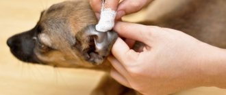
The open wound must be freed from dirt and bone fragments and treated with an antiseptic solution, for example Chlorhexidine, hydrogen peroxide, Furacilin solution, etc. The damaged tissue should be covered with a bandage to prevent infection.
First aid for a fracture
After stopping the bleeding and treating the open wound, the animal must be given complete rest and the injured limb must be immobilized. In the field, you can use improvised objects: a wooden board, a wide stick, a panel from a car. For small pets, a strip of thick cardboard will suffice. The diseased limb is fixed to the device using a belt, strap, scarf, etc.
A sick animal should be transported in the back seat of a car. Miniature and toy breeds can be carried in a small carrier or by hand, being careful not to dislodge the restrained limb. The use of ointments and attempts to independently reduce bone fragments are strictly prohibited.
To learn how to properly provide first aid to a dog with a broken paw, watch this video:
How to help your dog avoid breaking a nail
To reduce the chance of a broken nail, it is important to keep all of your dog's nails trimmed. Short claws break much less often than long claws
Ask your veterinarian about proper trimming techniques to trim nails at home.
A fracture is a break in the integrity of a bone.
. If the muscles and skin are damaged, the fracture is called an open fracture. While maintaining the integrity of the skin, the fracture is closed. Also, a fracture of a dog’s paw can be complete (with displacement of the fragments) or incomplete (without displacement, cracks).
The anatomy of the hind and front legs is very different, so they must be considered separately.
The structure of the forelimbs
Spatula
- a bone in the form of an elongated triangular plate that attaches the forelimbs to the body with the help of muscles. The wide base points upward. The narrow part of the scapula is directed downward, there is a glenoid cavity on it, into which the humerus is attached through tendons.
An important element is the spine of the scapula - a narrow but steep elevation that runs from top to bottom along the entire length of the scapula. The muscles that are responsible for the movements of the forelimb are attached to it.
Brachial bone
tubular, is one of the most massive and strong in the skeleton. It has two ends (epiphysis):
- the upper head is rounded, corresponds to the multiaxial shoulder-scapula joint, next to it is the large lateral tubercle - the place of attachment of powerful muscles;
- the lower head is in the form of two tubercles, which form a depression similar to a saddle.
The basis of the forearm is the ulna and radius bones
. On the ulna you can find a large olecranon process - the place of attachment of the powerful extensors of the elbow joint. And also a deep crescent-shaped fossa into which the lower head of the humerus enters.
The radius is thinner; together with the ulna, it allows the forelimb to rotate slightly around the longitudinal axis. This is necessary due to the fact that the elbow joint is a uniaxial block joint; its structure allows for movements with a wide amplitude, but does not provide the opportunity.
Wrist
in dogs it is represented by a number of individual asymmetrical bones. Five fingers are attached to these bones, four large ones form the metacarpus itself, and the fifth short finger is rudimentary, located behind the metacarpus in dogs. The bones that attach to the wrist are called metacarpals, and the bones that form the fingers are called phalanges:
- the uppermost phalanx is the fetlock;
- middle second – coronal;
- the lower third is claw-shaped (the claw itself).
The structure of the hind limbs
The belt of the hind limbs is
the pelvis
- a complex symmetrical structure.
Each half is called an innominate bone
, which in turn consists of three fused bones:
- ileal;
- ischial;
- pubic
The ilium is the largest bone, located above and in front, it rests on the pelvis and connects to it through the flat sacroiliac joint
.
The ischium in dogs is short and located at the back. Each half of the pelvis fuses below with the adjacent half through the pubic bone. At the site of fusion of the pubic, ilium and ischium bones there is a deep articular cavity
into which the head of the femur enters.
Femur
The massive tubular bone is one of the strongest bones in the body. At its upper end there is a spherical head, which fits into the cup-shaped articular cavity of the pelvis. This joint gives the greatest freedom of movement; for example, male dogs can move their leg far to the side when urinating.
The lower end of the femur is flatter, it is formed from two semicircular tubercles, between which there is a saddle-shaped fossa. It, together with the heads of the tibia and fibula, forms the trochlear joint. The joint also contains the kneecap, a bean-shaped bone with a base and an apex. The patella prevents the knee joint from bending forward.
Fibula
in dogs there is significantly less tibia. The latter is especially massive at the upper end, and at this end it bends quite strongly. The fibula at its lower end often fuses with the tibia. The structure of the metatarsus and tarsus of the hind limb is the same as that of the front paw.
Diagnosis of injury in a dog
At a specialized clinic, the sick animal will be given pain relief and a full clinical examination to look for injuries other than a broken limb. The most informative method of detecting a problem, its location, type and shape of the fracture is an x-ray examination. Diagnosis is most often made after the animal is sedated.
An X-ray is usually taken in two projections to correctly assess the complexity of the pathology. If necessary, the veterinarian may prescribe a computer or magnetic resonance imaging scan to assess the condition of soft tissues (muscles, ligaments, joints).
Impacted fracture
About treatment
As we have already said, in the simplest cases the “dropped out” hip can be realigned, but the chances of this are minimal. In addition, for a successful procedure, general anesthesia must be used. In addition, if the head of the femur has come out of the femoral socket at least once, the likelihood of recurrence is very high. The older the dog, the higher the chances. There are some factors that make relapse 100% possible:
Hip dysplasia- The dislocation occurred against the background of a serious injury
. - The dog is seriously overweight
. - After realignment, the limb never regained its former mobility.
As a rule, repeated dislocation
develops no later than two weeks after the first case. Over time, the frequency of this phenomenon will inevitably increase, and surgery will be required. If at least one repeated dislocation occurs, or, moreover, the reduction of the hip is impossible for some reason, a decision on surgical intervention must be made. Today, more than 12 different methods of operations have been developed, but many of them are only the development of ideas proposed back in the 60s of the last century; nothing more advanced has been invented since then. Here are a few methods that are used most often:
- Restoration of the round ligament (the same one that connects the head of the femur and the bottom of the acetabulum).
- Reparation (restoration) of the joint capsule.
- Transarticular fastening.
- Replacement of the joint capsule with a synthetic prosthesis.
The pathology is accompanied by a pronounced pain reaction. The dog transfers its body weight to its healthy limbs; it can no longer step on the sprained paw. The paw may be turned inward or outward, depending on the nature of the damage. In 90% of cases, the head of the femur moves forward from the acetabulum.
Partial displacement of the head is called hip subluxation and is common in dogs with severe hip dysplasia. This damage is most often observed on 2 joints, while with a traumatic dislocation one joint suffers.
Animal treatment
Given the complexity of the injury, veterinarians use both conservative and surgical techniques. Conservative treatment of long bone fractures in dogs is used mainly for uncomplicated closed fractures.
Without surgery
After pain relief and the use of muscle relaxants to relax the muscles, the veterinarian will realign the displaced bone fragments. To immobilize the damaged bone, special bandages are used to limit the movement of the limb. The animal is given complete rest. To limit movement, it is best to place a small dog in a crate for 3 to 4 weeks.
Removal of the immobilizing bandage is carried out after 30 - 40 days in young animals and after 45 - 50 days in older pets. For small dog breeds, the bandage is removed after 20-25 days. While the animal is in a special splint, it is necessary to ensure that it does not compress the soft tissues, causing the development of congestion.
To learn how to properly apply an immobilizing bandage for a fracture in dogs, watch this video:
Surgical intervention
In most cases, a veterinary specialist recommends that the owner undergo surgical treatment for limb fractures in dogs (osteosynthesis) on the first day of injury. This is due to the complex nature of injuries in animals and the development of severe traumatic edema, which complicates surgical manipulation and increases the risk of postoperative complications.
Osteosynthesis involves the use of special devices to stabilize bone fragments: metal plates, knitting needles, wires, screws, staples, pins. Recently, veterinary surgery has used not only metal tightening elements, but also polymer pins.
For femur fractures, the surgeon most often uses steel staples to fix the bone fragments. Spiral, longitudinal, and oblique fractures of the tubular bones of the extremities require the use of nickel-plated nails or staples, which the surgeon drives into a specially drilled hole.
In case of fractures of the ulnar and calcaneal tuberosity, the femoral bone, the fragments are connected, as a rule, using metal screws or screws. This method avoids bone splitting and securely fixes the bone fragment. The screw is removed after 35-40 days through a skin incision.
For complex comminuted fractures, as well as to prevent shortening of the limb, distraction splints are used in veterinary practice. Their use allows for osteosynthesis with elements of bone traction. This method with metal pins requires the selection of their size and configuration by radiography. Surgery is performed both under general and local anesthesia.
Why is it not recommended to use plaster for fractures in dogs?
A plaster splint does not allow bone fragments to be accurately connected and held for the required time. This leads to fracture nonunion, limitation and loss of limb function. The dog may limp or not be able to put weight on its leg if the fracture has not healed properly. In addition, the cast limits the dog’s support on his paw; he cannot walk normally in a splint throughout the entire required time of fixation of the fracture, which is 1.5-2 months. During this time, blood circulation in the paw is disrupted and the muscles atrophy. Because of this, the dog’s fracture heals poorly, it takes a long time to recover, or the support on the paw is not restored at all. After treatment with surgery, the dog can use his paw for up to a week after surgery. This allows you to maintain good blood circulation in the paw and speed up the healing of the fracture.
How long does it take to heal
Healing of bone injuries in dogs goes through several stages. After 10 - 15 days
A connective tissue callus is formed. The fusion of bone fragments occurs by 35–45 days.
Dogs begin to include the damaged limb in the support function at the moment when the formation of a bone callus occurs. Until this moment, sick animals protect their broken paw.
In order to prevent infection after osteosynthesis, a sick animal must be prescribed a course of antibacterial drugs. On the recommendation of a veterinary surgeon, the animal can be prescribed immunomodulators, vitamins, and minerals.
4-5 weeks after osteosynthesis, the animal is given a control radiography to assess the effectiveness of the operation.
Rehabilitation of a dog with a broken paw
Further treatment at home can take place in the absence of postoperative complications (blood clots, surgical infection). Usually, after making sure that everything is in order, the dog is discharged after 6-24 hours. The owner’s task is to provide peace for the pet, and to prevent the dog from gaining weight (due to inactivity), a diet should be prescribed . Special care consists of giving painkillers, calcium supplements, and daily treatment of sutures.
The pet begins to lean on the limb after 3-5 days, and the fracture heals completely within 2-3 weeks, after which time the dog begins to run and play. However, the strength of the bone at this time may be low, so it is recommended to protect the pet from excessive mobility and traumatic situations for another 2-3 weeks. In total, rehabilitation is 4-6 weeks; in the third or fourth week it is advisable to take an x-ray and make sure that the bone is healing correctly.
Jaw fracture
These fractures in dogs are extremely rare, but if a dog's jaw fracture does occur, such injuries require very close attention. Typically, such fractures are open; numerous bacteria from the oral cavity can penetrate through the mucosal defect into the thickness of the jaw, so such fractures must be treated as soon as possible. They usually happen during fights with other dogs, as a result of a car injury, or if the dog crashes its head into an obstacle. An additional risk factor is old age and poor oral health (gingivitis, periodontitis). With these diseases, the jaw bone becomes weak and can easily break with minimal impact. We must also warn that jaw fractures in small dogs sometimes occur when owners and unscrupulous veterinarians try to remove their baby teeth without anesthesia. We categorically do not recommend doing this; the animal will defend itself so energetically that it can harm itself.
Symptoms of a jaw fracture include severe pain, inability to close or open the mouth, inability to eat, an asymmetrical appearance of the muzzle, and bleeding from the mouth. To treat such fractures, surgery is required: osteosynthesis with a plate, knitting needles or wire sutures, depending on the location of the fracture and the size of the dog. Within a day after the operation, the dog will be able to eat soft food and will quickly recover.
You can contact our veterinary center regarding any type of fracture in your dog. To do this, you do not need to make an appointment with a surgeon or traumatologist. Bring your dog to any general practitioner every day from 10.00 to 22.00. He will conduct an examination, provide pain relief, evaluate associated problems, take x-rays, and record the fracture until the time of surgery.
If you have already been examined and want to undergo osteosynthesis in our clinic, you can sign up for the operation and ask all your questions by calling us at the Veterinary by phone.
Useful materials:
- Determine the breed of a dog Dog Breed Determiner What does this application do? Determines the breed of a dog from a photograph using the camera of your device...
- Where did dogs come from? Ch. Darwin's assumptions Ch. Darwin's expeditions on the Beagle ship allowed him to travel to different countries.…
- Cestal reviews for dogs AnaloguesCestal for dogs is a popular remedy that can be found in any veterinary pharmacy, but...
- Fiprist for dogs instructions Composition and release form Fiprist is available in the form of drops and spray, and the drops are produced in two…
Sample action plan for injury
Carefully and carefully examine the animal. If you find an open bleeding wound, apply a bandage. Take the animal to a veterinary clinic as quickly as possible for examination and further research. When transporting small dogs, use special hard carriers; in extreme cases, a simple box with a large animal will do; you can use a blanket or a piece of some dense fabric, for example, burlap or tarpaulin.
Dear owners of dogs and other animals!
Remember: timely visit to a veterinary clinic is the key to health and quality treatment, and as a result, the well-being of your pet.
A fracture is a break in the integrity of a bone.
. If the muscles and skin are damaged, the fracture is called an open fracture. While maintaining the integrity of the skin, the fracture is closed. Also, a fracture of a dog’s paw can be complete (with displacement of the fragments) or incomplete (without displacement, cracks).
The anatomy of the hind and front legs is very different, so they must be considered separately.
The structure of the forelimbs
Spatula
- a bone in the form of an elongated triangular plate that attaches the forelimbs to the body with the help of muscles. The wide base points upward. The narrow part of the scapula is directed downward, there is a glenoid cavity on it, into which the humerus is attached through tendons.
An important element is the spine of the scapula - a narrow but steep elevation that runs from top to bottom along the entire length of the scapula. The muscles that are responsible for the movements of the forelimb are attached to it.
Brachial bone
tubular, is one of the most massive and strong in the skeleton. It has two ends (epiphysis):
- the upper head is rounded, corresponds to the multiaxial shoulder-scapula joint, next to it is the large lateral tubercle - the place of attachment of powerful muscles;
- the lower head is in the form of two tubercles, which form a depression similar to a saddle.
The basis of the forearm is the ulna and radius bones
. On the ulna you can find a large olecranon process - the place of attachment of the powerful extensors of the elbow joint. And also a deep crescent-shaped fossa into which the lower head of the humerus enters.
The radius is thinner; together with the ulna, it allows the forelimb to rotate slightly around the longitudinal axis. This is necessary due to the fact that the elbow joint is a uniaxial block joint; its structure allows for movements with a wide amplitude, but does not provide the opportunity.
Wrist
in dogs it is represented by a number of individual asymmetrical bones. Five fingers are attached to these bones, four large ones form the metacarpus itself, and the fifth short finger is rudimentary, located behind the metacarpus in dogs. The bones that attach to the wrist are called metacarpals, and the bones that form the fingers are called phalanges:
- the uppermost phalanx is the fetlock;
- middle second – coronal;
- the lower third is claw-shaped (the claw itself).
The structure of the hind limbs
The belt of the hind limbs is
the pelvis
- a complex symmetrical structure.
Each half is called an innominate bone
, which in turn consists of three fused bones:
- ileal;
- ischial;
- pubic
The ilium is the largest bone, located above and in front, it rests on the pelvis and connects to it through the flat sacroiliac joint
.
The ischium in dogs is short and located at the back. Each half of the pelvis fuses below with the adjacent half through the pubic bone. At the site of fusion of the pubic, ilium and ischium bones there is a deep articular cavity
into which the head of the femur enters.
Femur
The massive tubular bone is one of the strongest bones in the body. At its upper end there is a spherical head, which fits into the cup-shaped articular cavity of the pelvis. This joint gives the greatest freedom of movement; for example, male dogs can move their leg far to the side when urinating.
The lower end of the femur is flatter, it is formed from two semicircular tubercles, between which there is a saddle-shaped fossa. It, together with the heads of the tibia and fibula, forms the trochlear joint. The joint also contains the kneecap, a bean-shaped bone with a base and an apex. The patella prevents the knee joint from bending forward.
Fibula
in dogs there is significantly less tibia. The latter is especially massive at the upper end, and at this end it bends quite strongly. The fibula at its lower end often fuses with the tibia. The structure of the metatarsus and tarsus of the hind limb is the same as that of the front paw.
What will the doctor do?
First of all, he will provide high-quality pain relief. After this, he will conduct a full examination of the dog and the injured limb. This is especially important for those who have suffered a car injury or fallen from a height, because in addition to the obvious problems for the owner - a broken paw - the dog in such a situation may have a chest or abdominal injury. These injuries may not be visible upon a superficial examination of the dog by the owner, but require more urgent and complex correction than a fracture. After the examination, the doctor will take an x-ray of the broken bone to assess the configuration of the fracture and plan treatment, and will also conduct additional diagnostics for other injuries, if any. Sometimes sedation is required for x-rays.
In veterinary practice, 99% of paw fractures in dogs require treatment in the form of surgery, and this operation is called osteosynthesis. This operation is performed as planned, usually 3-5 days after the injury. This is due to physiological features. The fact is that during an injury there is a massive release of blood into the fracture zone, and subsequently this blood and the parts of destroyed tissue that get into it become so-called “osteogenic elements” - substances that stimulate the bone to heal. If you surgically intervene in the fracture area immediately after injury, the entire contents of the hematoma will simply leak out and be lost, and healing will be slower and more difficult. An additional difficulty for manipulating bone fragments is created by swelling of the soft tissues, which disappears just 3-5 days after the injury. The exception is open fractures - due to the open gate for infection, these fractures require urgent (within 24 hours) surgery.
Before surgery, the doctor will apply a fixative bandage to the broken limb.
Of course, fractures of the jaw, pelvis, and spine require a special approach—we’ll talk about them a little later.