Causes
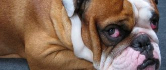
Entropion of the eyelid may have physiological causes caused by the specific structure of the muzzle in dogs with an abundance of folds of skin on it. Wool in constant contact with the sclera and mucous membranes injures them, causing irritation and inflammation. As a result, erosion first forms, and then ulceration. It can be accompanied by an infection, which causes conjunctivitis and other dangerous diseases.
Among them is keratitis, leading to clouding of the cornea, its vascularization, and weakened vision. Over time, without treatment, the dog may become partially or completely blind.
The causes of keratitis are:
- injuries of the eyelid, eyes;
- ophthalmological diseases, inflammatory processes;
- damage to the cornea, causing the dog to constantly squint;
- systemic diseases;
- congenital or acquired change (reduction) in eye size.
The number of diseases does not include age-related entropion of the eyelid, which occurs in older dogs due to weakening of the ligamentous apparatus.
Causes of entropy (turned eyelids) in dogs
Entropion is divided into primary and secondary, depending on the cause of eyelid deformation. Thus, primary entropion is hereditary in nature, the deformation occurs due to the presence of skin folds or lack of elasticity of the skin of the eyelids, which is typical for large breeds.
Secondary entropion develops as a result of inflammatory processes in the eyeball or deformation of the eyelid structure. When the cornea is damaged, dogs, in an attempt to reduce pain, squint their eyes, which causes muscle strain and the eyelid begins to turn inward. After treatment, his position may be restored.
Another cause of entropion in dogs is inflammation of the masticatory muscles, due to which the dog loses weight due to difficulty eating, or sudden weight loss for other reasons leads to loss of muscle tone around the eye.
Main symptoms
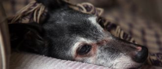
An inversion of the eyelid begins with attacks of photophobia. The dog suffers from a sharp reaction to light; its eyes become watery and inflamed. The discharge from them first looks like mucus, then becomes thick and purulent. The animal tries not to go out into the light, hiding in the shadows.
Caring owners will pay attention to the strangely changed look of their pet. He begins to look a little askance, because the falling fur causes acute pain. At this point, you need to go to the veterinarian as quickly as possible, since the next stage will be severe inflammation, ulceration of the cornea and keratitis. Treating the disease is extremely difficult and troublesome.
Causes of entropion
An important factor influencing the likelihood of entropion is heredity. Moreover, the evil irony is that most often this disease occurs in purebred individuals, especially in the case of inbreeding. However, the statement that the disease occurs solely due to genetic predisposition is also impossible, since it is simply not possible today to determine the gene responsible for the occurrence of bloat. The development of such a defect can be influenced by a number of important factors, for example, the shape of the skull, the location and shape of the eye, the presence of folds on the dog’s head, as well as the degree of elasticity of the upper and lower eyelids.
There are also cases when the occurrence of entropion in a pet is caused solely by mechanical action. The appearance of a defect can be caused by an injury to the eyelid or one of the chronic diseases of the eyeball. However, such cases are quite rare.
Volvulus can occur as a complication of other diseases, for example, inflammation of the conjunctiva, as well as previous eye surgeries.
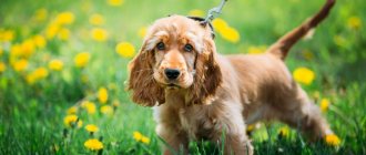
More often than others, dogs with short heads, bulging and sunken eyes, as well as pets with almond-shaped eyes suffer from bloat.
Often, the occurrence of entropion is promoted by microphthalmia, a disease characterized by underdevelopment of certain parts of the eye and a minimal volume of the eyeball, which can be completely or partially blind. Such a defect often goes along with agenesis.
Diagnostics in a veterinary clinic
Short-faced dogs often experience habitual lacrimation, so it is very easy to miss the onset of the disease. A careful examination and regular visits to the clinic will help.
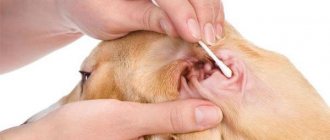
At the hospital, the dog is examined by dropping a special painkiller into the eyes. To detect erosions and ulcers, solutions with fluorescent properties are injected. By illuminating the surface of the eye with a source of ultraviolet radiation, any lesions and damage to the integrity of the membranes can be identified.
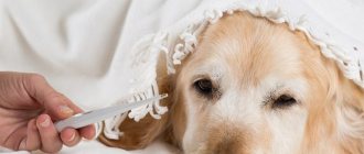
If the disease has progressed and an infection has appeared, the veterinarian will take a sample for bacterial culture. This will reveal the type of microorganisms that caused the inflammatory reaction and will help you choose the right drug for treatment.
Eyelid inversion surgery
The surgical method plays a leading role in treatment. Even in case of serious complications (keratitis, conjunctivitis), the operation will alleviate the general condition and allow for high-quality sanitation of infectious foci.
During the operation, entropion of the eyelids in dogs is stopped by straightening and cutting off the folded part. After this, supporting sutures are applied to fix the ligaments in a certain position. Sutures are often used, which dissolve over time and do not require removal. In adult dogs, several operations may be required to fully strengthen the ligamentous apparatus.
After the operation, regular antiseptic treatment of the orbital area is carried out. Additionally, a plastic collar is put on for 7-14 days, which prevents the paw from touching the seams. The decision to prescribe systemic antibiotics is made by the doctor.
Treatment method and prognosis
If the disease can be detected in the early stages, then it is enough to limit yourself to drug therapy. The veterinarian will prescribe drops with an analgesic and anti-inflammatory effect, as well as gels for the treatment of the eyelid, which have bacteriostatic and antiseptic properties.
A dog that is constantly trying to scratch its eyes should be fitted with a veterinary Elizabethan collar and monitor its behavior, not allowing the situation to worsen.
If there is a severe volvulus or an advanced disease, you will have to resort to surgical intervention. The surgeon excises the fold that folds inward and places sutures to prevent it from folding back and keep the ligaments in the correct position.
Most often, specialized suture material is used that does not need to be removed. It simply dissolves after the surgical wound heals, which helps to injure the animal less.
In older dogs or severely affected dogs, several surgical procedures may be required to completely resolve the problem or to improve the animal's condition.
Causes and consequences – keratitis, conjunctivitis.
There are two causes of entropion in dogs – primary and secondary. Primary entropion of the eyelid in animals occurs due to heredity. Purebred dogs are susceptible to this disease due to inbreeding. However, the statement about the exclusivity of the genetic innateness of entropion is impossible. At the moment, the gene that is responsible for the occurrence of this disease has not been identified. Therefore, primary entropion is often associated with the shape of the dog's skull, the location and shape of the eyes, the presence of folds on the head and the degree of elasticity of the lower eyelid.
Secondary entropion of the lower eyelid is associated with mechanical impact. This pathology appears as a result of injury to the eyelid, chronic disease of the eyeball, the presence of a foreign body in the eye, and inflammation of the conjunctival membrane. There are cases of entropion of the lower eyelid with significant weight loss and atrophy of the dog’s masticatory muscles.
The disease that provokes the occurrence of entropion of the lower eyelid in a toy terrier is microphthalmia. Underdevelopment of certain parts of the eye with complete or partially blind volume of the eyeball, which occurs together with agenesis, often becomes the cause of entropion.
The danger of this pathology lies in the bad consequences for the terrier, namely:
- chronic irritation of the cornea of the eye.
- in the successful development of secondary microflora.
- severe inflammation of the cornea, followed by perforation.
- corneal ulcer.
- conjunctivitis.
- development of keratitis.
- decreased vision.
Eversion of the eyelid in dogs
- a pathological condition of the eyelid, in which its conjunctival surface is turned outward and does not come into contact with the eyeball.
In combination with impaired tear production, this can cause corneal disease, leading to vision loss.
Rarely observed in cats. There is a genetic predisposition in dogs of sporting breeds (spaniels, hounds, retrievers), large breeds (St. Bernard, mastiff) and breeds with numerous folds on the muzzle (especially bloodhound). Progressive eversion of the eyelids is observed in dogs of the above-mentioned breeds, most often under the age of 1 year.
Eversion of the eyelid can be observed in dogs and other breeds, with the onset of age-related changes in the muscles of the muzzle and the development of laxity of the skin. Periodic eyelid ectropion caused by fatigue can be observed in animals after vigorous exercise or during sleep.
In most animals the disease is secondary, especially in breeds with a deformed facial region and poor eyelid support. The occurrence of ectropion is influenced by loss of muscle mass. A blank expression in the eyes in dogs with low thyroid function can also lead to the disease.
Scarring of the eyelid after injury or after overcorrection of entropion can cause cicatricial entropion.
Diagnostics
The disease is manifested by inversion of the lower eyelid and insufficient contact with the eyeball. The inner conjunctiva and third eyelid are usually visible.
Strong lacrimation is often observed: tear fluid passes through the nasolacrimal duct and causes mucopurulent discharge as a result of irritation of the conjunctiva and recurrent bacterial conjunctivitis.
Clinical manifestations of ectropion are usually obvious, but in dogs without a genetic predisposition to this pathology and in animals with senile changes, careful investigation must be carried out to identify the underlying cause. With myositis of the masseter muscle, eyelid inversion may develop due to loss of muscle mass around the orbit of the eye. The disease is observed with nerve paralysis, with simultaneous loss of muscle tone in the periorbital space.
Animals suspected of having myositis of the masseter muscle should be examined for the presence of autoantibodies to muscle fiber antigen 2M. It is also necessary to examine animals with palsy of the eyelid nerve or a blank expression of the eyes for hypothyroidism.
In cases of eyelid nerve palsy, a complete neurological examination and testing for hypothyroidism should be performed. If, according to bacteriological and cytological studies, secondary conjunctivitis is detected, local use of antibiotics is recommended. With the fluorescent method of examining the cornea or conjunctiva with fluorescein of the cornea or conjunctiva, ulceration of the cornea and the degree of its damage can be detected.
Treatment
Careful care and good hygiene of the eyes and the entire face of the animal in mild cases can achieve good results.
For bacterial conjunctivitis or corneal ulceration, topical treatment with broad-spectrum antibiotics is effective. To prevent excessive secondary drying of the cornea and conjunctiva, appropriate eye ointments are recommended.
In case of hypothyroidism or myositis of the masseter muscle, treatment of the underlying cause of the disease helps to correct the ectropion.
Surgical treatment by shortening the eyelid or radical skin tightening is indicated in severe cases, with chronic eye irritation. Surgical intervention is not performed for periodic eversion of the eyelid caused by fatigue.
In puppies, eyelid inversion may become more pronounced as they grow older. In animals cured without surgery, it is necessary to monitor facial hygiene and prevent the development of infectious conjunctivitis, keratitis, corneal ulceration and dermatitis in the periocular space.