All dogs are susceptible to bacterial infections, regardless of age and breed. The most susceptible to bacterial diseases are small puppies with weak immune systems, dogs with weakened body resistance, and malnourished animals.
Some types of pathogenic bacteria cause a wide variety of diseases in dogs, cats, and pets. Infection occurs by contact, airborne droplets (aerogenous), and nutritional routes. A dog can become infected not only through close contact with a bacterial carrier, an infected individual, but also by eating food contaminated with bacteria, through household items, and dog equipment. Some bacterial infections are transmitted transplacentally (through the placenta). Newborn puppies can become infected as they pass through the birth canal during birth.
Predisposing factors for the development of bacterial infections are poor unfavorable living conditions, an inadequate, unbalanced, poor-quality diet. Animals kept in groups and dogs kept in large groups in enclosures and kennels are at risk.
In adult dogs with a strong immune system, bacterial infections can occur in a latent, hidden form. At the same time, latent bacteria carriers pose a real threat to healthy dogs and other domestic animals.
Symptoms of bacterial infections in dogs
Symptoms of bacterial diseases and infections can occur with varying intensity. The intensity of their manifestation and the duration of the incubation period depend on the general physiological state, the body’s resistance, and the age of the dog. In addition, the symptoms of diseases depend on where exactly the pathogenic microorganisms are localized.
The most commonly diagnosed symptoms of bacterial infections include:
a sharp increase in general temperature;
fever, chills, lethargy, apathy, drowsiness. decreased response to external stimuli;
cough, runny nose, rhinitis;
serous, purulent, purulent-serous discharge from the eyes, nose;
anemia, pallor of mucous membranes;
deterioration of the coat, dermatitis, allergic reactions;
loss of appetite, loss of body weight.
In sick dogs, as bacterial infections progress, their behavior and behavioral habits change. A complete refusal of food and favorite treats is possible. Dogs become inactive, refuse to follow commands, take part in outdoor games, and quickly get tired even after minor physical exertion.
In case of damage to the digestive tract, indigestion, diarrhea, constipation, nausea, vomiting, and pain symptoms in the peritoneum are noted. Dogs lose weight quickly. Mucus is noticeable in the stool and vomit, and there may be blood clots.
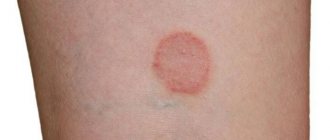
With bacterial skin diseases, hair loss, the appearance of ulcers, scars, hairless areas, spots, and scabs on the body are noted. Dermatological bacterial infections manifest themselves as allergic rashes and eczema. In protracted forms of the disease, inflammatory pathological processes from the superficial layers of the dermis pass into the deep structures of the epidermis.
Bacterial infections and diseases pose a particular danger to puppies, pregnant and lactating bitches.
Symptoms
The following symptoms will help recognize dysbiosis in adult dogs:
- loose stools, sometimes followed by constipation;
- mucus impurities in feces;
- change in color of stool;
- poor appetite;
- decreased activity;
- rumbling and bloating;
- weight loss.
Puppies show the same signs of intestinal microflora disturbance as adults.
Diagnosis of bacterial infections
To carry out diagnostics, based on the results of which the veterinarian can prescribe appropriate effective treatment, a series of laboratory, biochemical, serological studies, and instrumental methods are used. Conduct a visual examination of dogs with bacterial infections and palpation. Ultrasound, radiography, take into account clinical symptoms and anamnesis data.
Considering the similarity of clinical symptoms, differential diagnostics (PCR, ELISA), skin biopsy, and sensitivity testing (allergy tests) are performed. Laboratory tests of pathological material are also carried out using the culture method.
Infectious inflammation of the trachea and larynx in dogs
Inflammations of the pulmonary trachea and larynx in dogs are viral in nature, and are caused by a virus that spreads through direct contact with another dog or through human clothing and shoes. The inflammatory process occurs along with a strong cough and the release of thick sputum. The latter is especially dangerous, because a dog, if there is a lot of discharge, can simply suffocate on it. In this case, you need to urgently take the dog to a veterinary surgeon! There, the sick pet will undergo a tracheotomy, which is the insertion of a metal tube into the larynx and windpipe to allow breathing. This is also done in order to relieve the heart, which is overstrained during labored breathing. Because of all of the above, the mortality rate for herpesvirus disease is very high.
Treatment and prevention of bacterial infections
The treatment regimen and treatment methods should only be prescribed by a veterinary medicine specialist, based on the results of a comprehensive diagnosis. Treatment is aimed at eliminating the symptoms of bacterial infections, improving the general condition of sick animals, strengthening the immune system, and restoring impaired functions of organs and systems.
The duration of treatment depends on age, the form of the bacterial disease, and the severity of the infection. After treatment and the disappearance of clinical symptoms, a re-examination is required.
Dogs are prescribed antibacterial therapy, anti-inflammatory, symptomatic medications. As an additional treatment, a veterinarian may prescribe homeopathic remedies, immunomodulators, a therapeutic diet, vitamin and mineral supplements, and complexes.
To treat bacterial infections and diseases, veterinarians use complex antibacterial agents, penicillin and streptomycin antibiotics, cephalosporins, doxycycline, and clindamycin.
Given the resistance and habituation of bacteria to the effects of antibiotics, pharmaceutical manufacturers are constantly releasing new groups of effective drugs against various classes and types of bacteria.
After many bacterial infections, a specific immunity is formed in the dog’s body. Moreover, after some bacterial infections, dogs remain bacteria carriers for a long period of time.
To prevent your beloved dog from getting dangerous bacterial infections, in addition to proper, systematic care, you should not neglect preventive vaccinations. The vaccination schedule and the choice of preventive vaccines for your pet will be selected by your veterinarian.
Dog owners should closely monitor the general condition and behavior of their pets, and promptly carry out treatment against ecto- and endoparasites. When walking, do not allow your dog to come into contact with homeless people. stray animals. Do not allow your dog to pick up food, bones, or other “more dangerous” treats from the ground.
How to cure viruses in dogs?
Treatment of these types of pathologies is aimed at improving symptoms, for which rest, fluid and electrolyte replacement, non-steroidal anti-inflammatory drugs, antiemetics, probiotics and antipyretics are recommended. There are no specific medications that directly combat these viral diseases in dogs. Prevention is very effective because everyone has their own vaccine. Therefore, the best treatment in all cases is prevention.
Author of the article : Manuel F. Faneite P.
How is the disease transmitted from dog to person?
Contagious diseases of dogs can be transmitted in various ways, but the most common way is through fur. Play with the dog, pet it, and then you can pick up parasites that are transmitted through the fur. In particular, these are scabies, ringworm, and demodicosis. It is wool that becomes the main cause of human infection. Remember how many times a day you pet your dog, and how often do your children do this?
Another source of human infection from a dog is saliva. Of course - doctors say that dog saliva contains an active substance that helps blood clot. But this is if the dog is healthy. Dog saliva can contain very dangerous infectious agents - tuberculosis, helminthiasis, toxoplasmosis, leptospirosis, giardiasis in dogs, rabies. Owners simply squeal with happiness when their pet licks its face. The dog may also (as a sign of submission) lick his hands. Also, saliva remains on the toys she plays with.
Viruses and parasites can also be transmitted through food. This is a less common source of infection, but the fact remains. Of course, a normal person would not eat the food that a dog eats. But many owners have small children who do not yet understand that they cannot eat from a dog’s bowl. But in medical practice, there are cases where dogs stole food from the table and at the same time contaminated other food products. Therefore, it is strictly forbidden to eat foods that the dog has touched.
Stealing food is a favorite pastime for pets. And, in order to do their “dirty deed”, they can jump on the table, climb into the completely uncovered refrigerator. And there have even been cases when dogs opened the refrigerator themselves. Doctors recommend keeping food away from your pets, and if your pet gets creative, it's best to keep the kitchen locked.
Blood, just like food, is not such a common source of infection. But your dog's blood can get on your hands, face, and open wounds. Sometimes dogs that are sorting things out with each other can get into a fight and the owners then separate the pets.
Blood from dog fights can get on your skin. Also, during fights, one of the individuals may accidentally bite a person. But that is not all. Glass on the road, splinters and open wounds on a dog can become a source of infection. Through the same wound, an infection can enter the bloodstream, which, if handled carelessly, can infect a person.
Treatment options
Intestinal dysbiosis in dogs is not an independent disease, so before treating the syndrome, you should find out the cause of its occurrence and eliminate it.
If there is a disturbance in the functioning of the intestinal tract, first of all it is necessary to exclude infestation by intestinal parasites. The animal is dewormed twice with an interval of 2 weeks using anthelmintics (Drontal, Milbemax, Kanikvantel, etc.)
If staphylococcal or any other intestinal infection is detected, a course of antibiotics (Levomycetin, Ampicillin, Amptox sodium, etc.) is mandatory. Additionally, saline solutions and droppers are prescribed. Along with antibiotics, according to the doctor's indications, the animal is given antimycotics (Nystatin), which prevent the growth of fungi of the genus Candida. To restore damaged intestinal microflora, Hilak-Forte is prescribed.
Treatment of dysbiosis in dogs includes a set of measures aimed at:
- purgation;
- improving the functioning of the digestive system;
- restoration of beneficial microflora.
To cleanse the intestines of waste and toxins, enterosorbents (Smecta, Lignitin) are prescribed.
Enzyme preparations help improve the functioning of the digestive system. Veterinarians often prescribe Mezim and Cholenzym for this purpose.
If there is diarrhea, the dog is given decoctions of rice, chamomile or oak bark.
To restore the population of beneficial bacteria, long-term treatment with probiotics is carried out.
Diseases that can be contracted from a dog
There are two types of threats to humans that can be contracted from a dog. Any disease can affect either one type of carrier or several types. For example, fleas (Ctenocephalides canis) do not bite humans, just as human fleas (Pulex irritans) do not bite animals.
But there are parasites that are harmful to both humans and pets. Also, some helminths can travel on the same fleas. Therefore, timely removal of parasites from the dog’s body can save not only his life, but also your life. So what diseases can you get from a dog?
Tuberculosis bacillus (or Koch bacillus). A very common disease in our time, which many experience very hard, and the disease tends to be chronic. The disease itself manifests itself in the lungs and lymph nodes. This is an inflammatory process that is accompanied by swelling and a dry cough. The infection is transmitted through saliva and animal bites.
Helminthiasis (Helminths). Parasitic organisms that enter the body in the form of a cyst. The protective shell of helminths can withstand very low temperatures. And when the cyst gets into a suitable environment, it hatches into an adult, which attaches to the wall of the stomach and begins to reproduce.
Probiotic therapy
Dysbiosis in puppies and adults can be treated with human and veterinary probiotics. Among the most effective veterinary medications, doctors identify:
- Vetom 1.1;
- Fortiflora;
- Lactobifid;
- Lactoferon;
- Multibacterin Omega 10.
An alternative to veterinary medications are probiotics, which can be purchased at regular pharmacies:
- Linux;
- Bifiform;
- Bifidumbacterin;
- Bificol
- Probinorm;
- Baktisubtil;
- Lactoferon;
- Lactobifid.
The veterinarian determines which drug to prescribe after receiving the test results. The duration of therapy and dosage are also determined by the attending physician.
Prevention and treatment
If a person becomes ill with a viral infection or parasite that was transmitted through a dog, then each case must be considered separately. And while you are asking the question: “Can a person get infected from a dog?”, the disease is already taking over. The main thing is that if you or your pet have any suspicious symptoms, you should immediately consult a doctor.
To prevent this, you need to follow simple hygiene rules. Saliva, blood, fur - all these things can cause infection. Therefore, after every walk you should wash your pet. Now there are special anti-flea collars that repel or kill parasites. And, therefore, they also prevent other parasites that use fleas as “horses”.
You should also carry out regular deworming and give your pet all the necessary vaccinations so that the disease does not affect it, and then the owner himself. But there is one pitfall here. If the vaccination was carried out after the animal became infected with the virus, or was carried out at the wrong time, then the disease may manifest itself.
This method is the most effective against many parasites and viral infections. For example, shoes that we won’t hide anywhere. Shoes can contain many germs that end up on the animal after the pet has chewed or rubbed it.
After you have petted your animal, you should not grab its face or put your hands in your mouth. And even more so, cook food with these hands without first washing them. Thus, various cysts and other parasites can enter the body through dirt.
If there are children in the house
Pay special attention to children who squeal with joy when a dog appears in the house. There are several factors that increase the risk of infection. Firstly, children are much more irresponsible than adults. After contact, any adult will not rub their face or eyes, put their hands in their mouth, or take food with their hands.
But the child does not always realize what he is doing. And maybe, without washing your hands after the animal, you can take, for example, cookies or candy from the table. Also, a small child can imitate a dog while playing and put its toys in his mouth. In this case, you need to monitor the child who is playing with the pet. And teach older children to discipline and hygiene. Also, a child, unlike an adult, is less protected from diseases than an adult. This is due to the fact that his body is not yet fully formed; children usually suffer many diseases more severely.
Infectious canine hepatitis (Rubart's disease)
This disease is caused by canine adenovirus type 1 (ABC-1), and it differs significantly from adenovirus type 2 (ABC-2), which causes kennel cough. ABC-1 spreads easily, it has high preservation properties, so its pathogenic effect can pose a danger for up to 9 months. Most often, young dogs from 1.5 months to 1.5 years are susceptible to the virus.
The virus is released into the environment through the urine, feces and saliva of a sick animal, therefore the route of infection is predominantly nasal and oral.
The duration of the incubation period is from 2 to 5 days.
Severity of the disease: chronic, latent, subacute, acute, super acute.
Symptoms of Rubart's disease
The virus primarily affects hepatocytes (liver cells), so signs of hepatitis come to the fore:
fever;- anorexia;
- vomiting, diarrhea;
- polydipsia;
- apathy;
- pain when palpating the anterior abdominal wall;
- yellowness of the whites of the eyes.
The virus also infects vascular endothelial cells, so the following symptoms are observed:
- vasculitis;
- pinpoint hemorrhages;
- DIC – syndrome (disseminated intravascular coagulation);
- the formation of a “blue” eye, when the choroid of the eye and the cornea swell, which manifests itself in the coloring of the eye in a gray-blue color.
Sometimes neurological symptoms can be observed, which is associated with damage to the central nervous system and is expressed in the form of encephalitis. In hyperacute cases, the death of the animal can occur suddenly. However, most often symptoms of liver damage are observed, and all the complications arising from them, including damage to the central nervous system.
Drawing conclusions
Doctors give a clear and characteristic answer: “you can get infected with anything.” After all, a dog is a member of the family, just like your loved ones. And if someone gets sick, they isolate him and make sure that others do not come into contact with the sick person.
Therefore, if you decide to get an animal, you take full responsibility for its well-being. You will have to not only walk him, feed him and play with him, but also monitor his health. After all, any missed vaccination or carelessness can become a threat not only to your dog, but also to your entire family.
Bacteria are single-celled organisms that reproduce by division. There are “good” and “bad” bacteria. While bad bacteria cause disease and infection, good bacteria can benefit an animal's health. Pets, just like people, often contract bacterial infections, and if left untreated, these infections can lead to serious complications.
Symptoms and signs of bacterial infections:
- Fever
- Abscesses on the skin
- Wound infections
- Decreased appetite
- Cough
- Runny nose or watery eyes
- Vomit
- Infections of the skin, eyes, ears, urinary tract, or respiratory system
- Inflammation and redness
- Defeats
- Pain and discomfort
- Lethargy and weakness
Carnivore plague
It is one of the most dangerous diseases in carnivores. Even with proper and timely treatment, the infection can be fatal, and the clinical picture is varied.
Route of infection and duration of disease
The spread of infection occurs through the oral or airborne route, through the animal's bowl, the remains of its food, and through secretions. Sometimes an intrauterine route of infection is observed when the dog does not have an immune response. In this case, the central nervous system is most often affected (25%-75% of dogs have characteristic signs).
The incubation period of this disease is from 2 days to 2-3 weeks. The duration of the acute period of the disease is 2-4 weeks. Sometimes it becomes chronic, lasting up to several months.
Mortality from canine distemper is 80%-90% among puppies.
Symptoms of canine plague
Drowsiness, apathy, lethargy.- The dog is trying to hide in a dark place.
- Lack of appetite.
- Temperature up to 40-41 degrees.
- Purulent discharge from the eyes, ulcerative lesions of the mucous membrane.
- Mucopurulent discharge from the eyes.
- Impaired respiratory function, wheezing in the lungs.
- Vomiting, diarrhea, sometimes mixed with blood.
- Presence of pinpoint red rash (not always).
- Hyperkeratosis of the paw pads (keratinization) - observed at the last stage and is considered a malignant lesion.
- Signs of non-purulent meningoencephalomyelitis (symptoms of epilepsy, seizure, and other signs of damage to the nervous system are observed), and paralysis of the limbs is also observed, which indicates involvement of the spinal cord in the process. In this case, the main part of the nerve cells is already affected.
Treatment in all cases is symptomatic, since targeted therapy against plague has not been developed to this day. The best way to combat the distemper virus is timely vaccination of dogs.
Causes of bacterial infections
There are different types of bacterial infections that affect the skin, eyes, ears, urinary tract, kidneys, or nervous and respiratory systems, and they include:
Bacterial infections
skin lesions, also known as pyoderma, are typically caused by staphylococci and affect both dogs and cats. Staphylococci usually live harmlessly on the skin and cause infection only when the skin is damaged, for example by scratching. Common symptoms include yellow pustules, scabs, and redness. Fleas, ticks, and allergies weaken the local immune response. Stress, poor diet and illness can contribute to bacterial skin infections. This infection is not transmitted to people or other animals.
Streptococcal infections
are caused by streptococcus bacteria and usually affect both cats and dogs. They are most common among young and old animals, as they have the weakest immune systems. While older animals' defense mechanisms have weakened, younger animals' immune systems are not yet fully developed and strong enough to fight off infection.
Leptospirosis
caused by bacteria (spirochetes) and is highly contagious. Spreads through dirty water or soil and through the urine of animal carriers. Symptoms include fever, vomiting, lethargy, redness of the mucous membranes and conjunctiva. If left untreated, this infection can lead to kidney inflammation. Leptospirosis affects dogs much more often than cats, and regular vaccinations can prevent infection.
Actinomycosis
caused by bacteria of the genus Actinomycetes. Lesions are usually found in the oral cavity. Nocardiosis is caused by Nocardia bacteria, which are usually found in the soil. In both of these infections, bacteria enter through wounds in the skin, causing swollen lymph nodes. This infection may spread to the abdominal or chest cavity or lead to pyothorax.
Brucellosis
caused by the bacterium Brucella canis, found in dogs. This infection usually appears during the breeding season and is spread through contaminated urine, vaginal discharge from females, or semen from males. Male dogs may have fertility problems or develop prostatitis. When infected, females experience miscarriages and the birth of non-viable offspring is possible.
Bacterial dermatitis in dogs: features of pathogenesis and treatment tactics
Bacterial dermatitis in dogs occurs quite often, but only 20% of them are primary, the rest are based on some other diseases (flea allergic dermatitis, demodicosis, as well as hypothyroidism, hypercortisolism, hypo-, avitaminosis, less often diabetes mellitus or congenital immunodeficiencies ). The histological and pathomorphological picture of superficial and deep dermatitis is described, and the principles of treatment of the disease are discussed.
Bacterial dermatitis, especially local ones, are quite common in dogs. At the same time, it must be borne in mind that they are often preceded or accompanied by other diseases. Thus, according to our observations, the number of primary lesions is no more than 20% of all bacterial dermatitis, and in each such case, some other disease, most often demodicosis or flea allergic dermatitis, should be suspected as the underlying cause. Only when the underlying cause of the disease is not detected is dermatitis considered primary.
The term “primary” does not mean idiopathic. Primary - in the sense of the absence of background skin lesions, however, such dogs always have some systemic pathology that contributes to infectious skin lesions. If it is not detected, this means that we were unable to identify it: either due to reluctance to examine the dog in depth, or due to cost savings by owners who do not want to pay for additional research, or due to the insufficiency of the laboratory and instrumental equipment at our disposal. base, either due to the limited capabilities of modern veterinary science in relation to the diagnosis of these diseases. Such pathologies include hypothyroidism and hypercortisolism (spontaneous or iatrogenic), hypovitaminosis, and less commonly diabetes mellitus or congenital immunodeficiencies.
Bullous impetigo is a superficial pyodermatitis caused by staphylococci; in some cases, Pseudomonas sp. is also found in the pus. and E. coli. Unlike simple impetigo, which occurs with the presence of small pustules without the formation of erosions, this disease almost always develops against the background of hyperadrenocorticism, diabetes mellitus, hypothyroidism or other diseases and is accompanied by exhaustion and weakening of the dog’s body.
Histological examination of a skin biopsy shows that ulcers form in the thickness of the epidermis, mainly under the stratum corneum. Its rejection in the form of layers is accompanied by the appearance of extensive erosions. The hair follicles are not damaged. The superficial nature of the lesion determines the insignificance of the reaction from the mast cells located in the dermis, which is reflected in the absence of itching, and, consequently, the absence of deeper lesions, since the dog does not injure itself.
Deep pyodermatitis is characterized by damage to the entire thickness of the dermis, and sometimes by the involvement of subcutaneous fatty tissue. They can manifest as a general deterioration in the dog's condition and usually heal with scar formation. If the lesion is strictly localized, then it is possible that it is based on an infected wound. If the lesion is diffuse-focal in nature or is accompanied by damage to the skin of an entire anatomical area, then the presence of an underlying disease should be assumed. The affected skin areas with deep dermatitis usually look red and are almost always characterized by the presence of ulcers.
Histological examination of a skin biopsy is indicated in chronic cases when conventional treatment does not produce an effect, and there is a suspicion of demodicosis or other underlying skin lesions.
At the microscopic level, both in the area of the ulcerative lesion and far beyond it, extensive areas of inflammatory infiltration are determined, forming in places drainage fields. The infiltrate usually contains a mixture of neutrophilic leukocytes and plasma cells. Lymphocytes are represented to a lesser extent. A significant admixture of macrophages may indicate a possible fungal infection, especially if there is a tendency towards the formation of granulomas and the appearance of giant multinucleated cells. At the same time, there is an opinion that the macrophage reaction develops in response to the appearance of particles of degrading keratinocytes in the deep layers of the dermis. Deep dermatitis is manifested by folliculitis with the release of the inflammatory infiltrate beyond the follicles into the deeper layers of the skin, sometimes with the formation of boils, although abscesses, as such, are rarely observed. Not all authors agree with this point of view and describe abscesses with the formation of purulent fistulas that open to the surface of the skin.
In conditions of chronic inflammation, squamous metaplasia is usually observed in the sebaceous glands. Metaplasia is the persistent transformation of genetically related tissues from one to another. Traditionally, it is considered as a compensatory-adaptive reaction and is most often observed in epithelial tissues. At first glance, the epithelium of the sebaceous glands, which is considered as a multilayer cubic, has a phenotype similar to the epidermis - it is no coincidence that in case of IIIa degree skin burns, the epithelium of the sebaceous glands and other skin derivatives becomes a source of epidermal regeneration. Under conditions of aggression, a decrease in the intensity of apoptosis leads to a more significant production of kelons by mature cells, which inhibits the proliferation of basal cells and creates conditions for keratinization of epithelial cells. Epithelium with a more pronounced stratum corneum should better protect the underlying tissue. This is what happens with many chronic skin lesions, when acanthosis and hyperkeratosis of the epidermis are observed. It can be assumed that this is a typical reaction of many epithelia: respiratory in chronic bronchitis, secretory in the prostate gland in chronic prostatitis, transitional in chronic cystitis.
In cases of single long-term non-healing ulcerative lesions, histological examination reveals the growth of granulation tissue at the bottom of the ulcer without its characteristic division into layers and, in our opinion, indicates a disruption of the regeneration processes, thereby explaining the lack of the desired clinical effect.
Deep bacterial pyodermatitis is almost always accompanied by ulcerative skin defects and never by erosive ones. This appears to be a reflection of different defect formation mechanisms. In conditions of deep pyodermatitis, the vessels of the dermis are always involved in the pathological process, which leads to thrombarteriolitis, and this, in turn, is manifested by microinfarctions of the skin with subsequent formation of ulcers.
It should be borne in mind that in the development of bacterial dermatitis, figuratively speaking, it is not the bacteria that are to blame, but the animal’s body, since about 90% of all bacterial agents in such dermatitis are Staphylococcus intermedius and other microbes that are the normal microflora of the dog’s skin. Therefore, the treatment of bacterial dermatitis should consist not only of fighting bacteria, but also of eliminating the unfavorable background and maintaining the immunity and regenerative capabilities of the animal’s body.
The basic principles of therapy for bacterial dermatitis can be formulated as follows.
- Local use of antibacterial drugs can be effective only for superficial dermatitis; for deep dermatitis, systemic antibiotic therapy should be combined with local use of antiseptics, preferably in the form of warm lotions, which, in addition to the bactericidal effect, also cause dilation of skin microvessels and facilitate access of antibiotics circulating in the blood to the affected areas.
- The main microflora for such dermatitis is Staphylococcus intermedius, which is sensitive to most antibiotics of the penicillin, tetracycline and cephalosporin series, therefore, for initial use, the cheapest drug with a minimum of possible side effects should be prescribed. Eradication of the staphylococcal component can create unfavorable conditions for other bacteria included in the microbial association. If treatment directed against staphylococci does not lead to a cure, a bacteriological study should be performed to determine the sensitivity of the microflora to antibiotics and then the appropriate antibiotic should be used.
- Local use of antiseptic drugs reduces the microbial mass, reduces the antigenic and toxic load by removing bacterial breakdown products and tissues absorbed from the affected surface, facilitates the healing process, and reduces the itching and discomfort experienced by the dog. At the same time, crusts and scabs are also removed, which contributes to a more effective effect of externally used antiseptics.
- The visually observed healing and epithelization of erosions and ulcers is not yet an indicator of the animal’s recovery: palpation reveals nodular seals in the dermis. The disappearance of nodes in the dermis is not a reason to stop antibiotic therapy. According to some authors, after the disappearance of visual and palpation signs of damage, antibiotic therapy should continue for another 7-21 days, and the duration of such therapy after external normalization should be directly proportional to the period of time during which the external effect was achieved. Dog owners should be instructed that premature discontinuation of an antibiotic without a doctor's approval can lead to a relapse of the disease in the coming days.
- With deep dermatitis, usually at the beginning of treatment there is a rapid improvement, and then the process seems to freeze at one point. If no noticeable changes occur within another 2 weeks of antibiotic therapy, a biopsy is indicated. It can reveal either fibrosis at the site of inflammation - such nodes can be palpated in the skin for a very long time after clinical recovery, or ongoing inflammation, which usually acquires a granulomatous character - in this case, antibiotic therapy should be continued, or other factors that have not been reflected can be identified in the initial diagnosis (demodex, mycosis, etc.).
- During antibiotic therapy, glucocorticoids should, if possible, not be used. Being immunosuppressants, they prevent the animal’s body from resisting infection on its own, and in addition, they suppress inflammation and make it difficult to understand whether clinical improvement is a consequence of effective suppression of infection or suppression of the reaction to an infection that continues to persist in the skin.
- Relapses of bacterial dermatitis are common. If a relapse occurs 2-7 days after stopping antibiotic therapy, it means that it was stopped too early, before a true cure occurred. If a relapse occurs after a month or more, this usually indicates that the dog has some kind of systemic disease that determines the presence of an immune deficiency.
- In the treatment of deep chronic pyodermatitis, it is advisable to prescribe multivitamins and immunomodulators.
These provisions are especially applicable in the treatment of deep dermatitis. In some cases, antibiotic therapy can last up to 12 weeks (until clinical resolution of the disease) and at least another 2 weeks after visible recovery.
In the case of limited pyodermatitis (when there is no noticeable improvement within 10-14 days), surgical excision (if there are no obstacles) of the affected skin area is recommended. Firstly, experience shows the low effectiveness of therapeutic management of such limited pyodermatitis; secondly, the proposed approach saves owners money spent on antibiotics, reduces treatment time and is not accompanied by immunosuppression and intestinal dysbiosis, characteristic of long-term and intensive antibiotic therapy.
Prevention of infectious diseases in dogs
Timely vaccination will help to safely avoid most infectious diseases. Modern veterinary medicine has vaccines against diseases such as leptospirosis, infectious gastroenteritis, infectious hepatitis, plague and rabies.
At four months of age, animals must undergo mandatory vaccination against rabies, after which you will receive a certificate with the place and date of vaccination. All other vaccinations should be carried out at seven weeks of age, because it is during this time that the dog is quite capable of acquiring immunity to specific diseases. Before this, puppies are protected by antibodies that were produced by the mother's body.
The immunity acquired by dogs that have recovered from the disease is much stronger than that developed after vaccination. Therefore, throughout its life, at certain intervals, the dog should repeat vaccinations. You can see the vaccination schedule table below for information on when the next vaccination is due. It is especially important for your dog's health to undergo deworming. This is an important condition for the pet’s body to be able to develop immunity without harming its health, because the vaccine does not guarantee absolute protection.


