When breeding a breed, it is very important to follow the basic rules, since their violation can lead to serious anomalies in subsequent generations. Particular attention should be paid to cryptorchidism. To do this, you will need to find out what it is, who cryptorchids are, and which dogs have this problem more often than others.
What is cryptorchidism in dogs and how dangerous is it?
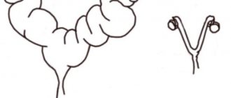
The first signs of cryptorchidism appear in puppies. At birth, their testes, or testicles, are located in the abdomen. They are connected to the scrotum by Gunther's ligament, which takes the main part in their subsequent descent.
By 6 months, it shortens and pulls the testes right into the scrotum. Under the influence of certain factors, one or both testicles may not descend even by this age. In this case, the animal experiences the following complications:
- Deterioration or complete loss of reproductive function.
The temperature inside the body reduces sperm activity. Because of this, a sick pet cannot conceive offspring.
- Tumor formation.
Overheating affects not only sperm production, but also the degeneration of healthy cells. Without timely help, the dog may die due to leydigoma, sertolioma or seminoma.
- Hormonal disbalance.
Instead of male hormones (testosterones), the body begins to actively produce female hormones (estrogens). This leads to the development of cystic prostatitis, anal gland hyperplasia and bone marrow pathologies.
- Inversion of the spermatic cord.
It is accompanied by acute pain, vomiting, nausea, bowel dysfunction, fever and chills.
The anomaly in question is a genetic disease, that is, it is inherited. For this reason, cryptorchid dogs must be excluded from further breeding.
Why does serous fluid appear?
Serous fluid usually appears for two main reasons:
- Fat deposits. During the operation, they peel off from the skin, resulting in the formation of cavities in which lymphatic fluid accumulates. It is believed that the presence of a large fat layer in a patient (more than 50 mm) is a reason for intensive weight loss before surgery or preliminary liposuction.
- The wound surface area is too large. It's simple: in this case, damage occurs to a large number of lymphatic vessels, which heal much more slowly than blood vessels. During the entire healing process, liquid oozes out of them, which accumulates in the cavities.
But doctors also consider “individual” reasons for the formation of seroma, depending on the nature of the injury.
Like post-traumatic, shin after injury
Post-traumatic seroma - for example, one that appears on the lower leg after an injury, is formed against the background of severe compression of tissue. It can be short-term, but at this time there is a deterioration or complete stop of lymph flow through the vessels. After medical assistance is provided, lymph rushes through the vessels with great force, so it immediately enters the tissues in large quantities.
Usually, after such injuries, seroma is immediately diagnosed in an advanced form. The reason for this is precisely the too large volume of lymphatic fluid poured into the cavity.
After breast surgery: mammoplasty, mastectomy
Seroma often occurs after breast surgery (mammoplasty, mastectomy), and the reason for this is simple - the mammary gland anatomically consists of glandular and fatty tissue, and the operation involves making large/extensive incisions that damage not only blood vessels, but also lymphatic vessels. The result is the accumulation of serous fluid under the skin already in the recovery period.
In 98% of cases, surgical interventions on the mammary gland end in the formation of seroma. Doctors warn their patients about this even at the stage of preparation for surgery.
Subcutaneous – the result of excessive electrocoagulation
Subcutaneous seroma is formed after electrocoagulation, a manipulation that allows blood vessels to be cauterized during surgery, which prevents the development of bleeding and most complications during the rehabilitation period. In essence, a burn occurs, in which the inflammatory process, necrosis and accumulation of fluids are inevitable.
Taking into account all the factors, doctors give a prognosis for the formation of seroma with 90% confidence. This can only be avoided by reducing the impact of electrocoagulators on tissue, but this is often impossible - the doctor works almost “blindly” and it is important for him to prevent bleeding.
After abdominoplasty, removal of hernia, appendicitis, gallbladder bed
There is a high probability of the formation of the pathological formation in question even after abdominoplasty, removal of a hernia, appendicitis and gallbladder bed, since during such surgical interventions the abdominal cavity is exposed to injury, in which not only a large number of lymphatic vessels are located, but also nodes of the same system.
Their damage is inevitable, and after stitching the wound and during the recovery/healing period, the lymphatic system simply does not have time to recover.
As a result, fluid from the vessels pours directly into the subcutaneous layers or soft tissues, and if rehabilitation is difficult, it accumulates in the cavities.
The second reason for the frequent diagnosis of seroma in these cases is the presence of both visceral and subcutaneous fat in the abdominal cavity.
Symptoms of postoperative seroma
The main symptom of postoperative seroma is the release of lymphatic fluid that occurs along the edges of the wound. It is necessarily accompanied by additional signs:
- increase in general body temperature - usually not higher than subfebrile levels, sometimes can reach 39C, which depends on the level of the patient’s immune system, the speed of the rehabilitation period, the presence of an infectious agent in the postoperative wound, the level of progression of the inflammatory process,
- pain at the site of seroma formation - it is aching in nature, can bother you for a long time, then stop for a short period of time and reappear,
- swelling of the surgical wound site - affects the nearest skin; upon palpation, puffiness of the subcutaneous layer is also felt, it is considered one of the first signs of the formation of seroma between the lipid and subcutaneous layers of the dermis,
- redness of the edges of the wound, which can spread to apparently healthy skin and have a variable shade from pale pink to distinct crimson.
The patient may also be bothered by a burning pain syndrome if he changes the position of his body and a sudden rush of blood and lymph begins to the wound. The secreted liquid has no odor, but if it is present (sour, with signs of rotting), then this indicates the addition of an infection or bacterial process.
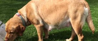
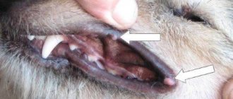
The main symptom is undescended testicle
The danger of the disease lies in the absence of pain and discomfort. Because of this, owners learn about the violation only during a routine examination by a veterinarian or when complications develop.
Despite this, a cryptorchid puppy can be recognized by its appearance. If by 6 months his scrotum is still empty, sign up for treatment.
In rare cases, the disease is accompanied by redness and swelling of the external genitalia, as well as abdominal pain. Hair may fall out in the scrotum area, and the skin adjacent to it may change color or become darker.
Prevention of seroma of the chest, pelvis, and abdominal cavity
To exclude the formation of seroma in the chest, pelvis, and abdominal cavity, doctors carry out a number of preventive measures:
- A weight is placed on the postoperative suture (immediately after the end of the surgical intervention). These could be sandbags or a heating pad with ice.
- The surgical wound is not immediately sutured, leaving a hole for installing a drainage system.
- Conducting antibacterial therapy in the early postoperative period.
- Refusal for a patient to undergo abdominoplasty if the subcutaneous fat layer is too large.
- The use of electrocoagulation is targeted, affecting only blood vessels without tension on soft tissues.
- Wearing high-quality compression garments in the early and late postoperative periods.
- Avoid physical activity for 3 weeks after surgery.
We recommend reading about complications after abdominoplasty.
From the article you will learn about local and general complications, aesthetic problems after surgery. And here is more information about rehabilitation and possible complications after lipofilling.
Seroma is not considered a dangerous complication of the postoperative period, but requires monitoring by medical professionals and therapeutic measures. With proper treatment, the problem is solved within 5-7 days; in especially advanced cases, therapy can last up to 60 days and be accompanied by severe complications such as necrosis of the skin flap, sepsis, and infection of the surgical wound.
Causes of cryptorchidism in puppies
The disease develops at the embryonic stage or shortly after birth. In the first case, the reason lies in:
- a gene passed on from the father;
- malnutrition of the mother, who did not receive the required amount of vitamins A and B9;
- anatomical features (large testis or underdeveloped scrotum, short spermatic cord, weak ligaments or too narrow inguinal canal).
In all other cases, infections of various etiologies, injuries and hormonal imbalances are to blame for the anomaly. As a result of their influence, the process of descent slows down or stops.
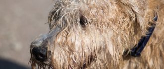
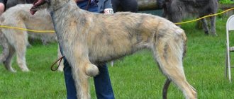
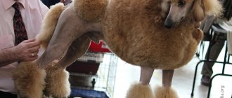
Purebred dogs get sick more often than mixed breeds. The risk group includes small and short-faced breeds: dwarf poodles and dachshunds, Yorkies, Chihuahuas, Spitz, Pekingese, bulldogs, boxers.
Types of pathology
Pathology is divided into several types. The classification is based on the time of development of the anomaly, the number and location of undescended testes, as well as possible ways to solve the problem.
Congenital or acquired
The main reason for the occurrence of a congenital appearance is poor heredity. The acquired form develops due to mechanical injuries, disturbances in the endocrine system and infection with fungi, bacteria or viruses.
Monolateral and bilateral
Monolateral, or one-sided, cryptorchidism in dogs is more common than bilateral, or two-sided. It rarely leads to infertility, since one of the testicles does descend. Sexual desire is also preserved.
With the second type the situation is reversed. Due to the stable heating of both testes, the dog loses its reproductive function and does not show interest in females.
True, false, risen
The true form is associated with anatomical abnormalities and can only be eliminated surgically. In contrast, the false one is notable for the free movement of organs between the abdominal cavity and the scrotum. After the provoking factor is eliminated, they return to their place, so surgery is not provided here.
The raised view is more unique. It occurs when the growth of the spermatic cord slows down, as a result of which the descended testicle rises back. It cannot migrate back without the help of a surgeon, so here, as with its true form, it cannot be done without surgery.
By location
The easiest way to diagnose is palpation, that is, palpation. According to the degree of palpability, pathology is divided into 2 types:
- Palpable.
Found at the top of the scrotum, on the inner thigh, or in another unusual location that can be felt.
- Non-palpable.
They can only be detected using special equipment. This is due to the size being too small, atrophy or an inconvenient location for palpation (abdominal cavity, inguinal canal). Also included here is agenesis - the congenital absence of both organs.
If you notice alarming symptoms, notice a discrepancy during examination, or confirm it manually, make an appointment with a veterinarian. After determining the type and cause of the disease, he will be able to choose an effective treatment for your pet.
What studies will confirm the pathology?
When diagnosing cryptorchidism in dogs, it is necessary to collect a detailed history, conduct a visual examination and palpation. The information obtained helps in determining the location of the unemergent testes and the truth of the disease. To do this, the veterinarian probes the entire scrotum, inguinal canal and adjacent areas, and if displaced organs are found, he tries to set them in the right place.
Taking blood for general and biochemical tests for this disease is not the main study. It is performed when infection and other conditions affecting the basic parameters of the hematopoietic system are suspected.
The main role in making a diagnosis is given to the following studies:
- Ultrasound.
Used to find non-palpable species. The results obtained are not always accurate, since they are affected by excessive intestinal gas contamination and poor quality of the apparatus.
- Laparoscopy.
The most accurate method for determining the position of undescended organs, which involves minimally invasive insertion of a laparoscope into the pelvic area. Does not require a long postoperative recovery period.
- Test with the hormonal drug Gonadotropin.
The testosterone level is measured for the four-legged patient and the drug is administered. An hour after the injection, the level of male hormone is checked again. If it turns out to be higher than the first time, the diagnosis is confirmed.
Based on the results obtained, it becomes clear whether it is worth resorting to surgery or whether conservative therapy is sufficient to eliminate the problem. When making a decision, the doctor takes into account the cause of the pathology and its type.
Prevention
You can avoid the accumulation of serous fluid in tissues by:
- Use of weights after surgery. It is placed on the seam immediately after the intervention is completed. Salt or sand can act as a weighting agent; its weight is no more than 1 kg.
- Use of traditional surgical drainage for 3 days after the intervention.
- Refusal to perform abdominoplasty if the thickness of the subcutaneous fat is more than 5 cm. Otherwise, liposuction must be performed first.
- Point impact on soft tissues during intervention. In particular, the surgeon should use electrocoagulation in isolation - only on bleeding vessels. Pressure and tension on soft tissues is not recommended.
- Use of high-quality compression garments. After surgery, you must definitely resort to such products. This helps to achieve good compression and fixation, and prevent displacement of skin and fat areas.
- Limit physical activity for 3 weeks after surgery.
Patients must adhere to all doctor’s recommendations for successful rehabilitation. This is the main way to avoid various complications, including seroma.
Treatment of cryptorchidism in dogs
The main and most effective method of treating pathology is castration. It is suitable in absolutely all cases, since otherwise the dog can pass the anomaly to its offspring or get cancer.
Does drug therapy produce results?
Drug therapy is effective in puppies under 6 months of age whose inguinal canal has not yet closed. It is also important that the pathology is palpable, and the reason for its appearance is a hormonal imbalance.
With this method of treatment, the four-legged patient is prescribed hormonal medications and massage courses. They stimulate the movement of organs in the right direction, promoting their prolapse.
Surgical prolapse of the testis
This operation is called orchiopexy. Under general anesthesia, the dog's peritoneum is cut and the testicles are lowered into the scrotum. After surgery, sperm quality improves in 30% of animals.
Despite this, the possible consequences should be understood. It is prohibited to breed a pet with this diagnosis, which is why many veterinarians refuse to perform orchiopexy.
The only convincing reason for operational omission is regular participation in exhibitions. Through surgery, you can achieve cosmetic improvements in appearance, which is very important for obtaining high grades.
Castration
Castration, or orchiectomy, involves the total removal of the reproductive organs. Depending on the location, the operation can be cavitary or non-cavitary.
Treatment
Treatment for a genetic abnormality is mandatory. The testis, located in the abdominal cavity, degenerates into a malignant formation and rapidly destroys the lungs, brain, liver, and spleen with metastases. The cause of testicular cancer is the temperature difference (up to eight degrees) between the peritoneum and the scrotum, where the physiological place of the testis is.
If the diagnosis of cryptorchidism is confirmed, castration is recommended for the animal, which has the best ratio of effectiveness to risk. Also, surgical intervention will help eliminate the risk of torsion of the spermatic cord located in the abdominal cavity.
Torsion of the spermatic cord can subsequently cause paroxysmal abdominal pain or other complications in a male dog.
Cryptorchid males are disqualified from breeding. Depending on the condition of the dog and the presence of serious contraindications to surgery, other treatment methods are used, which are conservative and do not give a 100% successful result.
These include:
- hormonal drugs, with large doses of testosterone administered to the animal, generally have a negative effect on the body;
- massage is most effective before the age of six months and when the testis is in close proximity to the scrotum.
At home, it is necessary to create adequate conditions for the pet’s recovery after surgery. In order for the animal to quickly return to an active life, it is necessary to follow all the doctor’s recommendations.
During this period it is important:
- prevent the dog from hypothermia, especially when recovering from anesthesia;
- place a diaper (oilcloth) on the dog’s resting place due to possible uncontrolled urination during the period of recovery from anesthesia;
- due to the dog’s disorientation when recovering from anesthesia, the sleeping place should be kept away from furnishings that could cause injury to the animal;
- You can feed the dog only after the effect of the drug has completely stopped;
- control that the pet does not “disturb” the seam;
- ensure the pet's drinking regime according to the doctor's recommendations;
Preoperative and postoperative care
To select a safe dose of anesthesia, a four-legged patient needs to undergo blood tests and have a cardiogram. During the postoperative period, the body's defenses are reduced, so before surgery, care should be taken for vaccination (20-30 days before) and antiparasitic treatment (10-14 days before).
You can wash the entire animal no later than one day before surgery. Water removes the protective layer, increasing the risk of infection in postoperative wounds.
You should also go on a fasting diet (12 hours before) and give up water (6 hours before). Otherwise, undigested food and water can cause vomiting, which can lead to aspiration pneumonia.
After waking up from anesthesia, the dog is returned to its owner. During the rehabilitation period you should:
- Take care to create a warm and comfortable place, protected from noise and other irritants.
- Strictly follow all doctor's recommendations for suture treatment.
- Avoid swimming before removing the suture material to avoid it getting wet and causing the wound to fester.
- Buy an Elizabethan collar or blanket that prevents wounds from licking.
- Avoid active games, long walks and contact with other animals.
If frequent vomiting, high fever, suspicious swelling, suppuration and other complications occur, you should consult a veterinarian. If the dog’s health is stable, then you should come for an examination only 2 weeks after the operation.
Complications
If detected early, seroma can be successfully treated, but in the absence of adequate correction, this condition can lead to:
- The development of infectious processes - inflammation and suppuration of the wound. A cavity filled with serous fluid is an ideal incubator for the activity of pathogenic bacteria. Moreover, they can get into the wound even with complete sterility during and after surgery. The infection easily spreads throughout the body along with lymph and blood, for example, from chronic lesions in the nasopharynx.
- Intensive growth of connective tissue at the site of intervention. As a result, a rough scar may remain on the skin, and if a seroma appears after implantation, the implant subsequently becomes deformed.
- The occurrence of a serous fistula. This condition, as we have already found out, increases the likelihood of secondary infection several times.
- Sepsis (blood poisoning).