Fecal analysis in dogs is one of the mandatory procedures when diagnosing many pathologies. Reasons for holding:
- If the body is suspected of being infected with helminths. In the feces of dogs and cats, not only the fact of the presence of parasite eggs is determined, but their species is also determined.
- Using a protozoa test, a specialist will detect endoparasites that cause diarrhea and other digestive problems.
- Conducting a virological and bacteriological study allows you to identify an infectious disease, for example, carnivore plague, parvovirus enteritis, leptospirosis.
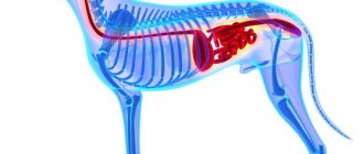
- A scatological analysis will show the presence of a pathological process in the small or large intestine and reveal hidden blood. The doctor can confirm or rule out diseases of the pancreas and liver, inflammatory and neoplastic processes in the large intestine.
A veterinarian may also recommend a stool test during a routine preventive examination of older dogs.
Preparation for delivery:
- collect the material in a sterile container for transportation. The best option would be to purchase special bottles with sealed caps at the pharmacy;
- 2-3 days before the examination, adhere to a certain diet: exclude animal feed that causes fermentation from the diet, and limit the amount of meat and fish;
- It is not recommended to undergo a scatological analysis after performing cleansing enemas, taking laxatives, or gastric lavages;
- if the dog underwent an X-ray examination the day before using a contrast agent, then the dog’s feces should be tested 2-3 days after the procedure;
- if medications are prescribed, the owner must notify the attending physician about this.
A dog's feces for scatological examination are usually collected during a walk. For this purpose, you must take with you a sterile container with a stick and gloves to maintain hygiene rules. After the dog has emptied the intestines of the contents, you should take a small amount of feces without impurities of blades of grass, dirt, sand, plant seeds, etc.
The collected material must be delivered no later than 12 hours after sampling. However, the container should be stored in the refrigerator. If stool analysis for protozoa is expected, then the time is reduced to 30 minutes , otherwise the result may be unreliable.
Decoding the consistency of stool:
- too liquid are observed with dyspepsia, ulcerative colitis;
- feces in the form of an ointment are often a symptom of impaired bile outflow;
- a mushy pattern is characteristic of dyspepsia and colitis;
- If the stool looks like sheep's beans, then the pet may have chronic constipation or colitis.
The color of feces depends on the animal’s diet, but changes in it can indicate pathological processes and diseases, for example:
- Black, tar-like stool may be a sign of life-threatening bleeding in your pet's digestive tube;
- dark brown color of feces often indicates the development of colitis in a dog, a violation of the enzymatic function of digestion and can accompany chronic constipation;
- a reddish color is observed in ulcerative colitis;
- the light yellow color of a dog’s feces may be a sign of a malfunction of the pancreas or malfunction of the small intestine;
- a grayish-white color may indicate blockage of the bile ducts with stones.
If a dog's feces smell putrid , this is a sign of pathologies such as colitis, dyspepsia, and impaired intestinal motor function. As a rule, feces become foul-smelling If fermentative dyspepsia develops, a sour odor is noted.
A general stool analysis in dogs involves determining the pH reaction. Normally, it is neutral or slightly acidic. Diseases such as colitis, constipation, and impaired secretory function of the pancreas are accompanied by a shift of the reaction towards the base. Tests have an acidic environment during the development of putrefactive and fermentative dyspepsia in dogs.
Normally, stercobilin is present in animal feces . Its increase indicates the development of anemia, and its decrease is observed in liver diseases. Bilirubin is not detected in healthy animals. Its presence indicates an increase in intestinal motor function and is often observed with long-term use of antibacterial drugs.
Soluble protein and muscle fibers are not contained in the feces of healthy pets. If protein is found in the samples, the veterinarian may suspect colitis, dyspepsia, bleeding and inflammatory processes in the digestive canal.
The detection of muscle fibers usually indicates the development of putrefactive dyspepsia, and may also accompany a disruption of absorption processes in the small intestine and a malfunction of the pancreas.
If mucus is detected , then such a symptom may indicate problems with the large intestine: constipation, colitis, neoplasms. The presence of red blood cells in the analyzes indicates that it is necessary to conduct a comprehensive examination of the large intestine and exclude polyps, tumor processes, and dysentery. Leukocytes found in a dog's feces can also indicate a malignant neoplasm in the intestines
Normally, no pathogenic microorganisms are detected in a healthy animal. In case of infectious diseases, the following bacteria can be detected in stool tests in dogs: C. perfringens, C. difficile, Salmonella spp., Campylobacter jejuni, Yersinia enterocolitica and strains of Escherichia coli. The detection of a particular bacillus in feces is not the basis for making a final diagnosis. The results of the study correlate with the clinical picture.
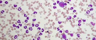
An example of a dog's stool analysis
After long-term use of antibacterial drugs, the number of enterococci, lactobacilli, and bifidobacteria is examined. Their significant decrease against the background of the detection of yeast-like fungi Candida, Proteus, Clostridia and Enterobacteriaceae indicates the development of dysbiosis in the four-legged pet.
In healthy animals, protozoan parasites are not observed in the feces. During a scatological examination, oocysts of Cystoisospora canis and Cystoisospora ohioensis, Giardia canis, as well as Cryptosporidium sp. can be detected in dogs. If pathogenic protozoa are detected, the sick animal is prescribed appropriate treatment.
An extended scatological examination includes indicators such as occult blood, hemosiderin crystals, calcium oxalate, Charcot-Leyden. The first ones appear when there is bleeding in the intestinal tube. The presence of calcium oxalate particles indicates insufficient secretory function of the stomach. With colitis and enterocolitis, Charcot-Leyden crystals appear in the stool of sick individuals.
The detection of occult blood in the studied samples indicates ulcerative processes in the stomach or small intestine, ulcerative colitis, the breakdown of neoplasms in the digestive system, if the intestinal wall is injured by helminths, or the animal has damaged the oral cavity or larynx.
Read more in our article about fecal analysis in dogs.
Reasons for stool testing in dogs
In rare cases, diagnostic measures in veterinary medicine do not require stool analysis in dogs. Fecal examination allows a specialist to answer many questions about the pet’s health. Scatological diagnosis of pets is usually carried out in the following cases:
- If the body is suspected of being infected with helminths. In the feces of dogs and cats, using various methods in the laboratory, not only the fact of the presence of parasite eggs is determined, but also their species is determined.
- In addition to helminth infections, by testing your dog's stool for protozoa, your veterinarian will detect endoparasites that cause diarrhea and other digestive problems in the animal.
- Conducting a virological and bacteriological examination of an animal’s feces allows a veterinarian to identify an infectious disease, for example, canine distemper, parvovirus enteritis, and leptospirosis.
- A scatological analysis will show the presence of a pathological process in the small or large intestine and reveal hidden blood. By examining a number of indicators, the attending physician can confirm or exclude diseases of the pancreas and liver, inflammatory and neoplastic processes in the large intestine.
A veterinarian may also recommend a scatological examination during a routine preventive examination of elderly dogs.
We recommend reading about why your dog has bloody stool. From the article you will learn about the reasons for the appearance of blood in feces during normal stools, diarrhea if vomiting occurs, diagnosis of the condition and treatment.
And here is more information about the causes of diarrhea and how to eliminate it in dogs.
Helminthological and bacteriological research
Helminthological research
is carried out to detect helminth eggs, protozoan cysts, segments, heads and other parts of adult parasites in the feces.
The most important and widely used is microscopic examination of stool for helminth and protozoan eggs. The protozoa most commonly found in our climate zone include Giardia, but other protozoa are also found. This is one of the most common causes of chronic diarrhea in dogs. It is important that the cycle of different protozoa differs, and their oocysts cannot always be detected in feces. Therefore, if nothing was found during the first study, this does not mean that there are no protozoa. A negative diagnosis is made after at least 3 negative samples taken at different (depending on the type of protozoa we suspect, most often 3 days) time intervals. The results of the treatment are also assessed - after a course of taking medications, feces are collected three times to make sure that all the protozoa have died.
Bacteriological research
carried out to establish dysbiosis or detect pathogenic bacteria when intestinal infections are suspected or to make a diagnosis of systemic infectious diseases. Dysbacteriosis is not currently an accepted diagnosis, but in some cases an overgrowth of various forms of bacteria in the intestine can be detected. most often this occurs against the background of a primary pathology, so diagnosis and treatment will depend on the primary problem.
A coprogram is a comprehensive study of human feces, its physical, chemical properties and various inclusions.
The coprogram allows you to evaluate the functioning of the gastrointestinal tract, detect dysfunction of the liver and pancreas, identify helminths in the intestines or diagnose inflammatory changes in the digestive tract.
Preparation for delivery
The correct interpretation of the results depends on proper preparation of the animal for the study. First of all, the owner must take care of a sterile container for transporting the dog’s excrement. The best option would be to purchase special bottles with sealed caps at the pharmacy. Their design makes it possible to collect the material under study and accompany it with the appropriate inscription.
2-3 days before the expected examination, veterinary specialists recommend following a certain diet. First of all, animal feed that causes fermentation should be excluded from the diet and the amount of meat and fish should be limited.
It is not recommended to undergo a scatological analysis after performing cleansing enemas, taking laxatives, or gastric lavages. If the dog underwent an X-ray examination the day before using a contrast agent, you should wait and have the dog’s feces tested 2-3 days after the procedure. If the animal is prescribed any medications, the owner must notify the attending physician about this.
A dog's feces for scatological examination are usually collected during a walk. For this purpose , you must take with you a sterile container with a stick and gloves to maintain hygiene rules . After the dog has emptied the intestines of the contents, you should take a small amount of feces without impurities of blades of grass, dirt, sand, plant seeds, etc.
After the tests are placed in containers, care must be taken that they are delivered to the veterinary institution no later than 12 hours after sampling. In this case, the container should be stored in the refrigerator.
If stool is to be analyzed for protozoa, the time from sample collection to laboratory examination is reduced to 30 minutes. This is due to the fact that vegetative forms of protozoa are quickly destroyed in the environment, and the result may therefore not be reliable.
Watch this video about preparing your pet for a fecal test:
What to do if there is blood in your dog's stool?
First and most importantly, do not self-medicate. A variety of reasons can cause bleeding, so it is impossible to identify them at home. Not only will you not help your pet, but you will also aggravate an already difficult situation.
Therefore, if you find blood in your stool, seek professional help immediately. Before coming to the veterinarian, prepare answers to several questions:
- What did you feed your dog for the last 2-3 days?
- Could your pet pick up an inedible object on the street or at home?
- Have similar situations happened before?
- Do you give your pet any medications?
If possible, take a photo of bloody stool. Do not throw away the stool; prepare it for laboratory testing. This will significantly save your time and allow the veterinarian to take all necessary measures in a timely manner.
If contacting a specialist is delayed for certain reasons, exclude dry food and solid food from the dog’s diet. This way you will be sure that it does not injure the mucous membranes of the gastrointestinal tract.
Under no circumstances should you apply cold/heat to your dog’s belly. Do not prescribe medications yourself. And most importantly, don’t panic. Drops of blood in the stool can be disposable and self-limiting, but, of course, this does not exclude the need for a full and professional examination.
Decoding the results
As a rule, in veterinary diagnostics, the physicochemical parameters of animal feces are examined and a microscopic examination is carried out.
Total information
First of all, the laboratory pays attention to such general parameters as the color and consistency of stool, and smell. Too liquid feces are observed with dyspepsia and ulcerative colitis. Feces in the form of an ointment are often a symptom of a violation of the outflow of bile. A mushy pattern is characteristic of dyspepsia and colitis. If the stool looks like sheep beans, then your pet may have chronic constipation or colitis.
The color of feces depends on the animal’s diet, but changes in it can indicate certain pathological processes and even diseases. For example, black, tar-like stool may be a sign of life-threatening bleeding in your pet's digestive tube. The dark brown color of feces often indicates the development of colitis in a dog, a violation of the enzymatic function of digestion, and can accompany chronic constipation.
A reddish color is observed in ulcerative colitis. The light yellow color of a dog's feces may be a sign of a malfunction of the pancreas or malfunction of the small intestine. If the stool is greyish-white in color, your veterinarian may suspect a stone blockage in the bile ducts.
The reason why a dog's feces smell putrid is due to pathologies such as colitis, dyspepsia, and impaired intestinal motor function. As a rule, feces become foul-smelling due to diseases of the pancreas or blockage of the bile ducts with stones. If fermentative dyspepsia develops, veterinary specialists note a sour odor from the test sample.
A general stool analysis in dogs involves determining the pH reaction. Normally, a neutral or slightly acidic reaction is observed. Diseases such as colitis, constipation, and impaired secretory function of the pancreas are accompanied by a shift of the reaction towards the base. Tests have an acidic environment during the development of putrefactive and fermentative dyspepsia in dogs.
Stercobilin is normally present in animal feces. Its increase indicates the development of anemia, and its decrease is observed in liver diseases. Bilirubin is not detected in healthy animals. Its presence indicates an increase in intestinal motor function and is often observed with long-term use of antibacterial drugs that suppress the natural microflora of the body.
An example of a dog's stool analysis
Soluble protein and muscle fibers are not contained in the feces of healthy pets. If protein is found in the samples, the veterinarian may suspect colitis, dyspepsia, bleeding and inflammatory processes in the digestive canal. The detection of muscle fibers usually indicates the development of putrefactive dyspepsia, and may also accompany a disruption of absorption processes in the small intestine and a malfunction of the pancreas.
If mucus is found in your pet’s stool, which is absent in a healthy animal, then such a symptom may indicate problems with the large intestine: constipation, colitis, neoplasms. The presence of red blood cells in the analyzes indicates that it is necessary to conduct a comprehensive examination of the large intestine and exclude polyps, tumor processes, and dysentery. Leukocytes found in a dog's feces can also indicate a malignant neoplasm in the intestines.
If you have been tested for bacteria
Normally, during a scatological examination, no pathogenic microorganisms are detected in a healthy animal. For infectious diseases, stool tests in dogs may reveal bacteria C. perfringens, C. difficile, Salmonella spp., Campylobacter jejuni, Yersinia enterocolitica and strains of Escherichia coli. However, the identification of a particular pathogenic bacillus in feces is not the basis for making a final diagnosis. The results of bacteriological examination in this case correlate with the clinical picture.
Feces for dysbacteriosis in a dog
After long-term use of antibacterial drugs, with an appropriate clinical picture, a veterinarian prescribes a dog stool test for dysbacteriosis. At the same time, the number of enterococci, lactobacilli, and bifidobacteria is examined. Their significant decrease compared to normal values against the background of the detection of yeast-like fungi Candida, Proteus, Clostridia and Enterobacteriaceae indicates the development of dysbiosis in the four-legged pet.
If you have been tested for protozoa
In healthy animals, protozoan parasites are not observed in the feces. During a scatological examination, oocysts of Cystoisospora canis and Cystoisospora ohioensis, Giardia canis, as well as Cryptosporidium sp. can be detected in dogs. If pathogenic protozoa are detected, the sick animal is prescribed appropriate treatment.
If an extensive stool test was performed
In some cases, a veterinarian may prescribe an extensive scatological examination. Decoding the analysis of dog feces includes indicators such as occult blood, hemosiderin crystals, calcium oxalate, Charcot-Leyden. Thus, hemosiderin crystals appear during bleeding in the intestinal tube.
The presence of calcium oxalate particles indicates insufficient secretory function of the stomach. With colitis and enterocolitis, Charcot-Leyden crystals appear in the stool of sick individuals.
The detection of occult blood in the studied samples indicates ulcerative processes in the stomach or small intestine, ulcerative colitis. Hidden blood in the stool can be due to the breakdown of neoplasms in the digestive system. In the event that the intestinal wall is injured by helminths, or the animal has damaged the oral cavity, larynx, hidden blood can also be detected in the feces.
We recommend reading about the signs of colitis in dogs. From the article you will learn about the causes and types of colitis in dogs, symptoms of the disease, diagnostic methods, treatment with diet and medications.
And here is more information about the symptoms and treatment of stomach ulcers in dogs.
A scatological examination helps a veterinary specialist understand the processes occurring in the digestive system of a furry patient. A general stool analysis is included in a comprehensive diagnostic examination of the animal. If necessary, an extended analysis can be prescribed, which includes the detection of occult blood, dysbacteriosis, and also allows one to exclude protozoal invasions and confirm a bacterial infection.
Therapeutic diet
If the cause of blood in your pet’s stool is improper feeding, frequent constipation or diseases of the gastrointestinal tract, then a specialist will prescribe a diet. Strictly adhere to the rules indicated by him, since dietary food is selected to suit the specific needs and characteristics of the dog.
Basic principles of a therapeutic diet:
- It is necessary to feed your pet in small portions, but at short intervals. Thus, the amount of food remains the same, but it is consumed at different periods of time. It is desirable that the intervals between feedings be the same (for example, 2 hours);
- Avoid low-quality dry food. In the first 5-10 days, give your pet low-fat meat broth, cottage cheese, rice with chicken and other foods with minimal fat content. This will reduce the load on the digestive system;
- Make sure your dog always has clean drinking water.
Useful video
Watch this video about what blood in a dog's stool indicates:
Similar articles
- Cholecystitis in dogs: symptoms, acute and chronic...
There are few reasons why cholecystitis occurs in dogs. It can be acute or chronic. Symptoms manifest themselves in the form of belching and gastrointestinal disorders. The treatment regimen is prescribed individually. What to feed, is it necessary and what diet... Read more - The dog has bloody stool: the main reasons if...
If your dog has bloody stool, this is a serious cause for concern. And it doesn’t matter whether it’s normal or liquid, with vomit and mucus, or minor discharge after. You should definitely show the animal to a doctor. Read more
- Candidiasis in dogs: symptoms, damage, incl. skin...
With weakened immunity, candidiasis can develop in dogs. The main symptoms of the lesion appear on the mucous membranes and skin. Treatment includes medications and a low-carbohydrate diet. Read more
- Nephritis in dogs: causes, symptoms, treatment, including...
A pathology such as nephritis in dogs requires emergency assistance from a specialist. After all, without treatment it can easily turn into a chronic form, which is not at all easy to cope with. And in order to consult a doctor in time, it is worth knowing the symptoms. Read more
- Cushing's syndrome in dogs: causes, symptoms, tests...
If there are problems with the adrenal glands or pituitary gland, Cushing's syndrome in dogs can develop. ... The symptoms are similar to many diseases; only a comprehensive analysis and diagnosis will help identify and select treatment. Read more
Blood chemistry
A biochemical blood test is no less important than the general one. The indicators will tell you about the functioning of many internal organs - kidneys, liver, gall bladder.
Elevated levels of alanine aminotransferase and aspartate aminotransferase will indicate that liver cells have been damaged. An increased amount of creatinine and urea is detected when the excretory system is disrupted. Based on biochemistry data, the doctor will determine the amount of protein compounds and mineral elements in the animal’s body. This study is done on an empty stomach; you cannot feed the animal for at least 8 hours before the manipulation. In some cases, the procedure is carried out before and after feeding.
Prevention
- Anthelmintic treatment. Typically, anti-parasite medications are given once every 3 months. However, if you notice worms in your pet, then treatment must be carried out 2 times, and the interval between them should not be more than 10-14 days;
- Keep an eye on your pet's stool. This will allow you to timely detect the problem and seek professional veterinary help to avoid complications and other dangerous consequences;
- Ensure proper feeding. A dog's diet should contain protein, fiber and fats. It is important that the amount of food and frequency of feedings correspond to the weight of your pet. Make sure he doesn't overeat and don't give your dog sharp bones;
- Special diet. This is an effective method of prevention, but only if the animal has a predisposition to diseases of the digestive tract. Therapeutic diets maintain healthy microflora and help avoid bleeding;
- Walks. Take extra care when walking your dog. It is important that the pet does not pick up anything from the ground: it can easily swallow small foreign objects that damage the mucous membranes of the stomach;
- Routine vaccination. Once a year, go to the veterinary clinic for a routine examination and antiviral vaccination. In the future, this will avoid dangerous viral consequences, the symptoms of which may be blood and mucus in your pet’s stool.
Remember that animals need professional veterinary care if they have blood in their stool. We strongly do not recommend self-medication, because the health of your dog depends on how promptly you contact the veterinary clinic!
Pets are prone to gastrointestinal diseases. Especially common in puppies under one year old. These may be internal non-contagious diseases, manifested by indigestion; infectious, caused by viruses and bacteria; invasive, caused by protozoan parasites and helminths.
Isosporosis is one of the diseases of the gastrointestinal tract that is caused by the parasite Isospora. This is a protozoan that infects the intestinal mucosa. It manifests itself as diarrhea, weakness, perverted appetite, and death.
Several species of isospores and coccidia can live in the intestines of dogs. However, only two cause the disease. These are isospora canis (canine isospore) and isospora felis (cat).