Anatomical parts of a dog's body
Features of the location of various parts of the body, physique and general appearance of the dog in accordance with their breed characteristics are called exterior. To assess the exterior, several parts of the dog’s body are anatomically distinguished:
- Head. The skull and muzzle, eyes, ears, and dental system are assessed.
- Neck.
- Torso. Along the top line look at the withers, back, loin, croup and tail. The chest and abdomen are assessed along the bottom line.
- Limbs. Presented front and rear.
Knowledge of exterior features is especially necessary for owners of purebred dogs. It helps control, preserve and develop dog breeds.
Skeletal system
The study of anatomy must begin with a consideration of the skeletal system. The skeleton is the bony basis of a dog's body. The development and productivity of the dog’s body as a whole depends on its condition. The skeletal system, together with joints, ligaments, muscles and tendons, makes up the musculoskeletal system. There are axial and peripheral sections of the skeletal system.
Axial section of the system
The structure of the axial skeleton includes:
- Scull.
- Spinal column.
- Chest with ribs.
Skulls are either dolichocephalic (long) or brachycephalic (short). The first type is typical for the breeds of shepherd dogs, Dobermans, collies, the second type of skull is for Pekingese, pugs, and bulldogs. A dog's skull has a cranial and a facial (muzzle) part. The bones of the skull, with the exception of the lower jaw, are fixedly connected. The mobility of the lower jaw is due to the need to grasp, hold and chew food. The dental system actively participates in this process. Adult dogs have 42 teeth, puppies have 28. There are incisors, canines, premolars and molars. The puppies are missing molars and one premolar.
Depending on the breed characteristics of closure, the front teeth (incisors) form a certain bite. The most preferred and most often mandatory for most breeds is the scissor cut, in which the upper incisors fit tightly behind the lower ones. In a straight bite, which is acceptable for some breed groups, the surfaces of the incisors are brought together by the cutting edges. Underbite is manifested by a strong protrusion of the upper jaw in front of the lower jaw, so that a large gap is formed between them. Underbite is characterized by protrusion of the lower jaw, causing the lower incisors to protrude in front of the upper ones and is a characteristic feature of breeds with a short muzzle.
The spinal column of a dog consists of seven cervical, thirteen thoracic, seven lumbar, three sacral and several caudal vertebrae.
The cervical region consists of seven cervical vertebrae, which begin with the first, the atlas, and the second, the epistrophea. The skull is attached to them and they allow the dog's head to be movable in different directions.
The thoracic region is represented by thirteen vertebrae, to which curved ribs of varying lengths are attached. The first four pairs of ribs close into the costal arch, the remaining nine pairs are shortened towards the lumbar region and bend freely. The ribs serve as protection for the dog’s internal organs and are involved in the breathing process.
The lumbar region consists of seven segments . The loin should not be long - this is considered a big drawback. Ideally, it is desirable to be short, convex and wide, reliably connecting the thoracic and pelvic spine and capable of acting as a spring. A long loin greatly affects the dog’s movements, the gait becomes slack, and the rear begins to wag.
Typically, dogs have 20–23 caudal vertebrae. There are also smaller numbers. To meet the standard, in some breeds the tail vertebrae are cut off (cropped), leaving several segments.
Peripheral skeletal system
The section is represented by the front and hind limbs of the dog.
The forelimb consists of a shoulder blade, preferably set obliquely, to which the humerus is attached through the glenohumeral joint. The shoulder is connected through the elbow joint to the bones of the forearm, consisting of two bones - the ulna and the radius. For most breeds, it is highly desirable that the lowest point of the costal arch reach or be below the elbow joint . Chest depth is one of the important parameters of the exterior. A fairly deep chest, with a moderate width, creates the basis for good development of the internal thoracic organs: heart, lungs, blood vessels.
The wrist joint consists of seven bones that connects the bones of the forearm to the five bones of the metacarpus. The forelimb ends with fingers, each of them is equipped at the end with a hard claw that cannot retract. Four fingers have three phalanges, and one has only two.
The forelimb is attached to the vertebral skeleton by very strong shoulder muscles. The protrusion of the obliquely set shoulder blade, rising above the thoracic vertebrae, creates a prominent withers. The measurement from the highest point of the withers to the ground of a dog standing calmly is a very important conformation parameter and is called “height at the withers” for evaluation. Depending on the accepted breed standard, the height at the withers has a different meaning. The fluctuation in height at the withers in various breeds sometimes simply amazes the imagination with the wonders of the selection work of breeders and breeders. So great is the difference in height between a miniature lap dog and the giants of the canine world, Great Danes and wolfhounds - from 6.5 cm to 111.8 cm in height at the withers.
The structure of the dog's body
Many dog breeders are interested in the question: what is the topography of internal organs in dogs? A dog is a mammal. Their skeleton has a typical structure and the same sections.
Mammals have 7 cervical vertebrae. Giraffes, which have very long necks, and whales, which have no necks at all, have the same number of vertebrae as dogs.
The thoracic vertebrae (depending on the breed of dog, their number varies from 12 to 15 pieces) together with the ribs and sternum are part of the chest.
The lumbar spine, which is responsible for protecting and fixing the dog’s internal organs (photo can be seen in this article), consists of six movable vertebrae. The sacral spine consists of 4 vertebrae connected to the pelvis.
Depending on the breed of the dog, the number of vertebrae in the tail of the skeleton can vary from three to several dozen. The length of the tail depends on the number of bones.
The skeleton of the forelimbs consists of two shoulder blades, which are connected by crow bones and two clavicles.
The supporting part of the hind limbs consists of the pelvic bone, which was formed from three fused bones. It is noteworthy that many mammals, including dogs, have well-developed muscles of the pelvis and limbs.
Internal structure of a dog's body
The internal organ system consists of the digestive, respiratory, excretory and reproductive systems.
Digestive
Its main purpose is to consume , promote, digest, assimilate food and water. Starting in the mouth with the teeth, it passes into the esophagus, which is adjacent to the stomach. In the stomach, food and water are mixed and, with the help of released hydrochloric acid, are broken down into nutrients (digestion process). Moving further, the food bolus enters the duodenum of the intestine.
The intestine is the main organ for further digestion and absorption of broken down particles - nutrients. The pancreatic secretions and bile, the pancreas and the liver and gall bladder, respectively, open their ducts into it and secrete the substances necessary for digestion. The intestinal section is very long, its length is from two and a half to seven meters. The intestine is divided into the small and large intestine, which ends in the anus.
Respiratory
The respiratory system is designed for gas exchange in the lungs. Oxygen enters the blood from the air, and carbon dioxide is removed back. By contracting and relaxing, the rib muscles cause the lungs to contract to remove carbon dioxide and inflate to suck in oxygen. The respiratory system consists of the nasal and oral cavities, larynx, trachea and lungs.
excretory
The system is represented by two kidneys with a ureter, bladder and urethra. The end products of metabolism from the blood in the kidneys pass through filtration into urine, which is collected in the bladder through the ureters and periodically removed from the body through the urethra.
Reproductive system
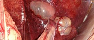
The organs of the reproductive system serve for procreation. Their structure is different in different sexes. In males, it includes the testes located in the scrotum, the vas deferens, and the penis, covered by the prepuce . In bitches, the reproductive organ system is internally located in the body and consists of the ovaries, fallopian tubes, uterus, vagina and external genitalia.
Types of fabrics
There are 4 main types of tissue in a dog's body:
- Integumentary. They form the outer layer of the skin and are also found on the inner surface of the nasal and oral cavities. They also envelop many internal organs, such as the esophagus, intestines, bladder and stomach. Integumentary tissues are needed not only to ensure reliable protection of vital organs. They also carry out metabolism, produce gastric and intestinal juices, saliva and tears.
- Support-trophic tissues. This type includes lymph, blood, fat layers, cartilage, bone and connective tissue. They differ significantly in structure and purpose, but their most important purpose is to create the skeleton of the entire organism. They are also responsible for connecting all organs to each other. One type of musculoskeletal tissue performs a protective function, preventing damage to blood vessels, nerves and other vital organs.
- Muscle tissue provides motor functions. They allow the animal to move and carry out the work of internal organs.
- Nerve tissues make up the dog's nervous system. It controls the functioning of organs, and also receives signals received from the external environment, processes them, and then sends information to the brain.
All of the above tissues are the so-called “building material” for organs. Typically, the predominance of one type of tissue in an organ determines its function. Thus, the brain contains the most nerve tissue.
Each organ in a dog’s body performs its primary function, but they all also perform secondary tasks, which are also important for the normal functioning of the body.
The main function of the skeletal bones is to allow the dog to move, but in addition to this function, the bones allow the body to be filled with nutrients and also provide blood flow to the limbs.
The animal's skeleton is involved in carbohydrate, protein, fat and mineral metabolism.
Management of the entire body as a whole
To control all body systems, the nervous, circulatory, immune, lymphatic, hormonal, skin and sensory systems are used.
The system is divided into central and vegetative. It consists of nerve fibers. Thanks to its high development, dogs have enhanced senses such as smell, vision and hearing. The central nervous system, together with the cerebral cortex, through congenital and acquired reflexes during life, regulates all systems of the dog’s body.
Blood
The cardiovascular system includes the heart and blood vessels: arterial, coming from the heart, and venous, coming to this organ. The main arterial vessel is called the aorta. The cardiovascular system is designed to supply all organs and cells of the body with oxygen and nutrients and remove waste products of metabolism. The location of the heart is the chest. It is located on the left side of it.
Sense organs and skin
External and internal influences are perceived and analyzed by the senses. A dog has five senses: visual, auditory, olfactory, gustatory and tactile. The optic eye consists of the eye with the pupil, eye muscles and nerves.
The auditory analyzer includes an ear, the structure of which is such that it not only perceives vibrations of sound waves, turning them into sound, but also has the function of correct orientation in space - balance. The sense of smell in dogs is very developed, its acuteness depends on individual characteristics and training. Taste buds are located on the dog's tongue and serve to analyze the composition and quality of substances that enter the mouth.
The cutaneous organ of touch represents primarily a barrier between the external environment and the internal system of the dog’s body. The tactile function protects the organs from adverse effects. Skin composition:
- Subcutaneous tissue.
- Epidermis.
- Wool is a derivative of leather.
Knowledge of the anatomy of the canine body allows us to better understand the reasons that prompt our pets to behave in one way or another.
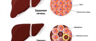
Treatment of ascites in dogs
The most important thing, of course, is to treat the underlying disease that has led to the accumulation of fluid in the dog's abdominal cavity. To properly treat the underlying disease, it is necessary to make an accurate diagnosis. Elimination of ascites, as such, is a fight against the consequence, not a treatment, but it can be a vital measure, and sometimes, unfortunately, practically the only possible measure, albeit temporarily, but radically improving the condition of a seriously ill animal. Such events could be:
- Therapeutic abdomenocentesis to remove large amounts of fluid from the abdominal cavity, especially when there is a lot of fluid and it creates compression of the internal organs and diaphragm, making breathing difficult.
- Diuretics (diuretics) to increase fluid excretion.
- Oxygen therapy for animals with breathing problems.
- Intravenous fluids in case of dehydration or shock.
- Blood transfusion.
- Antibiotic therapy if there is an infectious process.
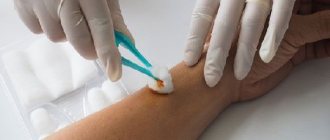
Suction with drained ascites fluid
It is important for owners of a pet with ascites to understand that the accumulation of fluid in the abdomen is a very serious sign, indicating that the pet needs urgent qualified help.
Features of the anatomy and physiology of dogs
Skeleton of a dog The skeleton plays an important role in the life of the body. It serves as a lever of movement, support for soft parts of the body, protection, a place for the development of hematopoietic organs, and also participates in metabolic and biochemical processes in the body. The skeleton of a dog is unique in its structure. Distinctive features of the skeletal system are strength and lightness compared to other tissues. Young animals have more elastic bones than older ones. As we age, bones become more brittle. The skeleton consists of 247 bones and 262 joints.
The skull consists of the facial and cranial bones. The lower jaw is attached to the skull by a joint, driven by powerful masticatory muscles. The upper and lower jaws contain teeth. An adult dog has 42 teeth, while puppies have 28 (up to 32) baby teeth. Sometimes in a set of teeth there are fewer teeth developed than 42 (oligodontia), sometimes there are more teeth (hyperdontia).
The thoracic limb begins with the scapula, then the humerus, forearm, wrist (7 carpal bones), metacarpus (5 metacarpal bones). The fingers are equipped with strong, non-retractable claws at the end.
The pelvic (hind) limb begins with the femur, passes into the tibia (tibia and fibula), then into the tarsus (consists of 7 bones). This is followed by the metatarsus (of 4-5 metatarsal bones), then 4 phalangeal toes, ending in claws. Sometimes a rudimentary (dewclaw) finger grows from the inside. It is usually amputated at a young age. The pelvic limb has an articular connection with the pelvis and is fixed by the muscles of the hip group.
Dog bones
The Dog Anatomy Atlas includes the skeleton of this animal. It consists of 29 types of bones. All of them are listed below:
- Upper jaw.
- Lower jaw.
- Thoracic vertebrae.
- Skull box.
- Metatarsus.
- Parietal bone.
- Cervical vertebrae.
- Lumbar vertebrae.
- Occipital protuberance.
- Tail vertebrae.
- Brachial bone.
- Bones of the forearm.
- Carpal bones.
- Pastern.
- Phalanges of the fingers.
- Shoulder blades.
- Ribs.
- Hip joint.
- Femur.
- Knee-joint.
- Sternum.
- Rib cartilage.
- Tibia.
- Hock joint.
- Tarsus.
- Fingers.
- Tibia.
- Hip bone.
- Heel bone.
Skin system
The skin that covers a dog's body consists of three layers: the epidermis, the skin itself and the subcutaneous tissue. The skin itself contains hair follicles, sweat, aromatic and fat glands with capillary vessels and nerve endings. The subcutaneous layer contains adipose tissue. Tufts of hair grow at the cuticle, each of which contains 3 or more thick and long hairs (guard hairs), which form the outer coat, and 6-12 short, delicate hairs (undercoat).
Wool
The fur covers almost the entire body (with the exception of the nose, toe pads and weak hair on the scrotum in males). Above the eyes, on the cheekbones, temples and upper lip there are long and very stiff hairs (tactile tentacles). In the spring it is subject to molting, and in the fall it grows warmer fur.
Sweat glands
Sweat glands are located in the skin of the paws, this is where sweat is secreted. That is why the dog does not sweat throughout the body, and evens out temperature deviations by accelerated breathing through an open mouth and evaporation of fluid from the oral cavity.
The skin also contains scent glands that produce the characteristic dog scent.
Dog body structure
Every owner who has a dog at home must have minimal knowledge about the structure of the body of this animal. This will help to promptly detect symptoms of a dangerous disease in your dog. In most cases, the life and health of their pet depends on the person. In addition, knowledge about the structure of a dog’s body will help the owner understand many of the animal’s behavioral features. This is especially important when a person wants to raise a small puppy to be an assistant in business, for example, in hunting or for guarding the house. It is necessary to raise a dog from the first months of life.
The dog's body consists of external and internal organs. All parts of the body are closely connected with each other, the work of each is directly dependent on each other.
Each organ consists of tissues that ensure its functioning. They are cells of various shapes, intercellular substance and fibers. Cells are the smallest structures in an animal's body; their shape and structure depend on their purpose. Cell size varies from 10 to 100 microns.
Dog nervous system
Dogs, like all mammals, have a developed nervous system and well-developed sensory organs.
Dogs' sense of smell is 48 times more acute than that of humans (these creatures naturally have more than 200 million olfactory cells; humans have only 5 million). It serves to search for food, navigate in space, is one of the means of communication, etc. A dog’s life is a world of smells.
Hearing also plays an important role in the life of canines. The upper threshold of hearing is almost 5 times higher than that of a person, which allows her to distinguish ultrasound.
Vision is less developed than smell and hearing. However, some breeds (for example, greyhounds) are characterized by greater vigilance: they are able to distinguish objects at a distance of up to 150 m. Dogs distinguish colors less clearly, but see better in the dark. They see the outlines of objects less clearly, but their field of vision is 50% wider than ours.
Touch. Tactile or tactile hairs (vibrissae) serve as organs of vision in dogs. They grow on the lower jaw, above the upper lip (mustache), above the eyes, on the neck. With their help, the animal navigates in the dark and determines the direction of the wind.
The taste organs are represented by taste buds located on the tongue.
Symptoms of ascites in dogs
The following symptoms are not necessarily always present, but may indicate ascites:
- Increased abdominal volume;
- Difficulty breathing or breathing with effort, since a significant amount of fluid can put pressure on the diaphragm and prevent it from participating in the act of breathing;
- Pain in the abdominal area;
- Lethargy of the animal, often the weight and volume of fluid is so great that it is simply difficult and difficult for the dog to move;
- Cough (rare);
- Vomit. Often it may not be a direct consequence of ascites, but may be one of the symptoms of the underlying disease along with abdominal dropsy;
- Fever. Inflammatory processes in the abdominal cavity, leading to ascites, often cause an increase in temperature;
- Anorexia (lack of appetite);
- Cachexia (wasting). Together with a large amount of water, a large amount of protein usually comes out into the ascites fluid, which is why the dog becomes thin;
- General weakness.
Reproductive system
The female reaches sexual maturity at 6-11 months (first heat). The maturation period for a female puppy usually varies depending on the breed. The maturation period for very large breeds can take up to 15-16 months. By 15-16 months, male dogs have completed puberty.
The optimal period for the first mating for a bitch is 1.5-2 years. Fertilization occurs over a period of several days, called estrus, after about day 10, when spotting ends. The best period is considered to be from 10 to 12 days after the start of estrus.
Females come into estrus 2 times a year (with an interval of 6 months); its duration can be up to 28 days (average - 14 days). Pregnancy (puppy) in dogs lasts 59-65 days. In a litter, as a rule, there are from 2 to 6 puppies.
Diagnosis of ascites in dogs
Detecting fluid, as such, in the abdominal cavity of a dog is not a difficult task. If ascites has developed significantly, then the enlargement of the abdomen is visible to the naked eye, and to the touch it is clear that there is fluid in the abdomen. To confirm ascites, a doctor at the clinic may do an ultrasound. In cases where there is little fluid, without ultrasound examination it may be impossible to say with certainty whether there is ascites. In addition, an x-ray can show the presence of fluid in the abdominal cavity. During radiography, the fluid creates the so-called ground glass effect - those organs that are usually clearly visible on an x-ray are not visualized at all or poorly visualized with ascites. The abdominal cavity appears as a gray spot. Therefore, an x-ray is taken after the fluid is drained.
In order to find out the cause of fluid accumulation in the abdominal cavity, the following studies are indicated:
- Detailed blood test - clinical and biochemical;
- Analysis of urine;
- X-ray of the abdominal and chest cavities;
- Abdomenocentesis (puncture of the abdominal wall), fluid removal and its analysis - cytological, bacteriological;
- Ultrasonography;
- Echocardiography (ultrasound of the heart).