Dysplasia in dogs often appears in puppies. Experienced breeders understand that the majority of large purebred dogs are prone to diseases of the musculoskeletal system. Animals that have a powerful body build, large body mass and constantly feel strong physical stress often have difficulties with their joints. Timely treatment of the disease can help eliminate severe consequences, especially immobility.
The structure of the dog's hip joint
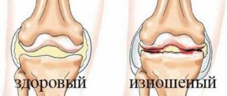
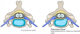
The dog's hip joint is not complex. This is a ball-and-socket joint consisting of the acetabulum of the pelvis and the head of the femur entering into it. The ligamentous apparatus of the joint is represented by the articular capsule and the round ligament, which is located at the bottom of the acetabulum of the pelvis. The round ligament connects the head of the femur and the acetabulum, providing stability to the joint. The acetabulum, except for the attachment point of the round ligament, and the femoral head are lined with cartilaginous tissue. The joint cavity contains synovial fluid. Movements in the hip joint can be performed in different planes. This is primarily due to its anatomical structure in the form of a ball-and-socket joint. Its mobility is controlled by several components: the round ligament, the articular capsule and the special shape of the surface of the acetabulum.
To perform its function normally, the joint must also be stable. Stability is ensured by the ligamentous apparatus (articular capsule, round ligament, muscles around the joint), as well as clear comparability of the articular surfaces - the presence of congruence. To reduce friction of the articular surfaces, the joint contains synovial or articular fluid. In addition to reducing friction, synovial fluid serves as a nutrition for cartilage cells on the articular surfaces.
For the proper functioning of the hip joint, the following aspects are important:
- anatomical structure of the acetabulum (take into account its size, depth and shape);
- anatomical structure of the femoral head (take into account its shape and size);
- congruence and degree of mobility between articular surfaces;
- angle of inclination and length of the femoral neck;
- hip joint capsule strength;
- structure and function of tendons and muscles.
Total hip replacement
Total hip replacement (THR) involves replacing the socket with a high-density polyethylene element and the head with a prosthesis made of a metal alloy of cobalt-chrome or titanium. Special bone cement (polymethyl methacrylate) is used to attach prostheses, although there are also cementless systems or combined systems that have proven themselves just as well. Essentially, the TZTS technique provides the dog with an artificial hip. For this type of surgery, it is necessary to wait until the dog stops growing, which usually occurs between 9 and 12 months of its life. As a rule, TZTS is applicable for animals weighing over 20 kilograms. A dog with hip arthritis, but not in severe pain and with normal hip function, is not a candidate for this surgery. TZTS is an expensive procedure, but it has a high success rate. Dogs feel more comfortable and their quality of life improves by more than 90%.
What does dysplasia mean in dogs?
The name of the disease - dysplasia - has its own functional justification and, when translated from Greek, means “pathological growth”. According to many data from foreign veterinary specialists, hip dysplasia is a hereditary disease that manifests itself during the dog’s growth period. Initially, a dog may be born with healthy hip joints, but later in the process of growth, weakness of the ligamentous apparatus of the hip joint appears and the process of disease development will begin. In puppies, changes in the load on the surface of the acetabulum or any other anatomical disturbances during the period of active growth can irreversibly change the shape of the articular surfaces and also lead to subluxation of the joint. This will greatly affect the functioning of the joint and leads to pathological stress on them. Over time, it develops to remodeling of the hip joint and the development of deforming arthrosis.
The cause of pathological weakness of the ligamentous apparatus of the hip joint in puppies is still not clear. According to some sources, it is believed that this is caused by a violation of the development of the head of the femur and acetabulum initially, according to others - by changes in the ligamentous apparatus of the joint itself.
In modern veterinary medicine, it is believed that the probable causes of the development of hip dysplasia in dogs are:
- changes in the anatomy of the hip joint: flattening of the acetabulum, changes in the neck-diaphyseal angle;
- changes in the anatomy of other joints of the pelvic limb;
- genetic factor;
- underdevelopment of muscle mass;
- obesity or too rapid growth of the dog;
- hormonal disorders of the reproductive system;
- neuromuscular diseases;
- lack of vitamin C.
In any case, regardless of the causes of dysplasia, the disease leads to overstretching of the joint capsule and subluxation. There is an excessive load on the joint capsule and it becomes damaged and inflamed. Swelling and subluxation lead to impaired joint mobility, irritation of nerve endings occurs and severe pain develops.
Reasons for development
Alas, poor heredity and genes do not always lead to hip dysplasia. Surprisingly, recent studies have shown that all puppies are born with normal joints, and the destructive process begins soon after birth.
Poor nutrition and lack of physical activity during the dog’s maturation period play an important role in the development of this disease. As you can see, the well-being of a pet almost always depends on its owner. Of course, there are animals with a predisposition to this disease, for example, large breeds or dogs prone to obesity.
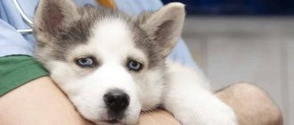
Clinical signs of hip dysplasia in dogs
Clinical signs of hip dysplasia depend on the age of the animal and the degree of dysplasia. In puppies, clinical signs develop gradually as the problem progresses. They become more noticeable from 4-9 months of age. Initially, when examining a puppy, signs of hip instability will not be noticeable even to the most experienced veterinarian. As you get older, the subluxation of the hip joint will increase, the joint capsule will begin to stretch and become inflamed, resulting in pain. Such puppies become inactive, have difficulty standing up, and pain may occur when the affected limb is abducted. In cases where the instability is significant, a click may occur in the hip joint.
Also, at the initial stage of the disease, puppies will have a noticeable “wobbly gait.” This strange gait is the result of instability of the hip joint along the transverse axis. The dog tries to walk normally, but due to the pain, it compensates for the stress on the joints by rocking its back from side to side. This helps the dog move forward without increasing the range of motion in the hip joint.
By reducing mobility in the hip joint, the dog also reduces the range of motion in the knee and hock joints, placing its paws at right angles. As a result, the dog walks on its paws extended at the joints.
In cases where the instability of the hip joint is severe enough, you can feel a click when you put your hand on the dog's hip joint while walking.
If pain occurs, atrophy of the muscles of the pelvic limbs will appear after at least 1-1.5 months. Visually, such a dog has a more massive front part of the body than the back. This occurs due to the transfer of body weight when moving to the thoracic limbs due to pain.
In dogs with dysplasia, the process of remodeling of the hip joint occurs. The peak occurs after about a year, when the dog’s body stops growing. The remodeling process is the body’s natural response to instability and consists of many mechanisms.
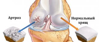
The final stage of the remodeling process is damage to the cartilage of the articular surfaces, stretching or rupture of the ligamentous apparatus of the hip joint, the formation of incongruity of the articular surfaces, the formation of bone spurs in the area of the edges of the acetabulum, and the final stage is the development of deforming arthritis of the hip joint.
In adult dogs, clinical signs will be observed as a result of degenerative changes in the hip joint. During the process of remodeling, the joint undergoes irreversible changes. As a rule, the joint becomes stable, but the articular surfaces will be irreversibly changed and susceptible to arthrosis. Such dogs experience pain, especially when getting up, and often such dogs refuse to get up. Upon examination, a decrease in the amplitude of mobility in the joint may be observed as a result of deforming arthrosis. Atrophy of the muscles of the pelvic limbs is also observed. As a result of the inability to move normally, these dogs are often overweight. An overweight dog with dysplasia practically cannot tolerate physical activity.
Treatment options
Conservative therapy includes prescribing a diet, chondroprotectors, antispasmodics and NSAIDs to the dog. Physiotherapy and exercise therapy may also be indicated. Conservative methods do not allow a dog to be completely cured. But with their help, remission of the disease is achieved and the process of joint destruction slows down. Surgery is considered the only effective treatment method.
Diet therapy
In the complex treatment of dysplasia in dogs, diet therapy plays an important role. It allows you to slow down the development of degenerative changes in the musculoskeletal system. The dog is prescribed a special diet in the form of industrial food:
- Pro Plan Veterinary Diets Joint Mobility JM;
- Brit Veterinary Diet Dog Grain Free Joint & Mobility;
- Hill's Prescription Diet Metabolic + Mobility;
- Hill's Prescription Diet j/d;
- Advance Articular Care;
- Royal Canin Mobility MC25 C2P.
Pro Plan Veterinary Diets Joint Mobility JM is enriched with vitamins and omega fatty acids, which are essential for maintaining healthy joints. The diet allows you to control your pet's weight.
Brit Veterinary Diet Dog Grain Free Joint & Mobility ─ food with chondroprotectors and Omega-3 fatty acids. Used in dogs with a genetic predisposition to dysplasia for preventive purposes, as well as to maintain joints in the presence of pathology or in the postoperative period.
Hill's Prescription Diet Metabolic + Mobility promotes weight loss in obesity and supports joint metabolism and improves joint mobility. Includes minerals, vitamins, antioxidants and omega-3.
Hill's Prescription Diet j/d contains chondroprotectors in the form of glucosamine, derived from crustacean shells, and chondroitin, extracted from pork cartilage. Facilitates the pet's condition when following a diet and is observed after 3 weeks.
Advance Articular Care ─ dog food with chondroprotectors in the form of hyaluronic acid, collagen, chondroitin and glucosamine. Reduces inflammation and reduces pain in dogs.
Royal Canin Mobility MC25 C2P is suitable for puppies over 8 months. Contains minerals, omega fatty acids, vitamins and collagen hydrolysate. Additionally, it can be used as a prevention of urolithiasis in dogs.
Chondroprotectors
Drugs with chondroprotectors are often prescribed in combination with NSAIDs and antispasmodics. Popular products in this category in the form of feed additives include:
- Artroglycan (for all breeds);
- Stride Plus (for all breeds);
- Kanvit Chondro Maxi (for large dogs);
- Gelakan Baby (for puppies).
Drugs with chondroprotective effects can also be prescribed by injection (Chondartron, Chionat).
Read more about chondroprotectors here.
Painkillers and anti-inflammatory drugs
Non-steroidal anti-inflammatory drugs are prescribed to dogs for severe pain and arthrosis that has developed against the background of dysplasia. Medicines in this category include Previcox, Rimadyl, Norocarp.
Travmatin injections for dogs are prescribed along with Chondartron injections. Refers to homeopathic medicines with anti-inflammatory, analgesic and regenerating effects.
To improve joint mobility and reduce pain, endoprosthetics of synovial fluid is sometimes performed using injections of Noltrex, Fermatron, Ostenil. They are injected every six months directly into the affected joint.
Physiotherapy
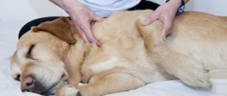
Physiotherapeutic procedures that are most widely used in the treatment of dysplasia include:
- massage;
- acupuncture;
- electrophoresis;
- physiotherapy.
An underwater treadmill is also used for joint therapy. Such physical therapy allows you to keep the posterior muscle group in good shape. Reducing the load on joints is achieved through water.
What does fitness mean for dogs, and how is it carried out for dysplasia, you can learn from the video below.
Surgery
The type of surgery depends largely on the age of the dog and the type of dysplasia. To treat pathology, operations are performed:
- juvenile symphysiodesis;
- pelvic osteotomy;
- arthroplasty;
- endoprosthetics.
Juvenile symphysiodesis is a low-traumatic method. Indicated for puppies aged 4-5 months.
Pelvic osteotomies are performed in dogs aged from six months to 7 months.
Arthroplasty involves resection (removal) of the head of the hip bone with further suturing of the articular capsule and the formation of a gasket from the deep gluteal muscle, which prevents bone friction. A long recovery period is required.
Endoprosthesis replacement is performed for animals weighing more than 20 kg. It involves replacing the joint with a titanium prosthesis. It is one of the most expensive types of surgical treatment. The price of the operation starts from 50,000 rubles, the cost of the prosthesis can reach 120 thousand rubles.
In case of dislocation against the background of developing dysplasia, the ligaments are also sutured in the clinic.
Diagnosis of hip dysplasia in dogs
Diagnosis of hip dysplasia in dogs consists of different research methods, since it is very important to determine the type of dysplasia and make the right decision about treating the animal. Owners, how important is an early visit to a veterinarian, even if a strange gait occurs or simply for prevention. When dysplasia is diagnosed at an early age, the effect of treatment is achieved better than in advanced stages of the disease. Also, early diagnosis will avoid expensive and quite traumatic operations.
Diagnosis of hip dysplasia consists of examining the animal, performing specific diagnostic tests for the hip joints, x-rays and, in some cases, a CT scan.
During the examination, the veterinarian will collect anamnesis, assess the degree of mobility of the hip joint, determine the presence or absence of pain in the joint, lameness or atrophy of the muscles of the pelvic limbs. In some cases, when the instability of the hip joint is significant, upon examination you can feel the moment of subluxation or dislocation.
A competent general examination will help in making a diagnosis, but only special tests and specialized research methods will help make it definitively. Special diagnostic tests in dogs are recommended to be carried out under sedation so that tension does not interfere. The essence of these tests is to determine whether there is instability of the hip joint (dislocation or subluxation) and to determine a special Barlow angle for further surgical treatment.
There are two common tests for hip dysplasia:
Ortolani test
The essence of the Ortolani test is to create a subluxation in the hip joint. This test is carried out in a lying position on your side. The veterinarian creates pressure on the knee joint with his hands, which leads to its subluxation. Without reducing the pressure, the veterinarian moves the dog's limb laterally and the hip joint snaps into place. A click is felt in the joint, which means the test is positive. Normally, pressure on the knee joint does not cause subluxation of the hip joint.
Bardens test
The essence of the Bardens test is also to achieve subluxation of the hip joint. This test is carried out in a lateral position. The veterinarian holds his fingers simultaneously on the ischial tuberosity and the greater trochanter of the femur, while with the other hand he shifts the femur to the mediolateral side, as if shifting the femoral head from the acetabulum downwards. With subluxation of the hip joint, a shift of the greater trochanter to the lateral side is felt. This symptom is positive.
For a complete diagnosis of hip dysplasia, an X-ray examination is performed. A prerequisite for this procedure is the use of sedation.
Radiographs take into account all the signs of hip dysplasia, namely:
- identify all signs of instability of the hip joint by displacement of the femoral head from the acetabulum: - Rhodes Jenny index - measurement of the lowest and highest points of the acetabulum; — Norberg-Olsson angle: determine the center of the femoral head using a stencil with circles applied and draw a line between them, then measure the angle formed by this line and a line drawn through the upper bony edge of the acetabulum. The norm is 105 degrees.
- The structure of the hip joint is assessed by the femoral head and acetabulum.
- identify signs of degenerative disease of the hip joint with dysplasia.
Sometimes Penn stress films can be done for hip dysplasia. With this method, joints are assessed under load. The assessment is based on hip instability only.
CT scans of the hip joints can be used in similar ways to x-rays, such as measuring angles and detecting instability. If we compare X-ray diagnostics and CT, then X-ray diagnostics is a cheaper and no less informative research method.
After a diagnosis such as hip dysplasia is made, its type is determined.
Hip dysplasia is divided into two types:
- Acetabular dysplasia (Dysplasiaacetabula). This type of dysplasia is caused by a normal neck-shaft angle (135 degrees) and weakness of the ligamentous apparatus.
- Cervical-diaphyseal dysplasia (Coxavalgaantetorta). This type of dysplasia is characterized by a change in the neck-shaft angle and the presence of a normal acetabulum. The angle for this pathology is more than 150 degrees.
Understanding the differences between types of dysplasia is very important when deciding whether to undergo surgical treatment.
To determine the degree of dysplasia, a special classification was created. It may differ in different countries, but the essence remains the same. In Russia, it is customary to classify dysplasia as A, B, C, D, E:
A - Normal joint; B - Joint within acceptable limits; C - Mild dysplasia; D - Moderate dysplasia; E - Severe dysplasia.
Main symptoms of the disease
Responsibility for promptly contacting a doctor lies entirely with the dog’s owner, because the sooner the animal receives qualified assistance, the greater the likelihood of stopping the course of this dangerous disease. Paying close attention to your pet and the following list of symptoms will help you recognize hip dysplasia in time:
- the dog gets tired quickly when walking;
- limps noticeably;
- has difficulty getting up after resting;
- there is obvious pain when walking;
- the dog’s movements are constrained;
- the arrangement of the limbs resembles the letter “X”;
- in advanced forms, complete immobility is possible.
One or more of the above signs is a serious reason to immediately contact a veterinarian.
Methods for controlling hip dysplasia in dogs
Hip dysplasia control methods should be implemented by breeders and owners of dog breeds at risk. At the moment, X-ray examination for dysplasia is carried out from the age of 12 months, when the dog has already grown. If such a diagnosis is confirmed, the dog should be discarded from breeding and sterilized.
If hip dysplasia is suspected, it is better to conduct an X-ray examination from 2-16 weeks of age. Research at an early age will significantly affect the dog’s recovery process and will help avoid radical surgical interventions.
Concept of triple pelvic osteotomy
The concept of triple pelvic osteotomy (TOP) is stabilizing reconstruction of the hip joint. It is usually used in young dogs with dysplasia up to 12 months of age, in which the head of the femur is insufficiently covered with hyaline cartilage (i.e. significantly damaged). Surgery helps stop subluxation and minimize hypermobility, which lead to severe arthritis. At the first signs of degenerative-dystrophic joint diseases (DDJ) in a dog, it is necessary to discuss the use of TOT surgery. TOT is typically used in dogs with hip misalignment. If a bilateral TOT procedure is necessary, then it is advisable to perform not one complex operation at once, but to divide them into two operations with an interval of 30-60 days. This technique is more acceptable and less traumatic for the dog.
Treatment of hip dysplasia in dogs
There are two types of treatment for hip dysplasia - conservative and surgical treatment.
Conservative treatment
aimed at reducing the load on the joint, especially in young animals. The dog's weight should be closely monitored with a balanced diet to avoid increased stress on the affected joint. It is also important to monitor your dog's physical activity in terms of frequency, duration and type. It is important that a dog suffering from dysplasia has a good muscular frame to provide support for the diseased joint. The best exercise is slow walking on a leash. For dogs with severe dysplasia, walking starts at 5 minutes a day, then increases by 5 minutes. If the lameness intensifies, then no time is added. If pain occurs, especially in older dogs with secondary signs of arthrosis deformans, non-steroidal anti-inflammatory drugs are prescribed for a long course.
Surgery
Hip dysplasia depends on the type of dysplasia and the age of the animal.
Juvenile symphysiodesis
This is the simplest surgical technique to prevent the occurrence of hip dysplasia. With coagulation of the pubic fusion of the pelvis, the growth of the pubic bone slows down, and the pelvis begins to grow in width. With this growth, the acetabulum rotates to cover the femoral head and make the joints stable. This surgical intervention is not painful for the dog and makes it possible to walk immediately in full. This procedure is performed on dogs up to 20 weeks of age. The best time to carry out this technique is up to 16 weeks.
Pelvic osteotomies (double and triple)
This type of surgical treatment is performed on dogs from 6-7 months of age for acetabular dysplasia, when juvenile symphysiodesis is no longer advisable. Also, this type of surgery is not suitable for dogs with damage to the dorsal edge of the acetabulum and signs of arthrosis of the hip joint. Although pelvic osteotomies are quite complex operations, they are used quite often in veterinary practice. The essence of the operation is to rotate part of the pelvis so as to rotate the acetabulum and cover the femoral head, as a result of which the joint becomes stable. After surgery, a mandatory rule is to limit the dog’s mobility for the period of pelvic fusion. The advantage of this operation is the preservation of the joint.
Intertrochanteric osteotomy
This type of surgery is performed on dogs with an irregular neck-shaft angle greater than 150 degrees. The operation is performed on the femur. The essence of the method is to change the angle and immerse the femoral head into the acetabulum.
Resection arthroplasty of the hip joint
This type of surgery consists of removing the femoral head and forming a pseudarthrosis. The use of this technique is possible only when the hip joint is destroyed as a result of deforming arthrosis. The operation is performed primarily to relieve pain.
Hip replacement
This is a total hip replacement in dogs - a technique that gives good results, but is expensive.
In conclusion of this article, I would like to say about the problems of breeding in our country. When purchasing a puppy of a breed prone to hip dysplasia, you need to look at documents confirming that the dog's parents have been tested for hip dysplasia. If you already have a dog and you notice a change in gait and other signs of dysplasia listed above, then do not delay in visiting a veterinarian. Remember! The sooner the disease is diagnosed, the easier it will be to treat your pet.
Femoral head ostectomy
Femoral head ostectomy (FHO), a hip replacement, is the most complex type of operative reconstructive surgery, usually performed on adult dogs suffering from severe osteoarthritis. Hip replacement is recommended for many dogs with dysplasia also because it has good results during the recovery period after surgery. The procedure involves removing the femoral head, its neck, and suturing the joint capsule. It is advisable to carry out the operation on an animal that is not overweight, and if the dog is still overweight, it is necessary to first prescribe a low-calorie diet and reduce it as much as possible. After surgery, comprehensive physiotherapy is necessary. Small to medium sized dogs generally do well after AMS. In large dogs, the outcome of the operation cannot be predicted, but it is still successful in 85% of cases.
Breeding animals with THD
Expert opinion says: it is important and necessary to carry out genetic selection and in every possible way prevent the spread of hip dysplasia. However, there are studies that prove that offspring obtained from parents with THD grow up absolutely healthy, subject to proper care and upbringing. This suggests a conclusion: growth rates, living conditions, and the presence of injuries received during growing up have no less influence on the development of THD than genetics.