Cataracts in dogs are one of the most common eye diseases. The most characteristic symptom of this disease is clouding of the lens. Why is this happening? We will tell you in our article how to recognize the disease and how to cure cataracts in a dog.
With cataracts, protein denaturation (folding) occurs. As a result, insoluble milky-white fibers appear inside the lens. Why do the proteins inside the “natural lens” fold, and what can cause cataracts in a dog?
Causes
Scientists are still trying to discover what exactly leads to clouding of the lens. They unanimously settled on one cause of cataracts in dogs – genetic predisposition. It has already been proven that in 8 out of 10 cases a puppy will have cataracts in the future if at least one of the parents had this disease. However, there are other reasons that lead to cataracts, so to speak, contributing ones. These include:
- Injury. Impact, scratch, constant mechanical irritation.
- Eye burns. Chemical, thermal (albeit less frequently), ultraviolet.
- Diabetes mellitus is the most common cause of cataracts in dogs.
- Problems with the immune system (weak immune response). Also, if the dog develops the same infectious disease more than once a year.
- Poisoning. Medicines and chemicals, but more often alcohol. You can't give your dog alcohol!
- Senile change. Well, who is not affected by age? Both people and animals suffer from a variety of diseases as they age. And one of them is cataract.
Breed Predisposition
Poodles (in particular, toy, dwarf and royal) suffer from this disease most often. Spaniels (American and English), miniature schnauzers, terriers, golden retriever. However, representatives of other breeds can also suffer from lens clouding.
Cataracts in dogs and their causes
If the transparency of the lens of the eye decreases and, due to its cloudiness, light cannot pass to the retina, it means that cataracts have begun.
The main reason is heredity, since it is genetic predisposition that most often causes vision diseases.
Cataracts in dogs can develop regardless of breed, progressing over the years.
But most of all, faithful golden retrievers , entertaining poodles , Yorkshire and Boston terriers , and cocker spaniels .
The critical age is 8 years, it is during this period that cataracts are most likely to occur.
In addition to heredity, cataracts can be caused by:
- eye diseases – glaucoma, uveitis;
- long-term drug treatment;
- endocrine disorders - diabetes, metabolic disorders;
- decreased immunity;
- due to injuries received;
- in case of concussion of an animal;
- as a consequence of illness;
- age.
Cataracts can develop in dogs regardless of breed, progressing over the years. But still, retrievers, poodles, Yorkshire and Boston terriers, cocker and Welsh springer spaniels are most susceptible to it.
Kinds
There are two types of cataracts in dogs:
- Primary. With this type, the disease is either congenital (a puppy is immediately born with this pathology) or acquired (it can develop after some disease). But how can a puppy be born immediately with cloudy eyes? Yes, it’s very simple, he could “get sick” of an infectious disease in the womb (together with his mother), suffer from toxins (if the mother dog was poisoned) or even injury (if a pregnant mustache was beaten, he fell, etc.). The primary cataract in a dog will also be one that develops as a result of aging. And it occurs in 4 stages. Secondary. Here, cataracts occur together with another disease. That is, the dog’s lens becomes cloudy, but at the same time the pet has some other disease. Secondary cataract is a symptom of another pathology developing in the body.
Detection of the disease - signs and diagnostic methods
A dog owner can suspect cataracts when the animal shows signs of blindness. There is a deterioration in the reaction, the dog bumps into objects. But blindness can occur due to various diseases, so it is important to conduct a diagnostic study. General symptoms do not allow making a reliable diagnosis and prescribing treatment for the dog.
The main symptom of cataracts is clouding of the anterior chamber of the eye, which is often detected during a simple examination of the dog. But opacities come in different shapes and degrees, so to diagnose them they resort to illuminating the eye and studying it with an ophthalmoscope. Thus, superficial opacification of the posterior wall, layered cataract, is revealed. It is easier to carry out an examination with a dilated pupil; atropine and phenylephrine are dripped onto the conjunctiva .
The naked eye can only make a preliminary diagnosis and only if the lens is severely damaged. If the clouding is weak, then ophthalmoscopy with lateral and focal illumination is used.
Examination can reveal the nature of the cataract - the color and thickness of the clouding. An ophthalmological examination allows for a differential analysis of the degree of corneal damage. In complete lesions, the second Purkinje image is enhanced, but the third is absent. At the same time, complete cataract is accompanied by miasm.
Differential diagnosis of the type and cause of cataract:
- with congenital opacification of the lens in a dog, a small plaque in the form of an asterisk or spindle is detected;
- cataracts caused by trauma change dynamically - in the early stages of the disease it is limited in nature, and in the late stages it is diffuse. Often wounds and damage to other parts of the eye are found - the cornea, sclera with hematomas;
- If the clouding is caused by other diseases, then a complex of symptoms is usually recorded in dogs. And the cataract itself takes on a gray tint, and its shape can be varied;
- Age-related changes in the lens are noted in dogs older than 10 years. They develop slowly in both eyes. Inspection reveals clusters of black dots.
Stages of disease development
Having recognized from the symptoms that your pet has problems with his eyes, you can begin treatment, thereby preserving your pet’s vision for a longer period. In total, there are 4 stages of cataract development in dogs:
| Initial | The very beginning. And it lasts for more than one year. Initial changes are invisible to the naked eye. To confirm that a dog has developed cataracts, it is necessary to undergo an examination at a veterinary clinic with good equipment. |
| Immature | The cataract is beginning to gain momentum, but the veterinarian will not perform surgery at this stage. It will be necessary to wait until the disease progresses to the next stage. Vision begins to gradually decline. |
| Mature | At this stage, the symptoms of cataracts are already noticeable. Vision almost completely disappears. It is at this stage that surgery must be performed. |
| Overripe | It is better not to reach this stage of the disease, otherwise the treatment of the dog will be complicated. Yes, they are operating on it. But the longer the animal owner delays, the less likely it is that the mustachioer’s vision can be restored. |
How quickly cataracts develop in a dog depends on the individual characteristics and health status of the animal, as well as on external factors.
Unfortunately, owners often notice too late that their pets have problems with eye health, and turn to the veterinary clinic already at the stage of a mature or overripe form of cataract, which makes drug treatment impossible. Therefore, the dog is sent for surgery.
Complications of the disease
Treatment of cataracts in dogs with drugs without a doctor’s prescription or folk remedies can lead to complications of the disease in the same way as a complete lack of treatment. Complications of the pathology are considered:
- blindness;
- development of glaucoma (sharp increase in intraocular pressure);
- the development of inflammation in the eye, which often leads to the death of the pet.
Treatment for cataracts in dogs must be carried out on time, otherwise it will cause complications for the pet and may also lead to its death.
Symptoms
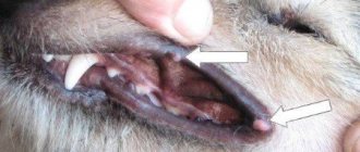
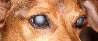
The most striking and reliable symptom of cataracts in a dog is clouding of the lens itself. When you look into the eyes of an animal, you can see how the circle in the middle (where the pupil was) is white, like a thorn. Such a whitish spot can appear on one or both eyes at the same time.
- At the beginning of the disease, such clouding may be insignificant. The dot will gradually increase in diameter. Over time, this “cloudiness” will occupy the entire space of the pupil. Essentially, the pupil is a “hole” that has the ability to increase and decrease in diameter. And the rays of light entering the eye through it cannot pass through the crystalline lens, which acts as a lens. Normally, the lens is transparent, so light passes through unhindered. And we see a black pupil.
- With cataracts, the light is “reflected” from the cloudy lens, thereby we see a white circle. This is how, even without special equipment, you can understand that your dog has problems with his eyes.
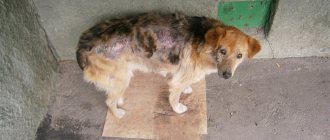
- Vision is impaired. And as a consequence of this cataract symptom, your dog starts bumping into things. He walks very carefully, taking his time. Refuses to play favorite games.
But don't discount the possibility that your dog may have another eye disease that may have symptoms similar to cataracts. Therefore, if you are not a qualified specialist in the field of canine diseases, it is better to consult a veterinarian regarding the tumor.
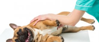
Using modern diagnostic methods and the latest medical equipment, specialists from veterinary clinics reliably identify the symptoms accompanying cataracts. The animal is examined both traditionally and using a new method, which makes it possible to make a more accurate diagnosis.
Below are examples of cataracts in dogs of different breeds:
Symptoms and diagnosis of the disease
At the initial stage of cataract development, it is difficult for the owner to recognize the disease, especially if the clouding begins in the peripheral parts. First, small cloudy areas appear on the lens, which periodically disappear and then appear again; the animal begins to frequently turn its head to catch objects in focus.
As the disease progresses, spots, stripes, and stars can be seen on the pupil of the affected eye - these inclusions are cloudy, whitish, and the eye itself acquires a bluish tint. Deterioration in vision is observed - the pet bumps into objects, becomes less active, and walks carefully. A healthy dog's pupils always have a uniform dark color.
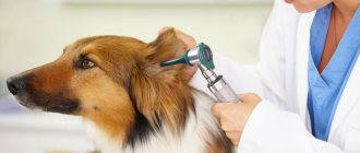
If you suspect the development of cataracts, the animal must be taken to the clinic; after examination and diagnosis, the veterinarian will be able to confirm or refute the diagnosis, tell you how to treat the pet, and what consequences may arise.
Research methods:
- examination of the eye using side lighting and transmitted light - allows you to identify areas of cloudiness and assess their size;
- Ultrasound of the eye;
- electroretinogram.
In young dogs, pathological processes in the lens develop much faster than in older pets.
Treatment
How to treat cataracts in dogs? Treatment of the animal in this case, unfortunately, is exclusively conservative - surgically.
Drug treatment for cataracts in dogs does not provide 100% hope of recovery; it only allows to slow down the process of clouding of the lens. Medicines are prescribed at the very beginning of the disease (when surgery is not yet required). Most often, vitamin and enzyme preparations are required. There are drops: there are a huge number of them. However, again, they cannot completely cure the animal. Scientists are still trying to invent a medicine that will stop the disease (stop the clouding process).
The sooner the owner recognizes the first signs of a serious pathology and takes the pet for treatment, the greater the chance of timely surgical intervention. As you know, cataract removal in dogs is one of the most technically difficult operations performed on the mucous membranes, and the best effect can be expected from it at the initial stage of the disease.
Preoperative preparation
If partial or complete removal of the lens of the eye is planned, it is necessary for specialists to conduct a complete physical examination of your pet approximately 4 weeks before the planned operation. Since it will be performed under general anesthesia, the veterinarian must check whether the animal can physically endure the operation. To do this, your pet’s blood and urine will be analyzed, possible changes in blood pressure will be monitored, and its medical history will be assessed.
If an animal has diabetes, it is extremely important to conduct a preoperative blood test for all parameters.
In addition, veterinarians may recommend giving your dog eye drops for 14 days before surgery to relieve inflammation. Typically, Flurbiprofen is used for these purposes - proven anti-inflammatory drops for cataracts for dogs.
Cataract surgery for dogs
If we talk about the surgical method of treatment, it is worth mentioning that there are 2 methods.
- One is the oldest. It involves removing the old clouded lens and replacing it with an artificial one.
- The second method gives positive results even at the initial stage. It's called phacoemulsification.
The name is long and somewhat scary, but in reality the method is not that complicated. And this is a more modern way of treatment. Its essence is this:
A small puncture is made in the cornea, and the lens is crushed using ultrasound. Through the same puncture (no more than 2 mm) the crushed contents are removed. And in its place, a rolled up special artificial lens is inserted (IOL, intraocular lens, intraocular, to put it in plain English). As soon as the IOL gets into the cavity of the lens bag (after all, nothing should remain from the lens itself), it straightens out. And the animal can see again.
During the operation, it is recommended to use viscoelastics - gel medicinal solutions injected into the eye and protecting it from damage and infection during surgery. The use of additional means of protecting the eye membranes, as well as the treatment of surgical instruments with a “viscous” substance, increases the safety of the phacoemulsification procedure and mitigates the pain of its implementation.
Without sufficient reason, sutures are usually not placed on the eye. The puncture, gradually “self-sealing”, heals in about 10 days.
The highest quality artificial lenses, as determined by experts, are produced by German companies.
The cost of the operation varies depending on the severity of the disease, the city, the qualifications of the doctor and the equipment of the veterinary clinic. However, you should not count on a price below 12,000 rubles for one eye. If both eyes of the dog are cloudy, the doctor will begin the operation with the one with less damage. After 3 months, the second one will be operated on. The fact is that in the initial and immature stage the lens is still elastic, and phacoemulsification takes place with a minimum of complications.
Treatment without surgery: fact or fiction
Unfortunately, treating cataracts in dogs without surgery is not possible. And, as already mentioned, the only way to get rid of cataracts is a surgical operation, during which, successfully or not (depending on various factors), the clouded lens is replaced with an artificial analogue.
Prevention
There is no specific prevention for cataracts in dogs. The fact is that no diet, super-high-quality food or constant intake of vitamins will save your pet from this disease. However, they significantly reduce the risk of developing diseases of the eyes and other organs and organ systems of the animal.
All you can do is examine your pet’s eyes and visit the veterinarian at least once a year (especially if the mustache has a breed or hereditary predisposition). It is very important to study the pedigree of your four-legged friend. If he had “relatives” in his family with cataracts, then there is a high risk that your dog will also have problems with his eyes. Alas, clouding of a dog’s lens is inherited.
Cataract treatment – therapy and removal surgery
There are currently no effective therapeutic treatments for ocular cataracts in dogs. Locally prescribed drops of withaioduroltriphos adenine, viceine, vitamin, catachroma, 1-2 drops 2-3 times a day for a long time. For traumatic cataracts, the use of tissue preparations and laser therapy is indicated. But these remedies do not eliminate the clouding of the lens, but can only restrain the further development of the disease.
Clients interested in cataract surgery should be referred to an ophthalmologist as early as possible. This will allow the specialist to examine the fundus of the eye for signs of progressive retinal atrophy or retinal detachment. Many cataracts, especially those with onset at a young age (1-5 years of age), and diabetic cataracts are accompanied by lysis and cause the development of phacogenic uveitis (FU). FU must be treated intensively to prevent the development of secondary glaucoma and to stabilize the patient before surgery. Since most cataracts today are removed by phacoemulsification, surgery is best performed in the early stages of cataract formation, before severe visual impairment develops.
The main difficulty in conservative treatment of cataracts is associated with the unclear etiology of age-related cataracts. Recently, the role of antioxidants in quenching free radicals and protecting lens proteins has been intensively studied. Research is underway aimed at elucidating the role of hereditary factors, environmental factors, the general condition of the body, and the state of hydro- and hemodynamics of the eye in the development of cataracts.
Surgical methods
Veterinary ophthalmologists can surgically remove cataracts and restore vision damage caused by cataracts in your pet. Most pets have no complications and return to normal activities, working and playing within a few days after surgery.
EEC with intraocular lens implantation
A through corneal incision was made in the dorsal part of the cornea from 10 a.m. to 2 p.m., 1 mm from the limbus. To achieve mydriasis, a 1% phenylephrine solution was injected into the anterior chamber of the eye. Then viscoelastic was injected. Anterior capsulotomy was performed using a “tin can” cystotome. After this, the nucleus was hydrodissected with an irrigation solution and evacuated using two spatulas. The lens masses were washed out using an automated irrigation-aspiration system.
Before intraocular lens implantation, the capsular bag and anterior chamber were filled with Provisc viscoelastic, and then the intraocular lens was implanted into the capsular bag using intraocular lens implantation forceps. The viscoelastic was removed. A continuous Pierce suture (8.0 nylon) was placed on the cornea.
Phacoemulsification with intraocular lens implantation
To perform FEC, we used a purely corneal tunnel incision 2 mm long and 2.5 mm wide. A 1% solution of phenylephrine and Viscoat viscoelastic were injected into the anterior chamber.
Using capsular forceps, a circular continuous opening of the anterior capsule (capsulorhexis) was performed. Next, hydrodissection of the lens nucleus was performed, its destruction and aspiration using an ultrasonic tip through the surgical wound. The capsular bag was filled with Provisc viscoelastic. The IOL was implanted using an injection system. The viscoelastic was evacuated, and 2 interrupted sutures, nylon 8.0, were placed on the surgical wound.
FEC with posterior capsulorhexis
The technique of performing the operation is the same as FEC with implantation of an intraocular lens. The difference is that before implantation of the intraocular lens, a continuous circular opening of the posterior capsule of the lens is performed.
Posterior capsulorhexis is performed with a high-frequency capsulotome according to Cloti from Oertli (Switzerland) as follows:
- ultrasonic fragmentation of the nucleus using a bimanual method;
- aspiration of lens masses;
- injection of Viscoelastic (Opti-med or Acrivet) into the anterior chamber.
Under the influence of viscoelastic, the posterior capsule straightens, and areas of opacification or stellate fibrosis are clearly visible on it, as well as the mooring between the capsule and the vitreous body. The tip of the high-frequency capsulotome of the Oertli phacoemulsifier in the Regular position is inserted into the posterior chamber until it comes into direct contact with the cloudy capsule. Then, in a circular motion, a capsulorhexis of the planned diameter of 4-6 mm is performed in one stage.
When performing capsulorhexis, great care and delicacy of movements must be observed to avoid damage to the anterior hyaloid membrane of the vitreous body. After capsulorhexis, the posterior capsule is removed using vitreal collet forceps. An artificial lens is implanted into the space between the anterior and posterior capsulorhexis, and the viscoelastic is completely washed out of the anterior chamber.
Cataract removal operations are performed at any stage of their development. But the less developed the disease, the easier it is to cure. Immature opacities do not harden the lens, so they are easier to remove without complications. Therefore, dog owners need to regularly visit veterinary clinics for examination.
Disease prevention
Avoid abuse:
- medications;
- additional vaccines;
- anti-flea shampoos;
- other chemically active substances.
Remember that some types of cataracts develop due to systemic toxicosis.
Did you know? Your pet spins around before going to bed not because he is not very comfortable, but because since ancient times, dogs have been doing this to scare away snakes and insects that might be in the thick grass.
Provide your pet with a diet rich in antioxidants. The eyes are damaged by free radicals. They attack protein molecules, causing them to harden and making the eye appear cloudy. Antioxidants prevent the oxidation of free radicals, restore damage already caused, and prevent the occurrence of new damage.
Powerful antioxidants include:
- blueberries;
- ginkgo biloba;
- wheatgrass extract;
- dandelion root.
Vitamins C and E are also antioxidants. They slow down the development and progression of cataracts. Complexes based on blueberries with the addition of vitamin E are effective means for preventing the disease.
Prevention of cataracts in dogs should include regular eye examinations (once a year). This will help to detect the onset of the disease in a timely manner. If there is an underlying disease that can trigger the development of cataracts, then its treatment will improve the prognosis of the course of eye diseases. Inoperable dogs with progressive cataracts can, with the help of their owner, learn to compensate for this deficiency with their sense of smell.
To help your pet, you need:
- Keep the environment stable. Do not rearrange chairs or other objects.
- Make sure that there are no objects lying on the floor that the dog could trip over.
- Do not let your pet off the leash while walking.
Did you know? Cataracts are more common in purebred dogs than in mixed breeds.
How is preoperative preparation carried out?
If your dog requires partial or complete removal of its lens, it is important that it is examined by a doctor a month before surgery. Since surgery is usually performed under anesthesia, the doctor must check whether the animal can actually undergo the operation. To do this, the dog's urine and blood are taken for analysis. If the dog has diabetes, it is given a full examination and drops are prescribed 2 weeks before surgery. For example, it could be Flurbiprofen, which has a powerful anti-inflammatory effect. Cataract surgery for dogs
The cost of cataract surgery depends on the method of surgical treatment. Today, the lens is removed in two ways. One of them is the oldest - the old lens is removed from the animal and replaced with an artificial one. In this case, photos of cataracts in dogs show that the operation leads to a complete cure, as well as restoration of vision. The price of this treatment is lower than the second surgical method.
The second surgical treatment option is phacoemulsification. During this procedure, a small puncture is made in the animal’s eye, after which the lens is crushed using ultrasound. After this, the contents are removed through the “hole”. Typically, such a puncture does not exceed 2 mm in diameter. Then a rolled lens is inserted through it, which will then open and replace the removed lens. After this treatment, the dog will begin to see again.
During phacoemulsification, it is important to use gel fluids that will protect the cornea from damage and infection. As photos of this treatment method show, after surgery the eye looks healthy again.
What is the cost of dog cataract surgery? Today, the average cost of cataract surgery for dogs is $200.