Dirofilariasis is the only helminthiasis in the temperate climate zone that is transmitted transmissibly through mosquito bites. The disease is recorded in warm-blooded animals and humans. In this article I will tell you how dirofilariasis develops in dogs, I will describe treatment, diagnostic methods, drugs for prevention based on the latest developments of parasitologists and infectious disease specialists.
- Laboratory diagnosis of dirofilariasis in dogs.
- Means for the destruction of adult heartworms.
- Drops.
What is dirofilariasis in dogs?
The disease is caused by round filamentous worms of the genus Dirofilaria. The name is translated from Latin as “evil thread”. Helminths reach a length of 15–30 cm and a diameter of 1.3 mm.
In the temperate climate zone, 2 types of heartworms are recorded:
- Dirofilaria immitis. Adult worms settle on the right side of the heart - in the ventricle, atrium, and lumen of the pulmonary artery.
- Dirofilaria repens. Sexually mature helminths live under the skin - in fiber or muscles.
With severe infestation, worms sometimes migrate to the abdominal cavity, brain, spinal cord, settle under the eyelids and even in the eyeball.
Dogs are very susceptible to heartworms, easily become infected and become the main carriers of helminths. One animal can be simultaneously parasitized by 1 to 250 mature worms. Intermediate hosts are mosquitoes of the genus Anopheles, Aedes, Culex. Insects carry larvae from sick animals to healthy dogs and humans.
Cats and wild canines are less susceptible to heartworms. They are less likely to have the most dangerous form of cardiopulmonary disease.
Life cycle of heartworm
The development cycle of helminths begins in the body of an infected dog. Viviparous female adult heartworms produce stage 1 larvae, microfilariae, daily. They circulate in the bloodstream of an infected animal before meeting a mosquito and live for 2–3 years.
Video from the channel of parasitologist Sergei Konyaev. Microfilaria Dirofilaria repens in the blood of a dog.
At the moment of the bite, the insect swallows the larvae along with blood. Inside the mosquito, the parasites mature to stages 2 and 3. They gradually migrate to the head section of the insect and become infectious to animals and humans.
The duration of maturation of larvae in the body of a mosquito depends on the temperature and humidity of the environment. The average period is 14 days. At 20 – 28℃ and above, the time is reduced to a week, at 18℃ and below it increases to a month.
When the temperature drops to 14℃, the development of microfilariae stops; when the air warms up, it resumes.
When an infected mosquito bites a healthy dog, stage 3 larvae burrow into the skin and penetrate the subcutaneous fat, where they remain for up to 21 days. The cardionematode Dirofilaria immitis then enters the bloodstream and reaches the heart 3 to 4 months after infection. Subcutaneous parasites Dirofilaria repens remain within the subcutaneous fat.
In the dog's body, the larvae molt twice. At the fourth stage they increase to 1.8 cm, at the fifth stage - to 12 - 16 cm. After 6 - 7 months from the beginning of infection, mature females begin to produce microfilariae in the blood. Adult heartworms live 5–7 years.
Is canine dirofilariasis dangerous for humans?
Humans are not typical carriers of heartworms. In Russia, 30–40 cases of infection are registered every year. Helminths settle under the skin, often localized in the area of the eyelids and eyes. They do not mature to the sexually mature stage. Throughout history, only 4 cases have been recorded when worms reached the heart.
The larvae become invasive to humans at stage 3, when they develop in the mosquito's body. Therefore, the only method of transmission is transmissible, through an insect bite. The owner cannot become infected directly from his dog. But animals influence the frequency of diseases in the region: the more dogs with dirofilariasis, the higher the risk of human infection.
What Causes Heartworm Disease in Dogs?
Heartworm disease, or heartworm disease, is a serious and potentially fatal disease in dogs and other animal species. Canine heartworm disease is caused by a parasite known as Dirofilaria immitis.
Adult heartworms are found in the heart and adjacent large blood vessels of infected dogs. Rarely, parasites can be found in other parts of the circulatory system. The female of this worm has a length of 15 - 36 cm and a width (about 0.5-1 mm). The male is approximately half the size of Sama. When conducting a pathological diagnosis, up to 300 worms can be found in one dog.
Dirofilariasis in Russia – statistics
The incidence of dirofilariasis depends on the number of days per year with an average daily temperature above 14℃. The screenshot shows a fragment of the guidelines on dirofilariasis of the Federal Service for Sanitary and Epidemiological Surveillance of the Russian Federation (2018):
Traditionally, foci of dirofilariasis are recorded in the south of Russia:
- in the Krasnodar and Stavropol Territories;
- Crimea;
- Rostov, Astrakhan, Volgograd regions;
- Republic of Adygea.
In 2021 - 2021, the study was carried out by employees of the Novosibirsk Institute of Animal Systematics and Ecology and the Moscow veterinary laboratory Vet Union under the leadership of the famous parasitologist Sergei Konyaev.
The research is based on an analysis of hundreds of publications from 1910 to 2019 and the results of a survey of practicing veterinarians. To collect information, a questionnaire was published on the website www parasitology ru and on social networks, which was filled out by 1039 doctors:
- 71.6% of veterinarians encountered the cardiopulmonary form of dirofilariasis;
- 36.4% rate it as a frequent disease - 10 or more cases per year;
- 10.8% were treated only with the subcutaneous form;
- 5.3% were diagnosed with cardiopulmonary dirofilariasis at least once.
Over the past decade, the range has expanded, the disease is recorded in the following areas:
- Vladimirskaya;
- Nizhny Novgorod;
- Ivanovskaya;
- Tula;
- Lipetskaya;
- Tyumen;
- Belgorodskaya;
- Tambovskaya;
- Kurganskaya;
- Tula;
- Sverdlovskaya;
- Jewish Autonomous Region;
- Kabardino-Balkaria;
- Tatarstan.
Full research results are published on the VKontakte page.
Symptoms of infection
Dirofilariasis does not have clear clinical signs that clearly signal infection and help to immediately make a diagnosis. The most common symptoms - increased fatigue, weight loss, weakness - accompany a lot of diseases.
During walks and training, the dog is reluctant to move, stop, sit down, or lie down. After physical exertion he breathes heavily and recovers more slowly.
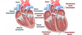
If helminths settle in the heart, pulmonary arteries, symptoms of respiratory and heart failure occur:
- Impaired capillary refilling. When you press on the gums, the normal pink color returns within 2 seconds.
- Pallor or cyanosis of the mucous membranes.
- Coughing.
- Foamy or bloody sputum.
- Dyspnea.
- Noisy breathing.
- Cardiopalmus.
During the examination, the veterinarian notices pathological changes in the heart: abnormal murmurs, rhythm disturbances, tachycardia.
Dirofilaria repens under the skin of a dog
Subcutaneous heartworms do not affect internal organs, but cause anxiety to the animal. In places where parasites are localized, the following occurs:
- seals;
- movable tubercles;
- rashes;
- sores;
- itching
Scratching causes fur to fall out and wounds to appear. If bacteria enter them, purulent inflammation develops. Indirectly, infection is signaled by symptoms of complications of dirofilariasis - an enlarged liver, free fluid in the abdominal cavity.
Dirofilariasis - diagnosis
The diagnosis is made taking into account the following studies:
- · X-ray. It is detected how dilated the pulmonary artery is, and the presence of right ventricular hypertrophy is also shown;
- · laboratory. Identifies larvae by examining a fresh drop of blood - they are clearly visible between red blood cells;
- · echocardiographic. The most used imaging technique for diagnosing cardiovascular diseases.
The analysis for dirofilariasis is carried out comprehensively. To the studies listed above, one-time express tests are added that show the presence of an antigen (potentially dangerous or foreign substance). These express systems have 100% specificity.
IMPORTANT! The sooner the animal owner contacts the veterinary hospital, the more accurately the doctor will make a diagnosis and prescribe the correct treatment.
Complications
Severe consequences occur in the cardiopulmonary form:
- Thromboembolism of pulmonary vessels. After natural death and during treatment, helminths disintegrate into fragments. Decaying parasites partially or completely clog the lumens of blood vessels and block the blood flow to the internal organs. Thromboembolism is the main cause of death of an animal due to dirofilariasis.
- Eosinophilic pneumonia. Occurs in dogs with a hypersensitivity reaction as an immune response to antigens of microfilariae circulating in the blood.
- Interstitial pneumonia is inflammation of the walls of the alveoli and connective tissue of the lungs.
- Ascites is an accumulation of free fluid in the abdomen.
- Glomerulonephritis is an immunoinflammatory lesion of the renal glomeruli. The complication arises as a reaction to the symbiont bacteria dirofilaria - Wolbachia. They accompany helminths, maintaining vitality and reproductive functions.
- Hepatomegaly is an enlargement of the liver due to circulatory problems.
- Respiratory distress syndrome is acute respiratory failure.
- Cardiogenic shock is an extreme degree of heart failure.
In the subcutaneous form, pathogenic microflora sometimes spreads at the site of worm localization and an abscess develops.
Cause of heart dirofilariasis
Heartworm disease, or heartworm disease, is a very serious and potentially fatal disease.
The disease is caused by the parasite Dirofilaria immitis. Adult worms in infected dogs are located in the heart and in the large blood vessels adjacent to it. Sometimes worms can be found in other parts of the cardiovascular system. An adult female worm reaches 15-36 cm in length and 5 mm in width. The male is approximately half the size of the female. One dog may have up to 300 worms at the time of diagnosis.
An adult heartworm can live up to five years.
Adult worms can live up to five years and during this time the female produces millions of offspring called microfilariae. Microfilariae live mainly in small vessels.
Forms of cardiopulmonary dirofilariasis
The severity of a dog’s condition is determined by 3 factors:
- Duration of carriage of helminths: the more time passes from the moment of infection, the higher the risk of complications.
- Intensity of infestation: the more worms settle in the heart, the more severe the disease.
- Individual resistance of the animal.
According to the symptoms, cardiopulmonary dirofilariasis occurs in three forms:
- Latent. The disease develops without clinical signs. The dog feels fine, and the owner does not even suspect the disease. Infection is detected accidentally when microfilariae are detected in a general blood test.
- Chronic. The disease develops slowly, with increasing symptoms. First, 1–2 clinical signs are noticed, and others gradually appear.
- Heavy. This form is diagnosed when a whole ball of worms accumulates in the heart, and the dog remains without treatment for a long time. The animal develops signs of cardiac and respiratory failure.
The course of the disease is influenced by the dog’s lifestyle. With dirofilariasis, the animal needs complete rest. If the disease is hidden, the owner is unaware of the infection and does not reduce physical activity. Against the background of an asymptomatic course, complications suddenly develop in the animal, and the condition sharply worsens.
Types of dirofilariasis
The disease, depending on the type of parasite, occurs in the cutaneous or cardiopulmonary form. The severity of the dog’s condition depends on the intensity of the invasion, as well as on the duration of the pathological process. The cardiac form poses a threat to the dog's life. Parasites provoke tumor-like growth of tissue inside the blood vessel. They are carried into the brain or spinal cord. They colonize the right ventricle of the heart and curl into balls.
Dead parasites pose the greatest danger. Their fragments contribute to the formation of blood clots. They clog the lumen of blood vessels and block blood flow. When worms decompose, toxic metabolites are formed and severe intoxication develops. Filamentous parasites interfere with the functioning of the heart. It increases in volume, and cardiac failure develops.
The cutaneous form of dirofilariasis does not threaten the life of a dog. Dermatitis develops, most often on the face. Scuff marks, baldness, and swelling occur.
Diagnostic methods
If dirofilariasis is suspected, it is necessary to identify the pathogen, determine the form of invasion, analyze the risk of complications, and exclude concomitant diseases. Therefore, a complex of studies will be required.
Laboratory diagnosis of dirofilariasis in dogs
In veterinary clinics, helminths are detected in three ways:
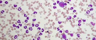
- Native smear method. Drops of fresh blood are placed between glass slides and examined under a microscope. When infected, mobile translucent filamentous microfilariae are visible. This is a simple, accessible and at the same time inaccurate method. If the degree of infestation is low, the larvae do not get into the samples, and the test shows a false negative result.
- Knott's method. Blood and 2% formalin are mixed in a test tube and rotated in a centrifuge. Then the supernatant liquid is drained and a dye, for example methylene blue, is added to the precipitate. This method is more accurate than the previous one. Microfilariae are stained and clearly visible under a microscope. The Knott method does not determine the type of pathogen. If there are few microfilariae, they may not be included in the samples at all.
Close-up of a stained microfilaria.
- Polymerase chain reaction (PCR) method. Enzymes are added to the venous blood that copy fragments of the isolated DNA of microfilariae. The laboratory technician then checks the results against the database. Larvae are easily detected due to the multiple increase in pathogen DNA in samples. The PCR diagnostic method is the most sensitive and accurate. Dirofilariasis is detected even if 1 microfilaria circulates in the blood, and the type of larvae is determined.
If the results are negative, but suspicions remain, then the studies are repeated up to 3 times at weekly intervals.
Express test
To quickly identify heartworms, an immunochromatographic analysis is performed. It detects an antigen protein in the blood that is synthesized by adult female heartworms.
The analysis is carried out using a test cassette. Blood is dripped onto designated areas. Then the result is assessed by the number of colored stripes: 1 line – positive, 2 – negative, no stripes – errors.
The studies take 5–15 minutes; dirofilariasis is detected with high accuracy only during intensive and long-term infection. False negative results occur in the following situations:
- less than 7 months have passed since infection;
- Only males are parasitic in the body;
- less than 5 females are localized in the heart;
- The dog receives preventive medications against helminths.
If microfilariae are detected in the laboratory using the Knott method or a native smear, and the tests show a negative result, then the subcutaneous form of dirofilariasis is diagnosed.
Additional Research
If infection with Dirofilaria immitis worms is confirmed, the heart and pulmonary arteries are examined to assess the risk of complications:
- Echocardiography – reveals pathological changes in the heart. Sometimes adult heartworms are visualized during the study.
- Electrocardiography - shows heart rate, rhythm disturbance.
- Chest X-ray - determines enlargement of the right atrium, ventricle, abnormal changes in the pulmonary arteries.
To assess the general condition, a clinical blood test is prescribed.
How is heart parasite disease diagnosed in dogs?
In most cases, one or a series of simple blood tests will diagnose the presence of heartworms in a dog's body. Further diagnostic tests are needed to determine whether the dog can safely undergo treatment for the condition. The following diagnostic procedures are recommended before starting treatment: Serologic testing for adult heart parasite antigens (antigen test, ELISA test, or SNAP test): This test is performed on a blood sample. The most widely used test detects antigens (proteins) produced by adult heartworms. It can be positive even if the dog does not have microfilariae in the bloodstream (about 20% of cases). However, it should be remembered that dogs with low levels of infestation (less than four to five adult parasites) may not produce sufficient levels of circulating antigens to produce a positive test result, so false negatives can sometimes occur in dogs with low parasite loads or in early stages. infestations. Since the detected antigen is produced only by female heartworms, a population of only males may also give a false negative result.
Microfilaria test result (microscopic blood spot test or modified Knott test). A native blood sample examined under a microscope for the presence of microfilariae. To improve diagnostic accuracy, the blood sample is centrifuged and then examined under a microscope to detect microfilariae. If microfilariae are visible, the test is considered positive. The number of microfilariae detected gives a general idea of the severity of the infection. However, microfilariae will be found in the bloodstream in greater numbers during the summer months and evenings, so the timing of sampling may affect this test. Approximately 20% of affected dogs do not test positive even though they have heartworms in their bodies because their immune system has acquired the ability to destroy the microfilariae. In addition, there are also similar blood parasites that are quite common in dogs, which are difficult to distinguish from microfilariae. For these reasons, the antigen test is the preferred diagnostic test. Other blood tests (OCA, blood chemistry, electrolytes) are also necessary for a quality diagnosis.
Treatment of dirofilariasis in dogs, drugs
Treatment of the cardiopulmonary form includes 3 areas:
- Destruction of adult helminths and larvae.
- Prevention of vascular blockage.
- Correction of cardiac disorders, prevention and treatment of complications.
There is no general scheme. Specific medications are prescribed only by the attending veterinarian after a comprehensive diagnosis, assessment of the animal’s condition and possible risks.
A drug for the destruction of adult heartworms
Now the only remedy prescribed against sexually mature helminths is melarsomin, trade name Immiticide. The drug was developed by American pharmacists in 1995. It is approved by the FDA, but is not licensed in Russia and is sold only in online pharmacies.
Before the advent of melarsomin, the drug thiacetarsamide was used to treat dirofilariasis. The drug caused severe complications from the liver and kidneys, and has now been abandoned.
Melarsomin is a toxic arsenic compound. It destroys sexually mature worms and does not affect larvae less than 4 months old. It is relatively safe for dogs in a therapeutic dose.
According to the instructions, the solution is injected deep into the lumbar muscles 2 times with an interval of 24 hours. A single dose of melarsomine is 2.5 mg per 1 kg of body weight. A total dose of 5 mg per 1 kg contains 0.75 mg/1 kg of arsenic.
Practicing veterinarians have developed their own administration regimens depending on the intensity of infection:
- 2 injections with an interval of 1 month.
- 1 injection, after 1 – 3 months 2 more at daily intervals.
With such schemes, the helminths die in parts, reducing the risk of blockage of the pulmonary artery due to the rapid mass death of parasites. Melarsomin is contraindicated in animals with renal or liver failure. The price of Immiticide is 24 thousand rubles.
After each injection, physical activity is completely excluded for 6-8 weeks, the space for movement is limited to the point of being kept in a cage, and the dog is walked on a short leash.
Drugs against microfilariae
The larvae are not as destructive as adult worms, but they also cause harm. They disrupt blood microcirculation and cause an allergic reaction in sensitive animals. To destroy microfilariae, macrocyclic lactones obtained from soil fungi are used:
- ivermectin
- milbemycin;
- moxidectin;
- dormectin;
- aversectin.
They do not destroy adult individuals, but reduce viability and suppress reproductive functions.
To destroy microfilariae, injection solutions with ivermectin and aversectin are often prescribed:
- Ivermek;
- Baymek;
- Novomek;
- Otodectin;
- Eprimek;
- Aversect K&S.
The solution is administered intramuscularly 1 to 5 times with an interval of 7 to 10 days. The exact regimen and dose are determined by the attending physician based on the condition of the animal. After the injection, microfilariae die within 4 to 24 hours.
Adjuvant therapy
Maintenance medications are prescribed before the administration of melarsomin and after the destruction of helminths:
- Doxycycline - reduces the population of Wolbachia bacteria, which complicate the course of the invasion.
- Alteplase - dissolves fibrin clots in the lumen of blood vessels. They are formed due to damage to the heart and poor circulation. Alteplase is administered once by intravenous drip.
- Prednisolone or Prednivet - suppresses the activity of the immune system against pathogen antigens, inhibits the allergic reaction.
- Heparin – inhibits blood clotting and prevents thrombus formation.
- Curantil - improves venous outflow, blood microcirculation in the heart, kidneys, stimulates the synthesis of interferon.
Previously, aspirin was prescribed for dirophyriasis to thin the blood and prevent the formation of fibrin clots. Now many veterinarians have abandoned it, considering the benefits to be insignificant and unproven.
Surgical treatment of cardiopulmonary dirofilariasis in dogs
Surgery is prescribed if the dog is brought to the clinic too late. When too many worms accumulate in the heart, the animal may not tolerate drug treatment. Due to the massive death of worms after administration of the drug, there is a high risk of complete blockage of the blood vessels of the heart and lungs.
During the operation, helminths are removed from areas accessible for extraction. The outcome depends on the degree of damage to the heart, lungs and arteries by parasites, the equipment of the clinic and the skill of veterinarians. If it is not possible to remove all the worms, drug treatment is prescribed after the operation.
Treatment of subcutaneous dirofilariasis in dogs
Dirofilaria repens does not threaten the life of the animal, so potent melarsomine is not used. More often, the dog is prescribed drugs to kill microfilariae. The drugs are prescribed in prophylactic doses for a course of 8 months to several years - until the natural death of the worms. Sometimes the worms are removed through cuts in the skin.
What happens in a dog’s body with dirofilariasis?
The disease proceeds hidden for a long time. This period usually takes several years before the dog shows clinical signs of infestation. Consequently, the disease is diagnosed mainly in dogs between two and eight years of age. The disease rarely occurs in dogs less than one year of age because the development time from microfilaria to macrofilaria takes five to seven months after infection. Unfortunately, by the time clinical signs are visible, the disease is at a very advanced stage.
Adult heartworms: Adult heartworms cause disease by blocking the heart and large blood vessels leading from the heart. They also interfere with the normal functioning of the valves in the heart. By clogging the main blood vessel, the blood supply to other organs of the body is reduced, especially the blood flow to the lungs, liver and kidneys, which leads to disruptions in the functioning of these organs.
Signs of heartworm disease depend on the number of adult worms, the location of the worms, the lifespan of the worms in the dog, and the extent of damage to the dog's heart, lungs, liver, and kidneys.
“Signs of heartworm disease are a soft, dry cough, shortness of breath... and loss of stamina.” The most obvious clinical signs of heartworm disease in dogs are a mild, dry cough, shortness of breath, weakness, agitation, lethargy, and loss of stamina. All these signs are most noticeable after physical activity. At times, some dogs may even become weak or disoriented. Your veterinarian may notice abnormal sounds in the lungs and heart when auscultated with a stethoscope. Congestive heart failure often develops with swelling in the extremities and an increase in the volume of the abdomen. In some cases, weight loss, poor general condition and anemia may develop. Seriously infected dogs may die suddenly during exercise or excitement.
Microfilariae (immature heartworms) circulate primarily in small blood vessels. Because they are about as wide as capillaries, they can block blood flow in these vessels. The cells that are supplied by these capillaries do not receive nutrients and oxygen. Microfilariae primarily affect the lungs and liver. Damage to the lung tissue leads to coughing. Liver damage can lead to cirrhosis, causing jaundice syndrome, anemia and generalized weakness. The kidneys may also be involved in the pathological process.
Preparations for the prevention of dirofilariasis
To protect against heartworm larvae, anthelmintic drops, tablets or solutions are used. Repellent preparations do not always protect against mosquitoes; they are used as an additional remedy.
In regions where dirophyariasis is intense, prevention begins in March-April, 1 month before the start of the mosquito season. Finish in October-November, a month after the end of insect activity.
Drops
The easiest way to protect your dog from infection is to apply drops on the withers. The following drugs have been approved for the prevention of dirofilariasis:
- Advocate. The drops contain a combination of moxidectin and imidacloprid. The first component enters the bloodstream and is excreted within 30 days, the second is distributed in the epidermis. Advocate destroys heartworm larvae at stages 3 and 4, when they penetrate the skin and subcutaneous tissue after the bite of an invasive mosquito. The animal is treated once a month.
- Stronghold. The drops contain selemectin and are available in a concentration of 6% for puppies and 12% for adult dogs. Dogs tolerate Stronghold safely; it is prescribed even to infected animals to reduce the concentration of microfilariae circulating in the bloodstream. Drops are applied every 30 days. The dog will have to be treated additionally for ixodid ticks; Stronghold cannot cope with them. In veterinary pharmacies you can find similar drops with selemectin - Selafort.
- IN-AP complex. The drug contains aversectin C1, which is active against heartworm larvae. The interval between treatments is longer than with previous drops - 6 weeks. IN-AP complex contains 2 more antiparasitic components: against ticks, fleas, tapeworms and roundworms.
If basement mosquitoes live in the house, then the dog is treated all year round.
Pills
To prevent dirofilariasis, tablets are given once a month.
- Nexguard Spectra. The drug consists of 2 active components: afoxolaner and milbemycin. The first protects against external parasites, the second protects against nematodes, including heartworm larvae. Tablets are available in different dosages for 5 weight categories of animals. The drug is contraindicated in puppies up to 8 weeks old and adult dogs lighter than 2 kg. See Nexgard prices here.
- Milbemax. The tablets contain a combination of praziquantel and milbemycin. There are 2 types available: for puppies and small dogs weighing up to 5 kg and for medium and large breeds. The prophylactic dose of milbemycin to protect against heartworms is 0.5 mg/kg. Milbemax can be given to puppies from 2 weeks of age.
- Helmimax. The tablets contain a combination of prazquantel and moxidectin. There are 3 types of tablets sold in pharmacies: Helmimax 4, 10 and 20. They are intended for small, medium and large breeds, respectively. Gelmimax can be given to puppies from 3 weeks.
- Dironet. The drug consists of 3 components: ivermectin, pyrantel and praziquantel. Puppies are allowed from 3 weeks. Contraindicated for pregnant dogs. The tablets are also available in 3 versions for small, medium and large dogs. Dironet contains a prophylactic dose of ivermectin: 0.006 mg/1 kg.
Solutions
For prevention, drugs with ivermectin are used, as for treatment, but in microscopic doses. Solutions are administered intramuscularly or taken once a month.
Most medications are difficult to dose. The prophylactic dose of ivermectin is only 6 mcg, and 1 ml of solution often contains 10 mg. It’s easier to calculate and fill the syringe with Otodectin. It contains 1 mg of ivermectin per 1 ml of liquid.
Treatment
In dogs, treatment of skin type diseases is carried out with medication and surgery. Surgery is required to remove an adult heartworm. Locally damaged areas are treated with a special solution for application to the skin: Imidacloprid 10% and Moxidectin 2.5%.
After a one-time use of the solution in the blood of the dog, the presence of microfilariae was not observed for 60 days. There were no traces of skin lesions either. Treatment of dogs with pulmonary-cardiac form of the disease is much more difficult. With this type of disease, microfilariae can be destroyed by using drugs such as Levamisole or Ivermectin.
This is usually enough to kill the larval stage. Sometimes such drugs are used in situations where treatment of dirofilariasis in dogs with the presence of mature worms poses a threat to the health of a weakened dog. If you immediately kill adult parasites, their remains will lead to a sharp deterioration in health.
If other pathologies are present, the dog may suffer greatly or even the consequences may be irreversible.
Only an experienced and knowledgeable veterinarian can select and prescribe medications for heartworms in dogs. All this is due to the fact that toxic arsenic compounds, melarsomine dihydrochloride, are used to destroy them. It may be sold under the name Immiticide.
This remedy effectively destroys adult helminths, but cannot be used if the dog has problems with the kidneys, liver, heart and lungs. In addition, this drug has another drawback - it is very expensive. In such a difficult situation, the dog is treated for a long time with Ivermectin.
Just killing parasites is not enough; this is just the first stage of therapy. After helminthiasis, animals are greatly weakened; they need to be restored and damaged organs and systems of the body must be treated. Each individual dog has its own treatment regimen depending on the severity and location of the lesions. Owners need to be prepared for the fact that treatment will be very long and complex.
Unfortunately, in some particularly serious and advanced cases, it is not possible to help the animal, and it dies. The greatest harm is caused by nematodes that cause damage to the heart and lungs; skin parasites can be dealt with much faster and more effectively, and with less damage to the dog’s health.
Forecast
The success of treatment is determined by 4 factors:
- number of sexually mature helminths;
- degree of damage to the heart, arteries, lungs;
- period from the moment of infection;
- restriction of animal mobility.
The most favorable outcome is with early diagnosis, when at the time of detection the disease is asymptomatic or with 1–2 clinical signs. In this case, dirofilariasis passes without a trace without complications. With delayed treatment and severe infestation, the dog develops irreversible changes in the lungs and heart, which shorten its life expectancy.
The article used materials from webinars of Doctor of Veterinary Sciences A.N. Shinkarenko, articles and recommendations on forums for dog owners by parasitologist S. Konyaev.