What it is?
Laryngeal paralysis is a disease in which the muscles of the larynx become too weak due to disruption of nerve impulses in the brain.
As a result, the laryngeal cartilage is destroyed and it becomes difficult for the dog to breathe, swallow and bark. Reference! As a result of research, it was possible to establish a direct relationship between laryngeal paralysis, degenerative changes in the central nervous system and hypothyroidism.
Juvenile
Juvenile laryngeal paralysis (polyneuropathy) is a mutation inherited in an autosomal recessive manner.
First of all, the disease affects the longest nerve endings of the larynx, and then spreads to the nerves innervating the hind limbs. Accordingly, you can track how the disease develops by progressing symptoms.
- The dog's voice changes and shortness of breath appears.
- There is difficulty in swallowing food.
- The hind legs weaken, and then the front legs.
Conventionally, laryngeal paralysis can be divided into four types:
- congenital:
- acquired;
- unilateral;
- bilateral.
Two-, left- and right-handed
Unilateral (left- or right-sided) paralysis is the inability of the arytenoid cartilage to drain air during inspiration. As a result, turbulence is created in the area of the glottis, which is expressed by wheezing, roughening or weakening of the voice.
Reference! With left- or right-sided paralysis, the dog begins to quickly get tired and out of breath during physical exertion.
If the disease progresses to a bilateral form, then in addition to the above symptoms, signs such as blueness of the tissues, severe shortness of breath and fainting are added. In some cases, bilateral laryngeal paralysis is accompanied by attacks of nausea, belching and vomiting.
Laryngeal paralysis in dogs symptoms
Diagnosis of laryngeal paralysis comes down to a clinical examination and additional instrumental studies.
It is mandatory to conduct an X-ray examination of the neck and chest cavity, ECHO KG (ultrasound of the heart). An x-ray evaluates the condition of the lungs, trachea and esophagus. ECHO CG is performed to exclude heart pathologies, which can give a similar picture of shortness of breath, cyanosis and fainting as with laryngeal paralysis.
The main diagnostic method will be laryngoscopy followed by tracheobronchoscopy. Laryngoscopy makes it possible to evaluate not only the structural features of the ligaments, cartilage of the larynx and trachea with bronchi, but also dynamic processes, namely the closing and opening of the glottis during the inhalation and exhalation phases.
When performing laryngoscopy, it is necessary to differentiate laryngeal paralysis from velum hyperplasia, laryngeal collapse and various neoplasms.
Causes
The primary cause of laryngeal paralysis is damage to the recurrent laryngeal nerve. The main factors in the development of this disease are as follows:
- congenital malformation;
- disturbance of nerve sensitivity and conduction;
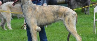
- “breed” predisposition;
- surgical interventions on the neck resulting in damage to the recurrent laryngeal nerve;
- neck injury during a fight;
- removal of the thyroid gland;
- poisoning with toxic substances.
Note! Wearing a collar that is too tight or too much tension on a leash or harness can also injure the laryngeal nerve.
Hello student
The larynx, larynx, is the protective part of the respiratory tube that regulates the flow of air into the trachea. The larynx serves as the vocal organ.
In carnivores, the larynx begins caudally from the branches of the mandibular bones and stretches with the head and neck extended to the level of the second cervical vertebra. Thus, it is easily accessible for palpation examination. Through the hyoid bone and its suspensory apparatus, the larynx is movably connected to the mastoid processes of the temporal bones. Its skeleton is made up of laryngeal cartilages, cartilagines laryngis, which, being hyaline cartilages, sometimes ossify in adult animals. Cartilages are connected to each other by connective tissue and/or joints, articulationes laryngis. Elastic ligaments, ligamenta laryngis, are also stretched between the cartilages. Proprietary muscles of the larynx, vol. laryngis, serve to displace cartilages relative to each other. Upper and lower (long) hyoid muscles, mm. hyoidei, change the position of the larynx. Inside, the larynx is covered with a mucous membrane, the relief of which is characteristic of each animal species.
LARYNGEAL CARTILAGE
The cartilaginous skeleton of the larynx consists of unpaired epiglottic cartilage, cartilago epiglottica, thyroid cartilage, cartilago thyreoidea, ring-shaped cartilage, cartilago cricoidea, and paired arytenoid cartilage, cartilago arytaenoidea. In the dog, on each of the arytenoid cartilages rostrodorsally there is a corneal process, processus corniculatus, which is absent in the cat. Only the dog has independent wedge-shaped cartilages, which in the rest are wedge-shaped processes, processus cuneiformes, connected to the arytenoid cartilages by ligaments, as well as a small intermediate interarytenoid cartilage, cartilago interarytaenoidea, lying dorsally between the arytenoid cartilages and the plate of the annular cartilage.
The epiglottic cartilage, cartilago epiglottica, is located rostroventrally at the entrance to the larynx. In carnivores, it is a more or less regular quadrangular plate of elastic cartilaginous tissue. It has a rostral apex, the lateral angles are curved caudally. At the base, the cartilage has a stalk in the dog, petiolus epiglottidis, which is absent in the cat. The epiglottis acts as a valve that closes the entrance to the larynx when swallowing food.
Thyroid cartilage, cartilago thyreoidea, hyaline, consists of two plates, laminae, connecting ventrally. Each plate has a pointed rostral horn, cornu rostrale, and a blunt caudal horn, cornu caudale. The caudal horn has an articular surface for articulation with the annular cartilage. The rostral horn is connected by a synchondrosis to the thyroid gland. Together with the cranial edge of the plate, it forms a wide thyroid fissure, fissura thureoidea, in carnivores. The outer side of the plate is divided by an almost horizontal oblique line, linea obliqua, into dorsal and ventral parts. On the caudoventral edge there is a caudal thyroid notch, incisura thyreoidea caudalis, deeper in the cat. Only in the dog, the junction of the cartilage plates bulges ventromedianally, forming a protrusion of the larynx, prominentia laryngea.
Rice. 1. Cartilages of the dog’s larynx, left side (after Nickel / Wilkens, 1987)
a cartilago epiglottica; b cartilago thyreoidea, b' lamina sinistra; with processus cuneiformis; d cartilago arytaenoidea; e cartilago cricoidea, arcus, e'lamina; f trachea
1 cornu rostrale, 2 cornu caudale, 3 cartilago thyreoidea, linea obliqua; 4 processus corniculatus; 5 cartilago cricoidea, crista mediana
Rice. 2. Cartilages of the cat’s larynx, view from the left side (after Nickel Wilkens. 1987)
a cartilago epiglottica; b cartilago thvreoidea, b'laminae; with left cartilago arytaenoidea; d'cartilago cricoidea, arcus, d' cartilago cricoidea, lamina; e trachea
1 cornu rostrale, 2 cornu caudale, 3 cartilago thyreoidea, linea obliqua; 4 cartilago cricoidea. Christa McDiana
Rice. 3. Dog laryngeal ligaments, median section (after Nickel Wilkens, 1987)
a os hyoideum; b cartilago epiglottica- with processus cuneiformis; d cartilago thyreoidea; e cartilago arytaenoidea; e' processus corniculatus; f cartilago cricoidea; g I tracheal cartilage
1 ligamentum hyoepiglotticum; 2 mcmbrana thvreohvoidea; 5 fibers related to plica aryepiglottica; 6 ligamentum vestibulare; 7 ligamentum vocale; 8 ligamentum cricoarytaenoideum, 9 ligamentum cricothyreoideum; 9' mcmbrana fibroelastica laryngis; 10 ligamentum cricotracheale; 11 ligamenta anularia
Ring-shaped cartilage, cartilago cricoidea, consists of a dorsal plate, lamina, and a ventral arch, arcus cartilaginis cricoideae, formed by hyaline cartilaginous tissue. On the plate along the midline there is a flat rounded ridge - the median ridge, crista mediana. At the rostral end there are articular surfaces for articulation with the arytenoid cartilage, facies articulares arytaenoideae. At the junction of the plate into the arch, on each side there is an articular surface for articulation with the caudal horn of the thyroid cartilage, facies articularis thyreoidea. The outer surface of the cartilage has flat pits for muscle attachment
Arytenoid cartilage, cartilago arytaenoidea. paired, due to its changing position, plays a special role in regulating breathing and producing sounds. It consists of hyaline and elastic cartilage tissue. In large domestic animals, its shape is more like a pyramid, but in a dog its shape is closer to square and has a more complex configuration than in a cat. At the rostral end of the cartilage in a dog there is an elastic corneal process, processus corniculatus, which is not found in a cat. The dorsal angle is elongated and forms a thin process, which, together with the interarytenoid cartilage, cartilago interarytaenoidea, communicates with the cartilage of the opposite side. The caudal angle of the articular surface articulates with the annular cartilage. The ventral angle is formed by the elastic vocal process, processus vocalis, to which the vocal cord is attached. The medial surface of the cartilage is smooth, and on the lateral surface in the caudal part, a muscular process, processus muscularis, rises in the form of a ridge.
The sphenoid cartilage (process), processus cuneiformis, in a dog is located rostral to the arytenoid cartilage, with the rostral end of which it is connected movably, but not through a joint. Rostrodorsally on the cartilage there is a caudally curved process, similar to the corneal process. Its second apex is directed rostroventrally to the stalk of the epiglottis.
Sometimes a dog has paired sesamoid cartilage, cartilago sesamoidea, which is located in the transverse arytenoid muscle or in the ventricular muscle.
Ligamentous joints and also joints provide the necessary mobility of the cartilages of the larynx relative to each other. In addition, they connect them with the hyoid bone and the trachea.
The hypoglossal ligament, ligaiientum hyoepilotticurn, connects the basishyoid and the rostral edge of the base of the epiglottis, and the thyroid-hyoid membrane, membrana thymohyoidea, connects the hyoid bone with the rostral edge of the thyroid cartilage. The epiglottic cartilage is fixed rostrally to the thyroid cartilage by means of the thyroid-epiglottic ligament, ligamentum thyreoe piglotticum, which in the dog arises from the stalk of the epiglottis. Also in the dog, the fibers of the sphenoid fold extend from the lateral edges of the epiglottis to the lateral surfaces of the sphenoid and arytenoid cartilages. In a cat that lacks the sphenoid and corneal processes, similar bundles of fibers extend to the annular cartilage. The space between the caudal edge of the thyroid cartilage and the rostral edge of the annular cartilage is covered by the annular thyroid ligament, ligamentum cricothyreoideum, which in the cat, in the form of an annular thyroid membrane, membrana cricothyreoidea, covers the deep caudal thyroid notch. In carnivores, the fibroelastic membrane of the larynx, membrana fibroelastica laryngis, extends lateral from it to the vocal cord. This membrane corresponds to the elastic cone in humans. The annular tracheal ligament, ligamentum cricotracheale, connects the caudal edge of the annular cartilage to the first ring of the trachea. The annular-arytenoid ligament, ligamentum cricoarytaenoideum, strengthens the joint capsule in the ventral part between the annular and arytenoid cartilages. The transverse arytenoid ligament, ligamentum arytaenoideum transversum, stretches dorsally over the arytenoid cartilage and in the dog covers the interarytenoid cartilage. Of the two ventral arytenoid ligaments, the cat has only a vocal cord, ligamentum vocale, which passes from the vocal process of the arytenoid cartilage rostroventral to the thyroid cartilage. Developed only in the dog, the short vestibular ligament, ligamentum vestibuiare, is stretched between the sphenoid and thyroid cartilages and forms the base of the vestibular fold.
The articular connections of the cartilages of the larynx are limited to the joint between the caudal horn of the thyroid cartilage and the ring-shaped cartilage, articulatio cricothyreoidea, as well as the joint between the arytenoid and ring-shaped cartilages, articulatio cricoarytaenoidea. While the first joint allows only deviation of individual cartilages relative to each other, in the second joint various displacements of the arytenoid cartilage relative to the annular cartilage are possible: flexion/extension, rotation and abduction/adduction. All these movements are carried out under the influence of the own muscles of the larynx. The hyoid muscles, muscles of the tongue and pharynx also take part in the movement of the larynx, especially when swallowing.
Rice. 4 and 5. Musculature of the larynx in dogs and cats, view from the left side (after Nickel Wilkens, 1987)
a cartilago cpifilottica; b carnlago thyreoidea; c processus cormculatus, c' processus cuneiformis (dog); d cartilago cricoidea (closed by muscles in a cat); e trachea; f ventriculus laryngis (dog); g - l os hyoideum: g basihyoideum, h thvreohvoideum, i ccratohyoideum, k cpihyoidcum, I stylohyoideum (in the dog it is represented)
1 m. ceratohyoideus; 3 m. thvreohvoidpus; 4 m. sternothyroideus; 5 m. cricothvreoideus; 6 m. cricoarvtaenoideus dorsalis; 7 m. arytaenoideus transversus; 8 m. cricoarytaenoideus lateralis; 9 m. thyreoarytaenoideus (cat), 9'm. ventricularis (dog), 9″ m. vocalis (dog)
Symptoms and diagnosis
So, you can suspect the presence of laryngeal paralysis based on the following signs:
- wheezing and whistling during breathing;
- change in voice (weakness, hoarseness);
- severe shortness of breath during exercise.
If your pet suffers from one or more of the above symptoms, take him to a veterinary clinic immediately. To make an accurate diagnosis, the doctor will use the following research techniques:
- Laryngoscopy. A visual examination of the larynx can reveal the presence of damage, tumors and inflammation. To relax the laryngeal muscles, the doctor administers a light local anesthetic.
- Radiography. Checking the condition of the chest organs. It is carried out to identify possible pathologies.
- Hormone analysis. It is carried out in cases where the dog is obese and has exercise intolerance.
Diagnostic methods
The main diagnostic method is a routine history taking and visual examination of the sick animal.
- Ultrasound and x-ray examination of the cervical spine are mandatory.
- It is important at the same time to exclude pneumonia, bronchitis, and other diseases of the respiratory system, as they can give the same symptoms.
- Be sure to evaluate the size of the esophagus, since its strong expansion (megaesophagus) also leads to the appearance of similar clinical signs.
- To make a final diagnosis, laryngoscopy is performed (using an endoscope or a specialized laryngoscope). The advantage of this procedure lies not only in the high value of the results obtained, but also in the absence of the need for full anesthesia. The animal is given mild sedatives and the movements of the laryngeal cartilage are assessed during inhalation/exhalation cycles.
Advice! Veterinarians advise not to waste time on x-rays, but to immediately perform an ultrasound, since the cartilaginous tissue of the larynx is visible much better and more clearly.
Forecast and consequences
If the operation is performed in a timely manner, the prognosis for recovery is favorable: the dog recovers quite quickly and returns to its normal life. However, in some cases postoperative complications may occur:
- infectious diseases, tissue inflammation;
- seroma – accumulation of fluid under the incision;
- cough while eating and drinking.
Note! The most dangerous possible complications are suture dehiscence, cartilage fragmentation, or aspiration pneumonia. Fortunately, they are quite rare.
Juvenile Laryngeal Paralysis (JLPP)
Laryngeal paralysis in dogs: data update based on the latest information https://infovet.ru/blog/paralich-gort...czii-5492.html If anyone is too lazy to read, then brief quotes: Quote: According to etiology, the disease is divided into congenital (hereditary laryngeal paralysis or hereditary polyneuropathy) or acquired (trauma, neoplasms, polyneuropathy, endocrinopathy) Quote: Etiologies and classification Laryngeal paralysis can be congenital or acquired and, depending on the etiology, unilateral or bilateral (Monnet & Tobias 2012; Stanley et al. 2010). A hereditary form of PG has been described in Huskies and Bouviers des Flanders (O'Brien & Hendriks 1986; Venker-van Haagen 1982). Bouviers de Flandres have a disease associated with the death of motor neurons in the nucleus ambiguus and is inherited in an autosomal dominant manner (Parnell 2010). This disease results in unilateral or bilateral paralysis and usually appears in dogs less than 12 months of age (Burbidge 1995; O'Brien & Hendriks 1986; Ridyard et al. 2000; Venker-van Haagen 1982). Quote: Congenital polyneuropathy with PG has been described in Rottweilers, Bouviers des Flanders, Bull Terriers, Dalmatians, German Shepherds, Afghan Hounds, Cocker Spaniels, Dachshunds, Miniature Pinschers and Huskies (Bennett & Clarke 1997; Braund 1994; Braund et al. 1994; Braund et al. 1994). al. 1989; Eger et al. 1998; Harvey & O'Brien 1982; Mahony et al. 1998; O'Brien & Hendriks 1986; Ridyard et al. 2000; Venker-van Haagen 1982). Clinical signs suggestive of polyneuropathy may be present, including weakened reflexes (in all four limbs), weakened postural responses, hypotonia and atrophy of the limb girdle muscles (Braund 1994; Braund et al. 1994; Davies & Irwin 2003; Gabriel et al. 2006; Mahony et al. 1998; Ridyard et al. 2000). Young Dalmatians and Rottweilers show axonal degeneration with loss of myelinated nerve fibers of the RLN and periglottic recurrent nerves (Braund et al. 1994; Braund et al. 1989; Mahony et al. 1998). In Huskies and German Shepherds, an association has been found between PG and certain phenotypic traits, such as white coat coloring, speckling and blue eyes (O'Brien & Hendriks 1986; Polizopoulou et al. 2003; Ridyard et al. 2000). Quote: Congenital PG in dogs is usually the result of a polyneuropathy complex and appears before 12 months of age (Monnet & Tobias 2012). This form of the disease is characterized by symptoms of PG in combination with cataracts and neurological symptoms such as tetraparesis (worst in the pelvic limbs), weakened reflexes in all four limbs, weakened postural reactions, hypotonicity and atrophy of the muscles of the limb girdles (Braund 1994; Braund et al. 1994 ; Davies & Irwin 2003; Gabriel et al. 2006; Mahony et al. 1998; Ridyard et al. 2000). There may also be comorbidities such as megaesophagus and aspiration pneumonia that are present initially or develop as the disease progresses (Braund 1994; Braund et al. 1994; Mahony et al. 1998; Ridyard et al. 2000). Quote: Prognosis and conclusions It is necessary to clearly distinguish between different forms of the disease. In cases of hereditary PG in dogs, the prognosis after surgical treatment is excellent. Congenital PH due to neuropathy has a poor prognosis and most dogs are euthanized within 10 weeks due to worsening clinical signs (Davies & Irwin 2003). The prognosis for acquired PH depends on the cause: in cases of injury, cure is possible; In case of PG as a result of a tumor, the prognosis depends on the type of tumor.
Treatment
The only effective way to treat laryngeal paralysis is surgery. Through surgery, the doctor restores space for air to pass into the lungs. However, in medical practice there are those rare cases when the disease could be eliminated at the earliest stages with the help of medications.
At home
Treatment of dogs with laryngeal paralysis is carried out in a hospital setting. The animal is sedated, additional oxygen is provided and, if necessary, the trachea is intubated. After the condition has stabilized, the doctor decides on the timing of the operation.
Traditional methods
The only thing that can save your dog from the consequences of the disease is a timely veterinary examination and surgery.
You should not self-medicate and experiment with various folk recipes. Even if they do no harm, you will lose precious time.
Nutrition after surgery
To make it easier and easier for your dog to swallow food, doctors recommend giving large, lumpy wet food without liquid. The plate should be on the floor. Equally important is constant access to clean water.
Treatment and care for a dog with paralysis
If the clinical symptoms are in the initial stage and the animal is not constantly worried, then it is necessary to make changes to the feeding diet (for dogs with increased body weight) and reduce physical activity.
During the hot season, the pet should be provided with a cool place to stay. In cold weather, walks should not be long.
In winter, it is recommended to take your dog for short walks.
Surgical intervention
Surgical treatment is recommended for severe paralysis in young and young animals. During surgery, the lumen of the glottis is increased.
In severe cases of paralysis, surgical treatment is prescribed.
Surgical intervention is performed using intralaryngeal access. Partial resection of the larynx is performed (with and without excision of the vocal cords) and longitudinal serrated excision (with and without excision), and it is also possible to install an implant.
Outside laryngeal procedures are aimed at restoring innervation.
Postoperative complications
Unfortunately, postoperative complications very often reach 50%.
Cough, pneumonia, residual noise, failure to restore normal voice, hematomas, respiratory distress and death of the animal. Thus, aspiration pneumonia is observed in 20% of postoperative patients. With resection of the larynx and partial removal of the vocal cords, complications reach 40%.
After the operation, the voice may not be fully restored.
Outcome of paralysis
The final result is influenced by:
- age;
- diseases of the esophagus;
- anatomical disorders of the respiratory tract;
- tumors;
- diseases associated with neurology.
The age of the dog affects the final outcome of paralysis.
Operational technology is constantly being improved all over the world. Specialists work to provide qualified assistance, but it takes a lot of practice to achieve pinpoint accuracy in performing such surgical interventions on dogs.
If left untreated, the disease progresses slowly, but gentle treatment can slow down the process somewhat. However, in all cases the prognosis is cautious.
>Conclusions
Attentive and careful attitude towards the animal can significantly alleviate the manifestation of symptoms.
To prevent the disease, you should treat your dog carefully.
> Diseases of dogs. How to treat a dog → Swallowing disorder
Swallowing disorder in dogs (dysphagia) is a difficulty in swallowing.
Prevention
To keep your pet healthy and protect him from throat problems, follow these rules:
- Don't put your dog under too much stress. Don't yell at her or scare her on purpose. Even the most harmless veterinary procedures (for example, x-rays or bandaging) are best performed after injection with sedatives.
- Do not expose your dog to overheating or hypothermia. If the weather conditions are not conducive to a long walk, it is better to refuse it or reduce the time you spend outside to a minimum.
- Limit your time in the water. This rule applies to swimming in chlorinated pools and natural bodies of water.
- Watch your weight : your body mass index may be slightly below normal, but not higher.
- Give preference to a harness : this will reduce the pressure on the dog's throat. Also, try not to keep your pet on a leash. The use of collars is strictly prohibited.
Diagnostics
- as soon as we hear a whistle while inhaling;
- voice change;
- refusal of physical activity or simply intolerance to it.
A change in a dog's voice is one of the symptoms of paralysis.
Moreover, if you apply light lateral pressure on the larynx, shortness of breath intensifies.
By comparing data on breed, sex, age and clinical signs, the diagnosis is made with an accuracy of up to 92%.
Inspection
To exclude an error, an examination of the larynx is necessary, which can reveal a tumor nature.
Diagnosis will require examination of the larynx.
In this case, laryngoscopy is a mandatory procedure. The event must be carried out only in a specialized clinic under local light anesthesia, which will help relax the muscles of the larynx to identify pathology.
Radiography
For diagnosis, it is also necessary to take a chest x-ray. This will make it possible to identify possible pathologies.
A chest x-ray will help identify possible diseases.
Hormone analysis
If your dog is obese, has skin discoloration, or has exercise intolerance, a hormone test is necessary.
If you are obese, you will need to do a hormone test.
Breath
Normally, an animal's breathing does not require much effort. The normal breathing rate at rest depends on the size of the animal: • for small breeds (Chihuahua, Yorkshire Terrier) this figure is 15-30 breaths per minute; • for medium breeds (Australian Shepherd, Boxer): 10-25 breaths per minute; • for large breeds (Labrador, German Shepherd): 10-20 breaths per minute; • for giant breeds (Great Dane, Malamute, Irish Wolfhound): 8-20 breaths per minute;
In hot weather or during physical activity, a healthy dog's breathing rate can increase to 200 breaths per minute. Puppies at 8-10 weeks of age breathe at rest at a rate of 60-100 breaths per minute.
Heart rate. In order to count the number of heartbeats (HR), you need to place your palms on the animal’s chest on both sides (for dogs of medium or large breeds), or clasp the chest from below with one palm (for dogs of small breeds) placing the palm immediately behind the elbows joints. Heart rate in dogs normally depends on the size of the animal: • for small and dwarf breeds (poodle, Chihuahua, Yorkshire terrier) 80-120 beats per minute; • for medium breeds (Australian shepherd, boxer) 60-120 beats per minute; • for large breeds (Labrador Retriever, German Shepherd, Golden Retriever) 60-110 beats per minute; • for giant breeds (Great Dane, Irish Wolfhound, Malamute) 50-100 beats per minute.
In athletic dogs, the heart rate may be slightly lower than indicated. In puppies up to 8-10 weeks of age, the heart beats at a rate of 150-200 beats per minute.
Symptoms of the disease
Symptoms of laryngitis in a dog are as follows:
- Change in voice - the dog’s barking becomes dry and hoarse, giving off a hoarse quality.
- Coughing attacks. Most often these are attacks of dry cough, which can occur without sputum discharge, less often - accompanied by pain and vomiting.
- The animal is depressed and lethargic, often loses appetite and refuses to eat, and when fed dry food, the symptoms intensify.
- When examining the dog's larynx and palpating this area, pain is revealed, provoking an attack of reflex cough.
- If at the initial stages the treatment of laryngitis was ineffective, or an advanced form of the pathological process was diagnosed, shortness of breath manifests itself. This suggests that inflammation affects the mucous membrane of the trachea and bronchi.
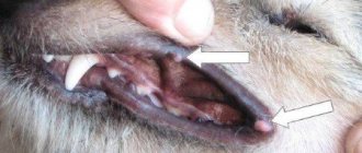
- If the catarrhal form of laryngitis is diagnosed, the animal’s body temperature remains within normal limits. In the case of the development of lobar laryngitis, the body temperature rises, attacks of muscle spasms appear, and cyanosis affects the mucous membrane.
Less often, discharge from the nasal passages and eyes may appear, but such symptoms are not typical for laryngitis. The main thing is to carry out timely diagnosis and select an effective drug.