Skeleton or skeletal system
The skeleton is a set of solid formations in the body that gives the animal’s body support and protects internal organs from damage. In addition, the bone marrow contains hematopoietic organs that are responsible for many biochemical processes.
The dog's skeleton consists of 289-292 bones (the difference is due to the number of tail vertebrae), and there are 262 joints in the body. The spine is represented by the following elements:
- Upper section, or neck. It includes 7 vertebrae, the first of which is called the “atlas” - it is responsible for supporting the skull and making a vertical posture of the head possible. The epistrophy (second vertebra) is necessary for horizontal movements of the dog's head.
- Thoracic department. It includes 13 vertebral bones, some individuals may have 12, which is not a developmental pathology. The ribs emerge from the dense transverse formations in this section to protect the internal organs and the heart. The ribs located closer to the chest are less mobile than the rest - this is important for the animal’s breathing.
- Lumbar region. It consists of 7 vertebrae, which are distinguished by their oval structure. The processes are very dense and long, located towards the head. Dogs also have a sacrum - this is the fusion of 3-4 sacral vertebrae into a single structure, which is necessary for a strong connection with the hind legs and hip joint. Bitches have a more powerful and wider sacrum, which is required for reproductive function.
- Tail. Depending on the breed, the tail consists of 15-23 vertebrae. The department is required to express the dog’s emotions and feelings, balancing the body during movements.
The skeleton also includes limbs. The front paws are formed from the shoulder, forearm and hand, claw (5 fingers with 3 phalanges each). The pelvic limbs are represented by the following elements: pelvic bone, femur, lower leg, foot. The femur itself is fixed using a movable head into the acetabulum on the pelvic bone. In large dogs, the femoral head is susceptible to disease with age, causing discomfort and pain.
Photo: wikimedia.org
Scull
The main function of the skull is to preserve the brain from damage; the upper and lower jaws are also located on this part of the skeleton. Based on morphological characteristics, the following types are distinguished:
- dolichocephalic (greyhound, greyhound, collie) – elongated shape with an elongated head;
- brachycephalic (Spitz, French bulldog, pug) – the head is short and wide;
- mesocephalic (Labrador, husky, husky) – moderately long and wide skull.
Photo: wikimedia.org
The connection of the jaw bones is movable, thanks to which the dog can chew even dense food. Puppies have 28 teeth, adult dogs have 42. Only breed standards affect the bite. Main types of bites:
- scissor-shaped (normal);
- pincer-shaped;
- straight;
- undershot;
- snack;
- bulldog-shaped (bulldog).
In the brain of a dog there are 3 paired bones and 5 single bones, the largest of which is the frontal. It forms the eye socket, temples and nose. And the cheekbones, auditory organs and chewing muscles are connected to the temporal bone.
Photo:
Benign prostatic hyperplasia in males
Roman Leonard, Ph.D., practicing veterinarian, President of the Russian Scientific and Practical Association of Veterinary Nephrologists and Urologists (NAVNU, www.vetnefro.ru)
Applied Anatomy and Physiology
The prostate gland (PG), or prostate, is the only accessory sex gland in males, tubular-alveolar in structure, androgen-dependent in regulatory mechanisms and exocrine in the type of secretion. The main function of the prostate is the production of certain components of the ejaculate.
The prostate surrounds the neck of the bladder and the underlying urethra just below the rectum. Normally, the prostate has a certain mobility and is also characterized by greater density relative to adjacent organs. The secretion of the pancreas is released into the lumen of the urethra through multiple ducts and an opening located at the top of the seminal tubercle (through which sperm are also released into the urethra).
Histologically, the central part of the prostate, directly surrounding the urethra, consists of many tubulo-alveolar glands formed by cubic or columnar multinuclear epithelium. The secretory part of the pancreas is surrounded by a stroma of dense fibrous tissue with a small amount (which is typical for males) of smooth muscle fibers. In general, the prostate is covered with a fibrous-elastic capsule, and the septa extending from it divide this exocrine organ into two lobes, which are relatively poorly distinguishable in adult males.
In uncastrated male dogs, the prostate usually increases slightly in size with age, which in some cases may not be a pathology.
The growth and function of the prostate is largely determined by androgens, and primarily by dehydrotestosterone (DHT), which is converted from testosterone under the influence of 5-reductase. It is the receptors for DHT that are abundantly represented in the pancreas, as well as the strength of receptor interactions with this biologically active form of testosterone is the most intense and significant for this organ.
Pathological enlargement of the prostate associated with its benign proliferation (benign prostatic hyperplasia (BPH), usually accompanied by pain during defecation and urination (including when marking territory) and/or can lead to partial or even almost complete infringement of these biological functions Therefore, BPH is often interpreted by animal owners as diseases of the lower urinary tract, or coprostasis. BPH is often the cause of discomfort and restless (aggressive) behavior in male dogs, as well as pain of varying severity and forced positioning of the body in space (“hunched over”, weakness/“ stiffness" of the hind limbs, changes in gait). In severe cases, this disease of the pancreas can even pose a threat to the life of patients.
Thus, BPH can lead to a significant deterioration in the quality of life of both the patients themselves and their owners, and can also cause the death of male dogs that have not reached physiological old age.
Castration always leads to a decrease in androgen levels, including DHT, which, in turn, causes pancreatic involution. This process can be registered within 3–4 weeks after surgery, and its maximum severity is usually observed after 9–10 weeks and persists until the end of life.
History taking and clinical examination
Since the anatomical ring of the pancreas surrounds the urethra and is located in close proximity to the rectum, the pathological processes in it, associated primarily with severe hypertrophy, will often have signs of diseases, to one degree or another characteristic of adjacent organs. Therefore, the pancreas of male dogs with signs of diseases of the lower urinary tract (frequent, painful, unproductive, “thin stream” urination, as well as incomplete emptying of the bladder with stagnation of urine) and/or coprostasis must be subjected to a medical examination.
The simplest and at the same time informative method of primary diagnosis of pancreatic diseases is its rectal examination. The prostate is located just above a horizontal line drawn between the neck of the bladder and the cranial part of the pubic bone. During a rectal examination, the pancreas in males can be easily detected by moving in the cranial direction from the anus along the penile part of the urethra located on the surface of the pubic bone, and then the pelvic part of the urethra (see Figure 1). In some male dogs, simultaneously with a rectal examination, palpation/fixation of the pancreas through the abdominal wall is also possible.
With hyperplasia, the pancreas usually acquires a more convex, close to spherical, shape, becomes denser and more voluminous (prostatomegaly), and is also, to one degree or another, displaced more caudally along the pubic bone.
In addition, both during hyperplastic processes and during infectious diseases, the prostate loses its mobility, which can also be detected during a rectal examination.
Visualization of the pancreas using radiographic examination (usually as part of a lateral view of the abdomen) is usually not very difficult. Normally, in uncastrated males, the prostate occupies about 70–75% of the volume between the sacral promontory and the cranial part of the pubic bone and has round, smooth contours of regular shape. Between the cranioventral side of the prostate and the caudoventral part of the bladder, an area of adipose tissue in the form of an isosceles triangle is also usually visualized, with an obtuse angle directed towards the lumbar spine.
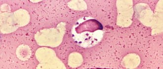
During ultrasound, the prostate in healthy, uncastrated male dogs is also visualized as a round formation with an even contour and a heterogeneous granular structure. And the prostatic part of the urethra should not differ in structure, wall thickness, or lumen diameter from the adjacent ones.
Since BPH is usually accompanied by episodes of difficult/unproductive urination and gross hematuria, the diagnosis of exclusion will primarily be urolithiasis (USD) and bacterial urocystitis/urethritis (the cause of obstruction in this case will be purulent-necrotic plugs, including a large amount of desquamated epithelium, and in some cases and components of the muscular syncytium of the bladder).
With urolithiasis, it is usually easy to visualize certain stones in the bladder and/or urethra, and a catheter inserted into the urinary canal even before its prostatic segment encounters a difficult (or insurmountable) obstacle.
And with urocystitis, an ultrasound will clearly show a significantly thickened double-circuit or even multi-layered (like a layer cake) wall of the bladder. The last group of uropathies, if untimely or inadequately treated, can be a predisposing or aggravating factor for bacterial prostatitis.
| Figure 1. Topography of the pelvic and external genitalia of a male dog (according to Boyd, JS; Paterson, C.; May, N., 1994): 1 - sacrum; 2 - descending colon; 3 - rectum; 4 - anus; 5 - pubic bone; 6 - bladder; 7 - prostate gland; 8 - pelvic part of the urethra; 9 - half-vesical part of the urethra; 10 - ischiocavernosus muscle; 11 - cavernous body of the penis; 12 - testis; 13 - scrotum; 14 - spermatic cord; 15 - superficial inguinal lymph node; 16 - rectus abdominis muscle; 17 - adductor muscle; 18 - penile bone; 19 - bulbous glands; 20 - long part of the gland; 21 - penile part of the urethra in the ventral groove of the penile bone; 22 - foreskin; 23 - jejunum |
Laboratory diagnostics
General clinical and biochemical (renal profile) blood tests for pancreatic diseases, as a rule, do not reveal abnormalities or are not very informative.
Urine examination is a mandatory laboratory test for suspected pancreatic pathologies that arise after collecting anamnesis and performing its imaging. A general clinical analysis of urine with examination of its sediment allows, among other things, to exclude/confirm/suggest concomitant diseases of the urinary system. If the urine has a pronounced inflammatory status and especially contains a large number of bacterial cells, a mandatory study is also a urine culture with an antibiogram.
Since BPH is often not accompanied by significant pathognomonic changes in analyzes of biological fluids and does not have clinical manifestations characteristic of this group of pathologies, there may be a need for examination (including bacteriological and cytological) of prostate secretion and/or ejaculate to exclude/confirm infectious component of the process.
Obtaining prostatic secretions for research is possible by massaging the prostate through the rectum. To evacuate the resulting secretion from the urethra, it is permissible to inject it through a pre-installed urethral catheter with a 0.9% sodium chloride solution. In order for the fluid to flow out and not into the bladder, simultaneously with the introduction of saline, it is necessary to compress the urethra at the level of the prostate.
Deviations in the functioning of the pancreas can also be identified by examining the ejaculate. The method for obtaining this biological fluid for spermogram can be easily found in the specialized literature.
Treatment
The most effective method of treating and preventing BPH is castration. After this procedure, usually within 4–6 weeks, the secretory epithelial cells of the pancreas atrophy, and their precursors also lose the ability to differentiate.
Previously used progestogens for the treatment of BPH have not shown any effectiveness, and estrogens (for example, megestrol acetate 0.50 mg/kg orally once daily for 4–8 weeks or medroxyprogesterone acetate 3 mg/kg subcutaneously once a week for for 10 months) although they were relatively effective, their therapy had a large number of significant side effects, especially pronounced when used for many months.
The standard treatment for BPH today is the use of 5α-reductase inhibitors (finasteride, dutasteride) and α1-blockers (α1-blockers) in monotherapy or in combination.
5α-reductase inhibitors. The mechanism of action of finasteride and dutasteride is based on the competitive inhibition of 5α-reductase, which converts testosterone into 5α-dihydrotestosterone (however, it should be noted that finasteride inhibits only type II 5α-reductase, and dutasteride inhibits both types I and II of this isoenzyme, which ensures it significantly more pronounced therapeutic efficacy).
As a result, the level of this highly active androgen in the blood and tissues of the pancreas significantly decreases within the first 24–48 hours after oral administration. Moreover, 5α-reductase inhibitors do not affect the level of testosterone itself, which helps maintain libido and an adequate level of spermatogenesis. This group of drugs is also characterized by a neutral metabolic profile, a wide therapeutic index and a low level of side effects.
Suppression of the formation of 5α-dihydrotestosterone is accompanied by a decrease in the volume of the prostate gland, an increase in the maximum flow rate of urine and a decrease in other symptoms of urinary tract obstruction. The full effect of using drugs in this group is realized after 3–4 months of continuous use. Another important characteristic of 5α-reductase inhibitors is that they have a significant effect on bladder emptying symptoms (obstruction), with little effect on storage symptoms (irritation). This not only normalizes the outflow of urine, but also the number of its passages during the day and generally improves the quality of life of both patients and their owners.
The author of the article proposes the following dosage regimen: finasteride 5 mg, and dutasteride 0.5 mg (in both cases this is 1 capsule) per animal weighing up to 50 kg once a day and 10 mg and 1 mg once a day, respectively ( 2 capsules) for males over 50 kg, for at least 6 months (and more often for life or until castration, since stopping the drug usually leads to a relapse of the disease).
α1-blockers (α1-AB). The basis for the use of α1-AB in patients with BPH is scientific data indicating a violation of sympathetic regulation in the work of the pancreas, which has an important role in the pathogenesis of this disease. It has been established that α1-adrenergic receptors are localized mainly in the neck of the bladder, the prostatic urethra, as well as in the capsule and stroma of the prostate. Hyperstimulation of α1-adrenergic receptors located in these areas, usually resulting from the progression of BPH, causes the development of chronic hypertonicity of their smooth muscle structures and, as a result, leads to the development of dynamic bladder outlet obstruction. The use of selective α1-AB makes it possible to normalize the smooth muscle tone of the pancreas itself and the structures of the urinary system surrounding it, therefore, reduce the severity of obstruction and normalize the outflow of urine.
In addition, it should be noted that blockade of α1-adrenergic receptors located in the blood vessels of the bladder stimulates blood flow and metabolic processes in the detrusor of this organ and, as a result, has a positive effect on its contractility. This ultimately leads to full-volume evacuation of urine during deurination and prevents intoxication of the body associated with stagnation of this biological substance.
Of the α1-ABs for the treatment of BPH, the author suggests using, first of all, a drug such as tamsulosin, and if the latter is unavailable, the following highly selective drugs from this group (the affinity for α1-adrenergic receptors is hundreds of times higher than for α2), such as doxazosin, alfuzosin, prazosin, silodosin, terazosin.
The use of selective α1-adrenergic blockers is primarily due to their greater effectiveness, as well as the fact that when they are used, patients develop side effects from the cardiovascular system (primarily due to blockade of α2-adrenergic receptors, arterial hypertension) much less often.
The use of tamsulosin as one of the first-choice drugs for the treatment of BPH is due to the fact that it selectively and competitively blocks postsynaptic α1-adrenergic receptors located in the smooth muscles of the prostate gland, bladder neck and prostatic urethra (subtype α1a), as well as α1- adrenergic receptors, predominantly located in the body of the bladder (subtype α1d). This leads to a marked decrease in the tone of the smooth muscles of the prostate gland, bladder neck and prostatic urethra and to an improvement in detrusor function. Tamsulosin's ability to act on α1a-adrenergic receptors is 20 times greater than its ability to interact with α1b-adrenergic receptors, which are located in the smooth muscle wall of blood vessels. Due to such high selectivity, the drug does not cause any clinically significant decrease in systemic blood pressure in both patients with arterial hypertension and in patients with normal baseline blood pressure.
The author of the article suggests using tamsulosin (for example, Tamsulosin-Teva, extended-release film-coated tablets) as a starting dose for the treatment of BPH at a dose of 5 mcg/kg for patients over 50 kg and 10 mcg/kg for dogs under 50 kg once per day. During treatment, the dose of the drug can be changed by the attending physician, either up or down.
Initial dose:
- doxazosin (Cardura, Artesin, Doxazosin-Teva, Doxaprostan, etc.) and prazosin (Prazosin) for dogs weighing less than 20 kg is 0.25 mg, from 20 to 50 kg - 0.5 mg and weighing more than 50 kg — 1 mg/animal once a day;
- alfuzosin (Alfuzozone) for dogs weighing less than 20 kg is 1.25 mg, from 20 to 50 kg - 2.5 mg and weighing more than 50 kg - 5 mg/animal once a day;
- silodosin (Urorek) for dogs weighing less than 20 kg is 1 mg, from 20 to 50 kg - 2 mg and weighing more than 50 kg - 4 mg/animal once a day;
- terazosin (Terazosin-Teva, Segetis, Kornam) for dogs weighing less than 20 kg is 0.5 mg, from 20 to 50 kg - 1 mg and weighing more than 50 kg - 2 mg/animal once a day.
With good tolerability and insufficient therapeutic effect, the doses of all of the listed α1-ABs can be gradually (with an interval of 7–10 days) increased by two or more times (usually with the use of drugs containing higher doses of active substances).
The greatest effect in the treatment of DHPS can be achieved as a result of the combined use of the two above-mentioned groups of drugs. In this case, the initial doses of drugs in combination therapy do not require adjustment. For large breed males, in this case it will be convenient to use, for example, combination drugs such as Duodart (modified release capsules: dutasteride + tamsulosin 0.5 mg + 0.4 mg) or Soniride Duo (set of tablets and capsules: finasteride + tamsulosin 5 mg + 0.4 mg).
Prevention
The most effective method of preventing BPH in male dogs is castration. If the animal is of breeding value and during dynamic observation there is a progressive enlargement/hardening of the pancreas, and the study of prostate secretions does not reveal signs of bacterial inflammation, then the first choice drugs for the prevention of BPH will primarily be 5α-reductase inhibitors (for example, dutasteride as a more effective drug from this groups). Subsequently, if necessary, low doses of α1-AB may be added to therapy.
Literature
1. Cohen SM, Werrmann JG, Rasmusson GH, Tanaka WK, Malatesta P., Prahalada S., Jacobs JG, Harris G., Nett TM, 1995. Comparison of the effects of new specific azasteroid inhibitors of steroid 5a reductase on canine hyperplasticity prostate: suppression of prostatic DHT correlated with prostate regression. The Prostate 26, 55–71.
2. Iguer-Ouada M., Verstegen JP, 1997. Effect of finasteride Proscar MSD on seminal composition, prostate. function and fertility in male dogs. J. Reprod. Fertil., Suppl. 51, 139–149.
3. Johnston SD, Kamolpatana K., Root-Kustritz MV, Johnston GR Prostatic disorders in the dog. Animal Reproduction Science 60–61 2000 405–415.
4. Juodziukyniene N. et al Effect of age, hyperplasia and atrophy on collagen parameters in dog prostates. Pol J Vet Sci 13(3):479–485, 2010.
5. Laroque PA, Prahalada S., Gordon LR, Noblot SM, Bagdon WJ, Duprat P., Peter CP, Van Zwieten MJ, 1994. Effects of chronic oral administration of a selective 5a-reductase inhibitor, finasteride, on the dog prostate . The Prostate 24, 93–100.
6. Lee KJ et al. Computed tomography of the prostate gland in apparently healthy entire dogs. J Small Anim Pract 52(3):146–151, 2011.
7. Nephrology and urology of small animals/edited by Joe Bartges, David J. Polzin. Willey-Blackwell, 2011.
8. Shidaifat F. Age-dependent expression of 5alpha-reductase and androgen receptors mRNA by the canine prostate. Physiol Res 58(1):155–158, 2009.
9. Wilson JD The critical role of androgens in prostate development. Endocrinol Metab Clin North Am 40(3):577–590, 2011.
10. Kirk's modern course of veterinary medicine/Trans. from English / In two parts. Part 1 (pp. 1–674). - M.: Aquarium Print LLC, 2014. - 674 p.: ill.
SVM No. 2/2017
Rate material
Like Like Congratulations Sympathy Outrageous Funny Thoughtful No words
1
Muscles and skin
The muscle system ensures the dog's mobility. There are 3 types of muscles in the body:
- smooth – located on the walls of all blood vessels;
- striated – attached to the skeleton; dogs have more than 180 single and group muscles of this type;
- cardiac.
All muscle fibers are penetrated by a huge number of nerve endings that transmit impulses to the brain. When contracting, heat is released, which is why in hot weather during activity the body overheats.
Skin provides many functions in a dog's body. It plays the role of a natural sensor that reacts to any external changes, and also protects muscles from injury and regulates body temperature. All layers of the dermis are penetrated by blood and lymphatic vessels, sebaceous and hepatoid glands, and on the surface layer - the epidermis - there are hair roots. The type of coat, its color and structure are determined by breed standards.
Photo: wikimedia.org
Internal organs
Various organs that are combined into systems depending on their location and physiological specificity. All of them are interconnected; when one of the structural elements is dysfunctional, serious pathologies and diseases are formed, for example, diseases of the gastrointestinal tract.
Photo: pinimg.com
Digestive system
With its help, the dog receives vital connections for the harmonious functioning of the whole body. Main organs of the digestive system:
- Pharynx. Outwardly, it resembles a funnel-shaped cavity with a complex structure. The pharynx connects the dog’s mouth to the gastrointestinal tract, and the nasal cavity to the lungs. Inside it is lined with soft mucous tissue and has powerful chewing muscles. After food enters the pharynx, receptors are triggered, reflexively closing the soft palate, relaxing the soft sphincter.
- Esophagus. This is a muscular tube through which food moves from the pharynx to the stomach. There are soft sphincters on both sides, pushing food through two waves of contractions (peristalsis).
- Stomach. This organ is located in the anterior abdominal cavity, next to the diaphragm and liver. It is a reservoir filled with hydrochloric acid. After eating, food enters the stomach cavity within 30-60 minutes, then is digested within 6-8 hours.
- Intestines. This is a muscular tube designed for the final stage of digestion and absorption of nutrients into the bloodstream. Dogs have two types of intestines - small and thick, with a total length of 2.3 to 7.3 m, depending on the size of the pet. Its duration is 5 times the length of the animal's body.
The food enters the mouth, where its chopping and grinding begins. Afterwards it is moistened with saliva and through the pharynx, the esophagus passes into the stomach. Absorption of nutrients is the main task of the intestines, and fibers and other undigested residues are exited through the rectum.
Respiratory system
It ensures a constant supply of oxygen compounds into the dog’s body and removal of carbon dioxide. Gas exchange takes place directly in the lungs, which are closed by the chest. As the air passes through the respiratory system, it is moistened, warmed and cleared of dust. During exhalation, most of the water, energy and a number of gases are lost in the form of steam. Sounds are produced in the air ducts. Main organs of the system:
- The structure of the nasal apparatus. The nose and mouth are located in the front part of the dog's skull - the muzzle. This is a steam cavity where the air is examined for odors, humidified and cleaned from dust. The nose is connected by the nostrils, to the pharynx through the choanae, and to the visual system through the conjunctival sac. The main mechanisms occur in the paranasal sinuses between the plates of paired skull bones.
- Larynx. The section of the respiratory system located between the pharynx and the cartilage of the trachea. In all dogs it is short and wide. This structure allows you to close the natural ingestion of food and make sounds. The larynx consists of 5 movable cartilages, and the inside is lined with soft epithelium. It is in this organ that the connections and muscles are located that allow dogs to make simple sounds.
- Trachea. It is required to transport air into the alveoli and remove it back. Externally, it is a small tube formed from 42-46 cartilages. It is located from the larynx to the heart, where it divides.
- Lungs. The organ is formed from vesicular tissue - alveoli. They are intertwined with many vessels, which ensures that oxygen enters the blood. With the help of connective tissue, the right and lung are combined into one structure. Normally, their mass should be 1.7% of the total body weight.
In a healthy dog, the total number of inhalations and exhalations ranges from 14 to 30 per minute. This wide range is determined by size, age and gender. Bitches of any weight breathe more frequently than males. Representatives of small breeds perform the breathing process more often. Air exchange occurs faster in hot weather, during muscle activity and in a standing position.
Circulatory system
It is necessary for the transport of substances, protection and exchange of necessary elements between all organs. Organs of the circulatory system of dogs:
- Heart. Located in the chest, the heart has four chambers - 2 ventricles, 2 atria. This organ connects all vessels into a single network; blood is pumped in small as well as large circles. The average time for a full circle is 13-25 seconds. In a healthy dog, the pulse varies from 70 to 110 beats per 60 seconds, which depends on the age and weight of the pet.
- Blood vessels. They are represented by arteries - they carry blood from the heart, veins - to the heart, as well as small vessels (capillaries). They are found in all organs, tissues and systems of the dog.
- Blood. It consists of a water base - plasma, which contains various types of cells. They differ in structure, composition and lifespan. The most important blood cells: red blood cells, leukocytes, platelets.
The hematopoietic organs are distinguished separately. In mammals they are represented by the spleen, red bone marrow and lymph nodes. There are 11 blood groups in dogs, but there is no Rh factor. The groups are not related to breeds, so when choosing a donor for transfusion, conformation is not a key factor. Blood volume is determined by total weight, accounting for 13% of body weight.
Excretory system
The main task of the urinary system is to cleanse the body of processed foods and remove excess water. It consists of the kidneys and the urinary system.
The kidneys are a paired organ located in the lumbar part of the dog’s body. Various mineral salts, decomposition compounds and toxins enter there through the blood vessels in the form of an aqueous solution. They accumulate in the renal pelvis, where they are transferred into urine. A healthy medium-sized dog excretes up to 2 liters of urine per day, emptying is controlled by the spinal cord and brain.
The bladder is responsible for the accumulation of fluid (it gets there through the ureters from the kidneys); this organ resembles a small ball capable of stretching. Urine leaves the body through the urethra in the penis in males, and the external genitalia in females.
Reproductive system
The reproductive and urinary systems are interconnected. In males, reproduction is ensured by the testes, which are located in a small leather sac (scrotum), and due to the prostate gland, they secrete seminal fluid through the penis. In bitches, the following are responsible for reproduction:
- gonads;
- ovaries;
- uterus;
- vagina;
- external labia.
The maturation of eggs occurs in the ovaries and is observed cyclically, starting from 8-10 months.
Sexual behavior is controlled by the brain and is especially pronounced in females. First, the hunting stage begins - the search for a male, the desire for sexual intercourse. During this process, estrus occurs - the release of clear fluid, which is accompanied by ovulation - the release of a finished egg from the ovary. The onset of this mechanism can occur before the animal matures, which is why the first mating is recommended no earlier than 1.5 years.
Nervous system
It performs the function of regulation and control of all physiological mechanisms in the body. Nervous tissue is formed by neurons and their processes - dendrites and small axons. Together they perceive reactions from other tissues and cells, redirect them to the brain, and then back. On the entire surface of the body there are sensitive endings - receptors that respond to any external factors.
The central nervous system of dogs is formed from the brain as well as the spinal cord. They create the peripheral and autonomic system, responsible for all functions in the body. Despite the complexity of this process, the average weight of a dog's brain is only 45-100 g, depending on the breed.
Photo: divineheartcommunications.com
The role of mating in the fertilization of a bitch
For some reason, many people think that during the mating process, a male dog releases only sperm. This is a misconception - during sexual intercourse, a male dog secretes three types of secretions:
- In the first stage, lubricant is released.
- In the second stage, sperm is released.
- At the last third stage, which occurs only during mating, secretions of the prostate gland are released.
Let's look at each stage in more detail.
This stage can be called preparatory. The male excretes the first portion of fluid almost immediately after entering the female’s vagina. There are no sperm in this portion - it is a clear liquid that is needed for lubrication.
This is the most important stage during which the male dog secretes a fluid (ejaculate) containing sperm. The second stage occurs after the penis is already sufficiently erect and its bulb has reached its maximum width. The volume of secretion is very small - only 2-3 ml, but it is with this portion that the male releases all the sperm - up to 600 million per 1 ml of ejaculate.
So, it turns out that conception can occur without mating . But it’s not for nothing that nature created the “lock” mechanism.
This is the last stage in dog mating, during which the male secretes prostate secretions with a volume of up to 80 ml. These secretions speed up the movement of sperm on their way to the bitch's uterus.
Sense organs
The sense system in dogs is better developed than in humans. Thanks to them, they feel and perceive the outside world. Regardless of the system, the process follows a single pattern - the receptor perceives the signal, transmits it to the brain, where the response of one or another organ is formed. Essential elements:
- Eyes. They are represented by eyeballs connected to the brain by a nerve bundle. The eyes are located in the sockets, anatomically they consist of several layers, inside of which there is a vitreous body. Dogs are best at distinguishing only shades of green and red, but cannot see an object located 25 cm from them.
- Ears. This paired sense organ consists of the outer, middle and inner ears. Sounds are directly captured by the external auditory canal, then the signal enters the brain. Dogs can hear in a very wide range and perceive ultrasound. They can distinguish low-intensity sound signals in a range of up to 50 m.
- Nose. Olfactory receptors are located in the nasal cavity. The cell layer is significantly larger in area and thickness than in humans. Dogs can distinguish odors at a distance of up to 1 km and differentiate up to 1 million aromas.
- Language. Receptors cover the surface of the tongue, which is a strong and mobile muscle. Outwardly, they resemble small papillae of various shapes. Unlike other senses, taste in dogs is less developed. They do not differentiate the taste of food well.
There are a huge number of recipes on the head and back of dogs. That is why the pet loves scratching and stroking in this place.
Species characteristics of the penis [ edit | edit code ]
Despite the variety of forms, studies in the field of embryology have shown that the penis in vertebrates developed once, and not several times, as previously thought. [1]
Mammals[ edit | edit code ]
The mammalian penis consists of a root, a body and a head, which in turn is distinguished by a corolla and a neck [2]. The penis begins on the tuberosity of the ischium of the pelvis with two legs. The legs form a body, which is located in the perineum. On the upper surface of the body of the penis there is a small groove in which nerves, arteries, and veins are located, and on the lower surface there is a groove for the genitourinary canal. The body of the penis ends with the glans, which is equipped with a large number of nerve endings, giving it increased sensitivity. In some animals, the penis forms an S-shaped curve that straightens during erection.
The basis of the penis is made up of three cavernous bodies. Two of them, starting from the tuberosities of the ischium, unite to form the body of the penis, the third is the cavernous body of the urethra, surrounding the urogenital canal and smoothly passing into the spongy body of the glans. The cavernous bodies consist of a dense tunica albuginea, forming numerous partitions inside, between which there are large communicating spaces that are heavily filled with arterial blood during sexual arousal. In some animals, at the base of the penis, a genital bone is formed from fibrous tissue, which performs a supporting function (carnivores, pinnipeds, cetaceans). The shape and structure of the glans, the size of the penis, the degree of development of its cavernous and spongy formations in different species of animals have characteristic differences, but in all animals the glans of the penis is in a calm state in the cavity of the preputial sac. In many mammals, the size of the penis when erect is larger than when the penis is not erect.
Typically, a mammal's penis is proportional to its body size, but it varies greatly between species—even between closely related ones. For example, the erect penis of an adult gorilla is about 4 cm long; The adult chimpanzee is significantly smaller (in body size) than the gorilla, having a penis size approximately twice that of the gorilla. By comparison, the human penis is larger than that of any other primate, both as a proportion of body size and in absolute terms [3] .
Horse-like [edit | edit code ]
The stallion's penis is very thick; the head in an erect state is a mushroom-shaped formation with a diameter of 12-15 cm due to the well-developed cavernous body of the head of venous origin. On the lower surface of the head there is a fossa with a process of the urogenital canal (length 1.5 cm). The length of the penis is 50-80 cm, there is no S-shaped bend. The prepuce sac is double, consisting of an outer and an inner prepuce (leaves). [4] [5]
Donkeys, zebras, and other members of the family have a similar structure with variations in size.
Proboscis [edit | edit code ]
Proboscis preserved the primitive method of reproduction of the first ancestors of mammals. Despite its impressive size, the elephant's S-shaped penis, sometimes reaching more than a meter in length, is not intended for insertion into the vagina, but only for leaning against its entrance. The sperm is sprayed with considerable effort and enters the vagina on its own (another archaic feature: the testicles of proboscideans are located inside the body, next to the kidneys).
Bulls [edit | edit code ]
In a bull, the penis is thin and long, with a point at the end, and has an S-shaped bend [6]. At the tip of the penis there is a neck of the glans, a process of the urogenital canal and a weakly defined glans. On the neck of the head there is a suture - a ligament twisted to the left. During erection, the diameter of the penis increases slightly, but when the bends are straightened, its length reaches 100-150 cm. During ejaculation, the tip of the penis bends and turns around its axis, describing an almost complete circle with a diameter of 12-14 cm. The terminal part of the penis is placed in the prepuce the sac located in front of the scrotum, closer to the navel. [5]
Goats [edit | edit code ]
In a ram and a goat, the penis is thin and long, at the tip there is a process of the urogenital canal 3-4 cm long (in a ram it is S-shaped, in a goat it is straight), which vibrates during ejaculation, spraying sperm into the vagina during natural insemination . During erection, the diameter of the penis increases slightly, but when the bends are straightened, its length reaches 30 cm. The body of the penis, without any special boundaries, passes into the head, which at its pointed end has a left-handed spiral twist. [4] [5]
Pigs [edit | edit code ]
The boar's penis is thin and spirally twisted at the end. The ligaments that retract the penis into the prepuce begin from the sacrum and pass to the penis near the back of the S-shaped bend. The length of the penis during erection is 46 cm [7]. In the upper wall of the anterior part of the prepuce there is a blind sac - the preputial diverticulum. [4] [5]
Canids [edit | edit code ]
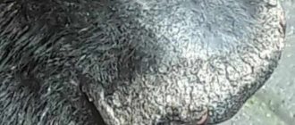
In a male dog, in the anterior part of the penis there is a bone 8–10 cm long, covered with the cavernous body of the glans. The upper edge of the bone is convex, at the bottom there is a groove in which the urogenital canal passes, and at the front it is built up with cartilage or fibrous tissue. The head of the penis is long and cylindrical with a pointed end. At the base of the male’s penis, during erection, the so-called “bulbs” swell, which increase in size (about 5 times compared to the normal state) and prevent the penis from being removed from the bitch’s vagina. The prepuce has two leaves on which lymphatic follicles are located at the base of the head. The length of the penis is up to 15 - 20 cm, depending on the size of the breed. [8] [5]
Similarly for wolves, foxes, and other canines.
Briefly about the main thing
- The skeleton consists of 290 bones, 262 joints. It supports the body and protects the organs.
- The jaw is one of the parts of the skull. An adult dog should have 42 teeth, and the bite depends on the breed.
- The internal organs are formed into the digestive, respiratory, circulatory, urinary, nervous and reproductive systems.
- Dogs' sense organs (with the exception of taste) are much better developed than those of humans. The main elements are eyes, ears, nose, tongue.
Do you know the location of your dog’s internal organs well? Share interesting facts about the structural features of these animals in the comments.