Cardiomyopathy in dogs is a serious heart disease. Elderly individuals and representatives of giant breeds are especially vulnerable to the disease. Statistics show that the gender of the dog plays a large role in the development of this cardiac pathology. Males are diagnosed with this disease more often than females. The article will provide detailed information about the types of cardiomyopathy, list its signs and most striking symptoms, and also discuss treatment methods.
general characteristics
Veterinarians usually mean by this concept cardiac pathology, which provokes dysfunction of myocardial contractility and disturbances in the normal functioning of the pumping function of the heart. The disease is accompanied by degeneration of the walls of the ventricles and expansion of the heart chambers, as well as congestive processes in the pet’s body and arrhythmia.
Cardiomyopathy is of primary and secondary types. The primary type occurs due to congenital defects of the heart muscle, but this occurs extremely often. Much more often, specialists are faced with a secondary type of illness that occurs against the background of diseases of viral, fungal or bacterial origin.
How does it affect the functioning of the body?
The left ventricle plays an important role in the large circle of hematopoiesis. It ensures blood flow to all organs and tissues of the body. The expansion of this department can threaten the development of serious complications. Here is what threatens an enlarged left atrium:
- heart failure - when a person's main organ loses the ability to pump a sufficient volume of blood to ensure normal functioning of the body;
- abnormal heart rhythm or arrhythmia;
- the development of a heart attack when the blood supply to the heart is interrupted;
- disturbance in the supply of oxygen to the tissues of the heart muscle;
- heart failure.
As you can see, left atrial hypertrophy leads to serious and sometimes irreversible complications. That is why, as soon as any sign of abnormal heart function is detected, you should immediately contact a specialist. It is important to identify pathology in the early stages of development to avoid severe consequences.
Types of disease
In addition to distinguishing by the nature of occurrence, veterinarians also distinguish varieties by what transformations occur in the cardiac tissue. These should include:
- Hypertrophic cardiomyopathy. It is characterized by a proportional increase in both the overall size of the heart and the thickness of the walls of the atria and ventricles. If the animal is elderly, then it begins to lack the strength to maintain the normal functioning of the grown organ. The reduction in the volumes of the atria and ventricles results in the dog’s blood receiving half as much oxygen and nutrients. In its advanced form, it can cause myocardial infarction or sudden cardiac arrest in the dog.
- Dilated cardiomyopathy. Experts are convinced that this type of pathology occurs most often in dogs. Sometimes dilation smoothly follows from the type of cardiomyopathy described above. In this case, the heart wall becomes amorphous, its texture loses its primary elasticity. This results in the organ being unable to contract normally. The dog develops severe shortness of breath and increased fatigue. The prognosis for cure is extremely unfavorable.
- Restrictive. In cardiac tissue, uncontrolled formation of fibrous fibers occurs, which are made from a vital organ - an unsuccessful analogue of cartilage. As a result, low myocardial contractility, leading to problems with oxygen entering the pet’s body. The symptoms are aggravated by the fact that the shaggy friend develops distinct painful sensations in the chest area.
- Mixed. It is characterized by a combination of signs of fibrosis of cardiac tissue with its hypertrophy. Variations in pathological processes may include concomitant expansion of the ventricles or atria of the organ.
Diagnosis criteria for canine dilated cardiomyopathy
Vladislava Illarionova, veterinary clinic “Biocontrol” (Moscow)
Dear Colleagues!
The article, written by an active member of our society, Vladislava Konstantinovna Illarionova, is the quintessence of the work done by a large number of veterinary cardiologists in the society in preparation for the recent conference (SVM No. 2, p. 13). The information in the article can be considered as the consensus Society Recommendations for the diagnosis of dilated cardiomyopathy. In the future, an article is planned on recommendations for the treatment of this disease.
Andrey Komolov, President of the Veterinary Cardiological Society
Abbreviations: DCM - dilated cardiomyopathy, CMP - cardiomyopathy, AHA - American Heart Association, BW - body weight.
Dilated cardiomyopathy is a myocardial disease manifested by dilatation of the ventricles of the heart and a decrease in their contractility [5]. Large and giant dogs are predisposed to this disease [18]. There are a number of predisposed breeds, which include: Dobermans, Newfoundlands, Boxers, Great Danes, Portuguese Water Dogs, Cocker Spaniels and Irish Wolfhounds [13]. Mestizos get sick less often than purebred animals. Males are more predisposed to developing the disease [11]. With age, the prevalence of the pathology increases.
Human dilated cardiomyopathy, as a separate pathology, was first described by W. Bridgen in 1957. With this term he designated “myocardial diseases of unknown etiology, not associated with atherosclerosis, tuberculosis and rheumatic heart disease, characterized by the appearance of cardiomegaly of unknown origin and the development of heart failure.” It took about half a century to study various forms of human cardiomyopathies, and in 2006 the AHA proposed a new definition, according to which CMP is a heterogeneous group of diseases of various etiologies (often genetically determined), accompanied by mechanical and/or electrical dysfunction of the myocardium and, in some cases, disproportionate hypertrophy or dilatation. Myocardial damage in CMP can be primary or secondary (with a systemic disease), accompanied by the development of heart failure and ending in the sudden death of the patient. In humans, all CMPs were divided into two large groups: ischemic and non-ischemic. The non-ischemic form, in turn, is divided into primary and secondary. The primary form includes a genetically determined pathology, which is often monogenic, has an autosomal dominant type of inheritance [25] and a prevalence of 35% [26]. The secondary form includes all specific (secondary) CMPs.
In veterinary cardiology, CMP has been considered since 1970. According to the classification, dilated, hypertrophic, restrictive, unclassified forms and arrhythmogenic dysplasia of the right ventricle are distinguished. In dogs, the most common variant is DCM. Arrhythmogenic right ventricular dysplasia is more often diagnosed in boxers and English bulldogs [42]. HCM acts as a rare form that develops secondary to arterial hypertension. All forms are divided into two groups: primary and specific (secondary).
Specific CMPs are considered within the pathologies that lead to their formation [6]. For example, nutritional CMP develops with a lack or disturbance of the metabolism of such nutrients as L-carnitine and taurine, the inflammatory form of CMP manifests itself as a complication of myocarditis [3; 4; 2], the toxic form is more often discussed within the framework of antitumor chemotherapy; ischemic CMP can extremely rarely accompany hypercholesterolemia in hypothyroidism and diabetes mellitus. There are no alcoholic or postpartum forms in dogs.
Thus, primary CMP is a diagnosis of exclusion. The etiology of primary CMPs remained unknown for a long time, so they were classified as idiopathic diseases. Numerous works in the field of genetics have shown the decisive role of hereditary predisposition in the development of primary CMP in dogs [8; 2; 9; 10]. In such cases, myocardial damage may be associated with a mutation in a gene (monogenic diseases) encoding three main classes of myocardial proteins: structural, regulatory, and those involved in the generation and conduction of excitation. The most common mutations in structural protein genes lead to changes in contractile, cytoskeletal and membrane proteins. Polygenic diseases arise from the combination of several unfavorable DNA changes (polymorphisms) that only partially affect the function of the encoded proteins. Each polymorphism changes the functions of the heart muscle to a small extent, but the combination of such molecular defects can lead to a weakening of the compensatory reserves of the system, especially in combination with other unfavorable factors, such as the administration of cardiotoxic drugs [14].
Monogenic diseases can be transmitted in a dominant or recessive manner and have several main modes of inheritance:
1. Autosomal dominant (one mutant allele is enough to develop the phenotype) - described in Newfoundlands, Pinschers [27], Boxers and Great Danes [32];
2. Autosomal recessive (the disease appears only in homozygous carriers) - described in Portuguese water dogs;
3. X-linked recessive (the mutant gene is located on the X chromosome and appears in males or females in a homozygous state) - described in Great Danes [9].
Clinical signs of the disease may appear to varying degrees in patients with the same genetic mutation. This is due to the different expressiveness of the disease, that is, the degree of manifestation of the pathology. Penetrance, that is, the frequency of phenotypic manifestation of a trait for a certain genotype, also plays a role in the occurrence of the disease. It is known that penetrance depends on time, since over the years, damage caused by a genetic defect accumulates in the body, and the risk of phenotypic manifestation of the disease increases. Gender can also influence the degree of manifestation of a pathological symptom.
DCM in dogs is divided into two histological types. The first type is associated with the formation of atrophied thin wavy myocytes, edema of the intercellular space and the development of subendocardial fibrosis. This type is the most common [19]. The second histological type is characterized by fatty infiltration, vacuolization and fragmentation of myocytes. It is found in such breeds as the boxer [20], 1983), the English bulldog [21] and the Irish wolfhound [22]. Changes similar to the second type have also been described in Doberman Pinschers [23; 24]. Sometimes histological examination reveals a mixed picture.
Autopsy reveals an increase in myocardial mass due to eccentric hypertrophy, predominant dilatation of the left or all four chambers of the heart (Fig. 1), relative thinning of the walls, smoothness of the papillary muscles, pale, soft myocardium [18]. The fibrous rings of the atrioventricular valves expand, but the leaflets do not change. Minimal myocardial fibrosis is detected. The coronary arteries do not undergo changes.
| Ill. 1. Macroscopic specimen of the heart of a Staffordshire Terrier diagnosed with dilated cardiomyopathy |
According to the results of one of the epidemiological studies (1986–1991), the prevalence of DCM in dogs is not very high and amounts to 0.5% [11]. However, in this work, only animals with clinical signs of CHF were taken into account. Further studies showed a greater predisposition in purebred dogs compared to mixed breeds and the existence of predisposed breeds with a very high prevalence of the disease. Thus, in prospective studies conducted in Dobermanns, the disease was detected in 63.2% of animals in the American population [15] and in 58.2% of dogs in the European population [16]. In Irish wolfhounds - 24.2%, in Newfoundlands - 17.6% [17], in Great Danes - 47% [32]. The data obtained take into account not only dogs with symptoms of CHF, but also animals with an asymptomatic stage of the disease.
When studying the nature of the progression of DCM, it was revealed that the pathology has a long latent period, which can last for several years [28; 29]. At this stage, dogs do not show any clinical signs and the disease can only be diagnosed through cardiac testing. This course is due to the fact that the development of pathology leads to the formation of CHF, in which compensatory mechanisms are mobilized in the first stages, which allows maintaining cardiac output at a sufficient level. The synchronous interaction of neurohormonal systems allows the heart to be in a state of adaptation to the emerging pathological conditions. Activation of protective forces makes it possible to compensate for lost functions, but over time leads to pathological changes in the body and ends in a “failure” of adaptation. From this moment on, a period of decompensation begins with the appearance of characteristic clinical signs.
The clinical picture often develops quickly - within a few days. Lethargy, exercise intolerance, tachypnea, shortness of breath and cough appear. As the disease progresses, anorexia, polydipsia, decreased body weight, ascites, hydrothorax and fainting occur. Clinical signs may be expressed to varying degrees and appear at different times. In Dobermanns and Boxers, fainting and sudden death may be the first manifestations of the disease [18], since dogs of these breeds have an arrhythmic stage, which is characterized by the development of fatal ventricular arrhythmias. A study of the European Doberman population showed that 37% of dogs with this pathology are at the arrhythmic stage of the disease. The same study found an increase in the prevalence of the disease with age. Thus, in dogs 1–2 years old, pathology was detected in 3.3% of cases, while in dogs over 8 years old - in 44.1% of cases. It also indicates a greater predisposition of females to the development of the arrhythmic form, and males to the formation of structural changes in the heart, determined by echocardiography [35]. Great Danes and Irish Wolfhounds are prone to developing atrial fibrillation [32; 22], which may precede or accompany the development of myocardial dysfunction. Animals with isolated atrial fibrillation may not have symptoms of the disease for a long time, but an increase in heart rate with this arrhythmia provokes over time the development of the arrhythmic form of DCM.
Clinical examination reveals expiratory or mixed shortness of breath of varying intensity, tachypnea. When examining the mucous membranes, there is pallor and enlarged SNK. The apex beat of the heart is diffuse, reduced in strength. The pulse in the femoral arteries is frequent with reduced filling. With concomitant arrhythmias, a pulse deficit is determined. Right ventricular failure is manifested by distension of the jugular veins, hepatojugular reflux, ascites, hydrothorax and hydropericardium. In the later stages of heart failure, cachexia, loss of muscle mass progresses, and hyperthermia may occur [33]. When auscultating the lungs, bronchial breathing and moist rales are heard. On auscultation of the heart, the sounds are slightly muffled, a third heart sound and a weak noise of relative atrioventricular valve insufficiency (usually not exceeding 3/6 loudness) may occur. With severe heart failure, sinus tachycardia develops with the disappearance of sinus arrhythmia. Clinical manifestations may vary depending on the stage of heart failure and the breed.
When assessing biochemical and clinical blood tests, it is usually not possible to identify any significant deviations from the reference values of the indicators. In severe heart failure, elevated urea levels can be detected as a result of prerenal azotemia, which develops with a decrease in cardiac output. Troponin I, a marker of myocardial damage, is usually slightly elevated. For Dobermanns, the limit for the value of this parameter was determined to be 0.22 ng/ml, the excess of which indicates DCM with a sensitivity of 79.5% and specificity of 84.4% [36]. In addition, the assessment of this indicator allows for a differential diagnosis of DCM with acute myocardial damage as a result of myocarditis, in which the level of troponin I becomes very high.
Electrocardiography is not an expert method for diagnosing DCM, since ECG changes in this disease are nonspecific. With pronounced dilatation of the heart chambers on the electrocardiogram, you can see signs of expansion of the left atrium (increased duration of the P wave) and left ventricle (increased amplitude of the R wave and increased duration of the QRS complex), signs of subendocardial myocardial ischemia (decrease in the S-T segment below the isoline by more than 0.2 mV). However, electrocardiography is of greatest value for diagnosing rhythm and conduction disorders. The most common rhythm disorder in DCM is atrial fibrillation (atrial fibrillation). This arrhythmia is characterized by the occurrence of rapid, uncoordinated contractions of the atria with irregular conduction to the ventricles. The most common is the tachysystolic form of atrial fibrillation with a heart rate of more than 180 beats per minute. Most Boxers and Dobermans experience ventricular rhythm disturbances: ventricular extrasystoles and ventricular tachycardia [34]. In Dobermans, extrasystoles and rhythms are more often formed in the left, and in boxers in the right ventricle, although this condition is not necessary. The development of ventricular tachycardia with a heart rate of more than 230 beats per minute can have fatal consequences, so timely diagnosis is necessary, including a five-minute ECG recording and Holter ECG monitoring (Fig. 2).
| Ill. 2. Fragment of a Holter ECG monitoring recording in a Doberman dog diagnosed with DCM. Episode of ventricular tachycardia |
In Dobermanns, a five-minute ECG recording with at least one ventricular extrasystole showed 64.2% sensitivity and 96.7% specificity [35] for identifying animals with 100 or more ventricular extrasystoles per day (such data were not obtained for boxers). This suggests that if at least one ventricular extrasystole is detected on the electrocardiogram in five minutes, the probability of an arrhythmic stage is very high.
The 24-hour Holter monitoring method is the gold standard in detecting arrhythmias. This study is of particular value for diagnosing the arrhythmic stage of DCM in boxers and Dobermans, since it is the only method that can provide information during this period. When conducting Holter monitoring in boxers, a rhythm containing more than 50 ventricular extrasystoles per day, especially with the presence of couplets and triplets, is considered pathological [37]. In this breed, the arrhythmic stage can last for years and be combined with minimal structural changes, as has been shown in the American boxer population [38]. In European dogs of this breed, arrhythmias are often complicated by pronounced morphological changes in the heart [39]. Conducting Holter monitoring is a mandatory condition for screening studies of Dobermanns participating in breeding. An animal that has more than 100 ventricular extrasystoles per day is considered sick. In addition, this method is used to register arrhythmias and evaluate the results of antiarrhythmic therapy in dogs of any breed at various stages of DCM.
Changes in radiographic examination of the heart are not specific. This method is used mainly to determine signs of venous stasis, pulmonary edema and pleural effusion. With venous pulmonary stagnation, the expansion of the pulmonary vein of the cranial lobe of the lung and the strengthening of the vascular pattern of the lungs due to the pulmonary veins are determined. With interstitial pulmonary edema, the interstitial pattern of the lungs intensifies; with alveolar edema, “cotton wool” shading appears in the roots and in the caudal lobes of the lungs (often with a picture of air bronchograms). With the development of right-sided heart failure, dilation of the caudal vena cava and signs of hydrothorax are noted. Detection of cardiomegaly indicates significant changes in the heart. As a rule, the left chambers of the heart enlarge first, and as the disease progresses, the heart acquires a rounded shape due to dilatation of all chambers.
Echocardiographic examination makes it possible to detect myocardial dysfunction of a predominantly systolic nature and dilatation of the left or all four cardiac chambers, while excluding other congenital and acquired heart pathologies. For research, B-mode, M-mode and Doppler modes are used. In M- and B-modes, measurements are taken of the linear dimensions of the cavities and the thickness of the walls of the heart. To obtain average values of these parameters, it is recommended to carry out measurements in five cycles for sinus rhythm and in ten cycles for atrial fibrillation. DCM is characterized by eccentric hypertrophy of the left and often right ventricles and a diffuse decrease in the amplitude of myocardial movement (Fig. 3.).
| Ill. 3. Echocardiogram of a Doberman dog diagnosed with dilated cardiomyopathy. Reduced amplitude of myocardial motion (M-mode) |
In this case, an increase in the end-diastolic size of the left ventricle indicates its volume overload, and an increase in the end-systolic size indicates a decrease in myocardial contractility. For Doberman Pinschers, an excess of the GDV of more than 48 mm (for males) and more than 46 mm (for females) and a GFR of more than 36 mm is an indicator of DCM [36]. The wall thickness remains within normal values. With pronounced dilatation of the left ventricular cavity, the relative thickness of the walls decreases significantly.
A change in the geometry of the left ventricle leads to an increase in the short radius of its cavity and the degree of sphericity. In this case, the sphericity coefficient becomes less than 1.65 (Fig. 4.).
| Ill. 4. Echocardiogram of a Doberman dog diagnosed with dilated cardiomyopathy. Calculation of the sphericity index |
This parameter was assessed in Dobermanns with DCM and showed 86.6% sensitivity and 87.6% specificity [40]. In addition, according to one-dimensional echocardiography (M-mode), it is possible to determine the distance from the peak E of the movement of the anterior mitral valve leaflet to the interventricular septum (EPSS), which showed a very high level of sensitivity (100%) and specificity (99.0%) for diagnosis DCM in Dobermans with a value greater than 6.5 mm [40].
With spectral Doppler ultrasound, a decrease in peak blood flow velocity in the outflow tracts of the left and right ventricles is noted, which is associated with a decrease in their contractility. Often a restrictive type of mitral flow is recorded with a high E wave and a decrease in the deceleration rate of flow, a shortening of the left ventricular isovolumic relaxation time (IVRT), which reflects an increase in end-diastolic pressure in the left ventricle. With a significant increase in the volume of the heart cavities and expansion of the fibrous rings of the atrioventricular valves, relative insufficiency of the mitral and tricuspid valves is recorded. DCM is characterized by the central position of the regurgitant flow on the atrioventricular valves (Fig. 5.).
The size of the left atrium increases significantly as the pathology progresses (Fig. 6). Right side enlargement may be associated with cardiomyopathy or result from the development of pulmonary hypertension.
| Ill. 5. Echocardiogram of a Doberman dog diagnosed with dilated cardiomyopathy. Regurgitation on the mitral valve. Color Doppler mapping |
| Ill. 6. Echocardiogram of a Doberman dog diagnosed with dilated cardiomyopathy. Significant dilatation of the left atrium (short axis at the level of the aortic root) |
To assess left ventricular systolic function, shortening fraction measurements obtained in M-mode at the chordal level along the long or short axis of the left ventricle are routinely used. At the preclinical stage of DCM, this indicator decreases, becoming less than 25%, and when the disease passes into the symptomatic stage - less than 15%. However, it must be borne in mind that while maintaining the normal size and geometry of the cavities, a reduced shortening fraction (20–25%) may be a normal option for some large dogs. It should be noted that the assessment of contractility by the shortening fraction has a number of disadvantages associated with the possible measurement error in the presence of zones of hypokinesia, since this method uses the determination of the degree of anteroposterior shortening of the left ventricle in a one-dimensional mode. In addition, there is a possibility of overestimation of values with severe mitral insufficiency. To overcome the described limitations, the volume of the left ventricle is calculated from two-dimensional echocardiography data using the Simpson disk method. This method is based on obtaining a long-axis (or apical) image of the left ventricle during systole and diastole, after which the images are automatically divided into 20 disks of equal height. Disc volumes are summed to give end-diastolic and end-systolic volumes, which are used to calculate ejection fraction. To obtain average values, it is recommended to measure in five cycles for sinus rhythm and ten for atrial fibrillation. In DCM, the ejection fraction should be below 40% [1], but there are no studies to support these recommendations. End-systolic and end-diastolic volumes of the left ventricle are normalized to the body surface area, obtaining the corresponding indices. A number of studies were conducted to obtain reference values for these indicators for Boxer and Doberman dogs. For boxers, normal values of EDOi are 49–93 ml/m2, ECOi 22–50 ml/m2 [41]. For Dobermanns, the upper limit of normal has been determined to be 95 ml/m2 - EDOi and 55 ml/m2 - ECOi [36].
Thus, an integrated approach to the diagnosis of dilated cardiomyopathy in dogs allows not only to diagnose the disease when characteristic symptoms appear, but also to suspect the disease at earlier stages, which solves the problem of removing sick animals from the breeding program. High alertness towards this pathology can reduce the spread of the disease and improve the quality and life expectancy of sick animals.
Literature
1. Dukes-McEwan, J. Proposed guidelines for the diagnosis of canine idiopathic dilated cardiomyopathy. ESVC Taskforce for Canine Dilated Cardiomyopathy / J. Dukes-McEwan, M. Borgarelli, A. Tidholm, AC Vollmar, J. Haggstrom // J Vet Cardiol. — 2003 Nov. - V. 5. - N. 2. - R. 7–19.
2. Oyama, MA Decreased triadin and increased calstabin2 expression in Great Danes with dilated cardiomyopathy / MA Oyama, SV Chittur, CA Reynolds // J Vet Intern Med. — 2009 Sep-Oct. - V. 23. - N. 5. - R. 1014–1019.
3. Higgins, R. Canine distemper virus-associated cardiac necrosis in the dog / S. Krakowka, A. E. Metzler, A. Koestner // Veterinary Pathology. - 1981. - N. 18. - P. 472–486.
4. Knee, B. W. Evidence for the role of myocarditis in the pathophysiology of dilated cardiomyopathy. In: Proceedings of the 11th ACVIM Forum/ BW Knee // Washington DC, 1993 - P. 565–567.
5. Richardson P, McKenna W, Bristow M, et al. Report of the 1995 World Health Organization/International Society and Federation of Cardiology Task Force on the definition and classification of cardiomyopathies. Circulation 1996; 93:841–842.
6. Cobb MA. Idiopathic dilated cardiomyopathy: advances in aetiology, pathogenesis and management. J Small Anim Pract1992; 33: 113–118.
7. Schönberger J, Seidman CE. Many roads lead to a broken heart: the genetics of dilated cardiomyopathy. Am J Hum Genet 2001; 69: 249–260.
8. Meurs KM. Insights into the heritability of canine cardiomyopathy. Vet Clin North Am Small Anim Pract 1998;28:1449–1457.
9. Meurs KM, Magnon AL, Spier AW, et al. Evaluation of the cardiac actin gene in Doberman Pinschers with dilated cardiomyopathy. Am J Vet Res 2001;62:33–36.
10. Stabej P, Imholz S, Versteeg SA, et al. Characterization of the canine desmin (DES) gene and evaluation as a candidate gene for dilated cardiomyopathy in the Doberman. Gene 2004;340:241–249.
11. Sisson DD, Thomas WP. Myocardial diseases. In Ettinger SJ, Feldman EC ed. Textbook of Veterinary Internal Medicine, 4th edition. . Philadelphia, W. B. Saunders; 1995: 995-1032.
12. G. Wess, J. Maurer, J. Simak, and K. Hartmann. Use of Simpson's Method of Disc to Detect Early Echocardiographic Changes in Doberman Pinschers with Dilated Cardiomyopathy. J Vet Intern Med 2010;24:1069–1076.
13. Martin MWS, MJ Stafford Jonsjn, G. Strehlay, JN King. Canine dilated cardiomyopathy: a retrospective study of prognostic findings in 367 clinical cases. Journal of Small Animal Practice (2010) 51, 428–436
14. Fatkin D, Graham RM. Molecular mechanisms of inherited cardiomyopathies. Physiol Rev 2002;82:945–980.
15. O'Grady MR, Horne R. The prevalence of dilated cardiomyopathy in Dobermann pinschers: a 4.5 year follow-up. Proceedings of the 16th ACVIM Forum 1998: 689.
16. Wess G., A. Schulze, V. Butz, J. Simak, M. Killich, L. J. M. Keller, J. Maeurer and K. Hartmann. Prevalence of Dilated Cardiomyopathy in Doberman Pinschers in Various Age Groups. J Vet Intern Med 2010; 1–6.
17. Dukes-McEwan J. Echocardiographic / Doppler criteria of normality, the findings in cardiac disease and the genetics of familial dilated cardiomyopathy in Newfoundland dogs. PhD thesis. University of Edinburgh. 1999.
18. Tidholm A., Jönsson L. A retrospective study of canine dilated cardiomyopathy (189 cases). J Am Anim Hosp Assoc1997; 33: 544-550. PhD thesis. University of Edinburgh. 1999.
19. Dambach DM, Lannon A, Sleeper MM, Buchanan J. Familial dilated cardiomyopathy of young Portuguese water dogs. J Vet Intern Med 1999; 13: 65–71.
20. Harpster NK. Boxer cardiomyopathy. In Kirk RW ed. Current Veterinary Therapy. Small Animal Practice VIII. Philadelphia, W. B. Saunders; 1983: 329–337.
21. Santilli RA, Bontempi LV, Perego M, Fornai L, Basso C. Outflow tract segmental arrhythmogenic right ventricular cardiomyopathy in an English Bulldog. J Vet Cardiol. 2009 Jun;11(1):47–51. doi: 10.1016/j.jvc.2009.03.006. Epub 2009 May 23.
22. Vollmar AC, Aupperle HJ. Cardiac pathology in Irish wolfhounds with heart disease. Vet Cardiol. 2021 Mar;18(1):57–70. doi: 10.1016/j.jvc.2015.10.001. Epub 2015 Dec 11.
23. Calvert CA, Hall G, Jacobs G, Pickus C. Clinical and pathological findings in Doberman Pinschers with occult cardiomyopathy that died suddenly or developed congestive heart failure: 54 cases (1984–1991). J Am Vet Med Assoc1997; 210:505–511.
24. Everett RM, McGann J, Wimberly HC, Althoff J. Dilated cardiomyopathy of Doberman pinschers: retrospective histomorphologic evaluation on heart from 32 cases. Vet Pathol 1999; 36:221–227.
25. Mestroni L, Rocco C, Gregori D, Sinagra G, Di Lenarda A, et al. (1999) Familial dilated cardiomyopathy: evidence for genetic and phenotypic heterogeneity. J Am Coll Cardiol 34:181–190.
26. Grünig E, Tasman JA, Kücherer H, Franz W, Kübler W, et al. (1998) Frequency and phenotypes of familial dilated cardiomyopathy. J Am Coll Cardiol 31:186–194.
27. Meurs KM, Fox PR, Norgard M, Spier AW, Lamb A, et al. (2007) A prospective genetic evaluation of familial dilated cardiomyopathy in the Doberman pinscher. J Vet Intern Med 21:1016–1020.
28. Calvert CA, Pickus CW, Jacobs GJ. Unfavorable influence of anesthesia and surgery on Doberman pinschers with occult cardiomyopathy. J Am Anim Hosp Assoc 1996; 32:57–62.
29. Calvert CA, Meurs K (2000) Doberman pinscher occult cardiomyopathy. In: Bonagura JD, Kirk RW, editors. Current Veterinary Therapy XIII. Philadelphia: W. B. Saunders. pp. 756–760.
30. Wess G, Schulze A, Butz V, Simak J, Killich M, et al. (2010) Prevalence of dilated cardiomyopathy in Doberman Pinschers in various age groups. J Vet Intern Med 24:533–538.
31. Meurs KM, Miller MW; Nicola A. Clinical features of dilated cardiomyopathy in Great Danes and results of a pedigree analysis: 17 cases (1990–2000), JAVMA, Vol 218, No. 5, March 1, 2001
32. Stephenson HM, Fonfara S, Lopez-Alvarez J, Cripps P, Dukes-McEwan J. Screening for Dilated Cardiomyopathy in Great Danes in the United Kingdom. J Vet Intern Med 2012;26:1140–1147.
33. Tidholm A, Jönsson L. Dilated cardiomyopathy in the Newfoundland: a study of 37 cases (1983-1994). J Am Anim Hosp Assoc 1996; 32:465–470.
34. Harpster NK. Boxer cardiomyopathy. In Kirk RW ed. Current Veterinary Therapy. Small Animal Practice VIII. Philadelphia, W. B. Saunders; 1983: 329–337.
35. Wess G., Simak J., Mahling M., Hartmann K. Cardiac troponin I in Doberman Pinschers with cardiomyopathy. J Vet Intern Med. 2010 Jul-Aug;24(4):843–9. doi: 10.1111/j.1939-1676.2010.0516.x. Epub 2010 Apr 16.
36. Wess G, Schulze A, Geraghty N, Hartmann K. Ability of a 5-minute electrocardiography (ECG) for predicting arrhythmias in Doberman Pinschers with cardiomyopathy in comparison with a 24-hour ambulatory ECG. J Vet Intern Med. 2010 Mar-Apr;24(2):367–71.
37. Borgarelli M, Tarducci A, Tidholm A, Häggström J. Canine idiopathic dilated cardiomyopathy. Part II: Pathophysiology and therapy. Vet J 2001; 162:182–195.
38. Meurs KM, Spier AW, Miller MW, et al. Familial ventricular arrhythmias in Boxers. J Vet Intern Med 1999; 13:437–439.
39. Wotton PR. Dilated cardiomyopathy (DCM) in closely related boxer dogs and its possible resemblance to arrhythmogenic right ventricular cardiomyopathy (ARVC) in humans. Proceedings of the 17th ACVIM Forum 1999: 88–89.
40. Holler PJ, Wess G. Sphericity index and E-point-to-septal-separation (EPSS) to diagnose dilated cardiomyopathy in Doberman Pinschers. J Vet Intern Med. 2014 Jan-Feb;28(1):123–9. doi: 10.1111/jvim.12242. Epub 2013 Nov 13.
41. Smets P, Daminet S, Wess G. Simpson's method of discs for measurement of echocardiographic end-diastolic and end-systolic left ventricular volumes: breed-specific reference ranges in Boxer dogs. J Vet Intern Med. 2014 Jan-Feb;28(1):116–22. doi: 10.1111/jvim.12234. Epub 2013 Nov 1.
42. Santilli RA, Bontempi LV, Perego M. Ventricular tachycardia in English bulldogs with localized right ventricular outflow tract enlargement. J Small Anim Pract. 2011 Nov;52(11):574–80. doi: 10.1111/j.1748-5827.2011.01109.x. Epub 2011 Oct 10.
SVM No. 3/2016
Rate material
Like Like Congratulations Sympathy Outrageous Funny Thoughtful No words
Causes
Experts find it difficult to accurately determine the factors that can lead to cardiomyopathy. Studies have shown that one of the reasons may be a lack of L-carnitine and taurine in the pet’s body. Some medium and large breeds of dogs are also prone to this pathology, in particular boxers, German shepherds, Newfoundlands and Dobermans. In puppies of these breeds, it can be transmitted through the genes of the parents. Cocker spaniels are the only small breed that is vulnerable to this disease.
Veterinarians identify the following causes of the disease:
- Diseases of infectious etiology.
- Serious metabolic disorders.
- Dysfunction in the coronary circulation in a pet.
- Inflammatory processes occurring in the body.
- Poisoning and side effects of drugs.
- Heart valve pathologies and myocarditis.
Doctors agree that cardiomyopathy is in many ways the final stage of pathological processes and metabolic defects progressing in the dog’s body. The weakened immunity of the dog and the lack of vitamins in its body, in particular selenium and vitamins E and B12, provide fertile ground for the occurrence of the disease.
Symptoms of the disease
In the early stages, the pathology does not show any signs of its presence. This is an extremely indolent disease that can progress over many months and sometimes years. There are often cases when it is detected quite spontaneously, during an ultrasound of the pet’s heart to determine if it has any other ailments. Clinical symptoms can develop either quickly or slowly. The sudden death of a pet, which was not preceded by any pronounced signs, is quite possible.
This disease can manifest itself in a dog as follows:
- The slightest physical exertion or minor excitement causes severe shortness of breath in the animal. The dog gets tired quickly and sleeps a lot;
- dry, hacking cough;
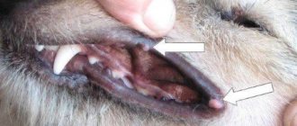
- the mucous membranes in the mouth turn pale;
- the weight of the pet is significantly reduced, while the volume of the abdomen increases (in veterinary medicine this is called abdominal dropsy);
- the dog may lose consciousness, respond sluggishly to commands, and lack interest in games and walks;
- convulsive seizures periodically occur at night;
- if the dog has developed pulmonary edema, then gurgling sounds will appear when breathing;
- low blood pressure.
The development of the disease in the preclinical (latent) stage complicates its timely diagnosis. Obvious symptoms appear too late for conservative treatment methods to be used. This is what complicates the future treatment of cardiomyopathy in dogs and makes the prognosis for recovery less optimistic.
The veterinarian can make a diagnosis only on the basis of anamnesis and physical examination, which must be supplemented by chest x-ray, ultrasound and ECG. Attentive owners should immediately take their pet to the veterinary clinic if they notice that they become tired much faster while walking, and even the slightest obstacle causes the dog to experience severe shortness of breath and coughing.
Symptoms of an enlarged heart in dogs
(Photo: Getty Images)
Symptoms of an enlarged heart in dogs can vary depending on how advanced the condition is. Sometimes veterinarians may miss signs of a dilated heart if dogs show no symptoms or only show mild signs of dilated cardiomyopathy. However, many subtle signs can be detected during a routine physical examination that would otherwise not be noticeable, so it is always important to keep up with normal checks. Here are some signs of an enlarged heart in dogs.
- Irregular or weak impulse
- Muffled breathing or crackling sounds when breathing
- lethargy
- Weakness
- Loss of appetite
- Weight loss
- Fast breathing
- Irregular breathing
- Exercise intolerance
- coughing
- Heart murmurs
- arrhythmia
- vomit
- Abdominal distension (ascites)
- Fainting (fainting)
- Sudden death
Treatment of the disease
Unfortunately, treatment of cardiomyopathy in dogs almost never produces a lasting effect. In the case of primary type pathology, it is generally impossible. The dog owner should not count on a complete recovery. The only thing therapy can help with is increasing the length and quality of life of the animal, as well as mitigating clinical symptoms. The treatment regimen largely depends on the stage of development of the disease process.
As a rule, experts advise starting therapy with the dog taking diuretics. They prevent the risk of developing congestion in the body, including such dangerous consequences as pulmonary edema, which can lead to the death of a pet. Furosemide is perfect for these purposes.
The inotrope Digoxin will help prevent atrial fibrillation, and ventricular extrasystole - Procainamide, which belongs to the group of antiarrhythmic drugs. In an emergency situation, when a pet’s heart loses the ability to contract synchronously, veterinarians use Dobutamine.
Recently, several enzymes have found widespread use in the treatment of cardiomyopathy. With their help, blood supply to the heart itself can be improved, and the risk of developing a heart attack is halved. In addition, they are able to nourish and restore heart tissue affected by the disease. The most famous among them is L-carnitine. The substances it contains have a beneficial effect on the metabolic process and also help neutralize harmful toxins in tissue cells.
It is important to understand that in addition to drug treatment, you also need to try to put less stress on the animal; stress and anxiety are strictly not recommended for it. The intensity of training and exercise should be significantly reduced during therapy. A necessary measure for a quick recovery is a proper diet. It is necessary to exclude heavy and fatty foods from your pet’s menu, and give more foods containing fiber and vitamins. Veterinary pharmacies sell special foods prescribed for use by pets suffering from cardiomyopathy.
Emergency treatment
In case of acute heart failure, the background of which is cardiomyopathy, the veterinarian carries out the following sequential procedures:
- Injection of Furosemide into a vein.
- Use of nitroglycerin ointments or their analogues (spray or infusion).
- Inotropic support is provided.
- The dog is placed in an oxygen chamber.
- If the patient is too worried, then light sedatives are indicated.
- If there is fluid in the pleural cavity, the specialist performs thoracentesis followed by pumping out the effusion.
Emergency care for heart failure requires experience and skill from the doctor. The slightest delay can lead to complications of symptoms and subsequent death of the pet.
Long-term therapy
This implies lifelong use of medications, with regular monitoring of the animal’s condition by a qualified specialist and rationing of dosages. Immediately after the start of treatment, the owner must come with the pet to the veterinary clinic at least three times a week, and when the animal’s condition stabilizes - 3-4 times a month.
The dog is prescribed medications such as:
- Taurine, Omega-3 fatty acids, L-carnitine;
- ACE inhibitors (use with extreme caution in combination with potassium supplements, they can cause hyperkalemia);
- Furosemide, combining it with potassium supplements;
- beta blockers (Metoprolol);
- for atrial fibrillation - Digoxin;
- for supraventricular tachyarrhythmia - Vetmedin.
Surgeons have developed special devices for the treatment of cardiomyopathy that can improve myocardial contractility and prolong its functional activity. Using them, the owner of the animal will extend his life for many years.
Forecast
The prognosis for recovery depends entirely on the stage of the pathology and the general condition of the dog. If the disease is complicated by pulmonary edema, then life expectancy depends on how quickly the dog was taken to the veterinarian. In the case where the owners did not react in time, you need to understand that the animal will live for 2-3 months at most. Prompt diagnosis and a good response to therapy will give your four-legged pet a life span of 6-7 months to 1 year. Determining the pathology at the very initial stages increases this period to 3-5 years or more.
Finally, I would like to say that cardiomyopathy is a terrible and insidious disease that can quickly kill a dog if it is not detected in time. Therefore, owners need to be extremely attentive and caring with their four-legged friend, and respond to the slightest symptoms of this pathology - fatigue, coughing and shortness of breath. There are no specific preventative measures for this disease. Monitor your dog’s diet, give him vaccinations on time, prevent him from getting hypothermic, and regularly take him for checkups to a specialist. All this will protect the dog from heart problems.


