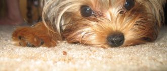
Pets require high-quality health care, and eye diseases account for most of the diseases. Glaucoma in dogs occupies a leading position among all eye diseases; about 80% of vision problems are caused by it. So, owners should pay special attention to the health of their pets’ eyes. If any abnormalities are diagnosed in time, it will be possible to return the dog to healthy vision with the least effort.
What is glaucoma in dogs?
This is a disease characterized by increased intraocular pressure. It is possible to determine the pressure level using a device - a tonometer. The cause of glaucoma is insufficient removal of fluid from the membrane of the eye. In this case, the secretion of fluid is not disturbed, that is, its amount is at the same level.
The eye is formed from various parts, but its shape, size and elasticity are directly controlled by the fluid contained within the eyeball. In this case, the liquid is under pressure, which in front is called intraocular. At the same time, it increases with a large accumulation of fluid, which causes the eye to bulge. Production occurs on an ongoing basis; the ciliary body is responsible for this function.
The fluid that is inside the eye contains the necessary nutrients for the membrane, as well as oxygen. When there is excess pressure, that is, a lot of fluid accumulates, it flows into the gap between the cornea and the iris. Constant pressure is maintained as long as fluid production and removal or drainage occur in equal proportions.
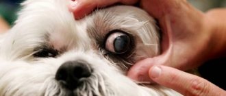
Diagnostics
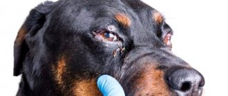
The owner is required to give a complete medical history: how it all started, what contributed to the development of the disease, and whether there were any potentially sight-threatening injuries. Be sure to tell your doctor about all cases of keratitis, conjunctivitis, or anything similar. The main prerequisite for making a diagnosis of glaucoma is increased intraocular pressure, measured using a special tonometer. Gonioscopy is very important. This is the name of the procedure in which the fundus of the eye is examined with a special device (gonioscope), revealing the degree of nerve damage. The results of these studies are very important for subsequent treatment.
Classification of glaucoma
Glaucoma is divided into several types: primary and secondary. Primary is characterized by the formation of high pressure, but the eye itself is healthy. However, certain breeds of dogs are more susceptible to the disease compared to others. The reasons are hereditary, as they contain certain anomalies in the structure of the iridocorneal angle, where excess fluid drains. So, at a narrow angle, the outflow of liquid becomes somewhat more difficult, which provokes an increase in pressure. Secondary glaucoma is caused by damage to the eye itself, which consequently led to the disease. This may include injury, illness, or damage.
The classification also includes another variety - according to the angle of the anterior chamber, which can be closed, open, or narrow. Moreover, both types of classification exist in parallel and occur in different forms.
Therefore, in the case of a hereditary predisposition to such a disease, you will have to especially carefully monitor the pet’s health. If the first signs are detected, examination and subsequent therapy will be required.
Primary open-angle glaucoma is considered a purely hereditary disease, which is more often observed in beagles and poodles. The disease is characterized by a chronic form, so the pressure increases gradually and takes a long time. Moreover, the dog is able to see even in the last stages of the disease.
Goniodysplasia is a variation of the form of the disease just described. It primarily affects Great Danes, Cocker Spaniels, Samoyeds, Beagles and Labradors. At the same time, it is typical for the disease to have a recurrent manifestation in the form of severe attacks. So the eye swells, the pupils tend to dilate due to a change in the angle of refraction of light, and hyperemia is noted. Sometimes it even leads to blindness in the pet.
Causes and symptoms of glaucoma
Glaucoma in dogs, in the very first stages of development, is characterized by the absence of specific symptoms. As the pathological process progresses, the signs increase and become noticeable to the animal owner.
At the slightest sign of ill health in your pet, it is recommended to bring the animal for a preventive examination to a veterinarian. A group of dogs has been identified whose risk of developing glaucoma is significantly higher than that of their relatives. Thus, the risk group includes:
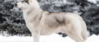
- representatives of breeds - spaniels, huskies, hounds, golden retrievers, Dalmatians and beagles;
- animals with chronic eye diseases not treated in a timely manner, such as conjunctivitis, keratitis, eversion of the eyelids;
- dogs that have been treated with steroid medications (in such cases the risk of glaucoma progression increases);
- Pets over the age of 7 years, against the background of senile changes in the body, more often suffer from diseases of the visual organs;
- dogs with a genetic predisposition to diseases of the visual organs;
- animals with a history of serious eye injuries.
The cause of the development of glaucoma can be diseases of a systemic nature - pathologies of the endocrine system (diabetes mellitus), high blood pressure, atherosclerosis.
There are several forms and types of glaucoma in dogs. Depending on the factors that provoked the pathological process, glaucoma is divided into primary and secondary.
The primary type of pathology develops in a clinically healthy animal, and secondary glaucoma occurs against the background of inflammatory eye diseases and changes in the position of the lens.
Glaucoma is classified depending on its location. The disease can be closed, open or narrow.
Symptoms of glaucoma do not appear for a long time. As a rule, dog owners turn to veterinarians when the process is in an advanced stage and conservative treatment methods do not produce positive results.
In this regard, veterinarians strongly recommend regular preventive examinations of pets, at least once every 6 months. General diagnostics makes it possible to timely identify not only eye diseases, but also other systemic pathologies that do not manifest characteristic symptoms for a long time.
Progressive glaucoma in dogs is characterized by the following symptoms:
- increased formation of tear secretion and its constant flow;
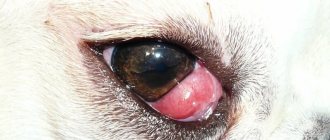
- the occurrence of hyperemia of the mucous membranes associated with edematous phenomena;
- disturbances in the general condition of the pet - the animal refuses to spend active time, looks lethargic and apathetic;
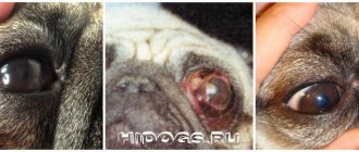
- when examining the eyes, cloudiness of the sclera is noted;
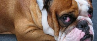
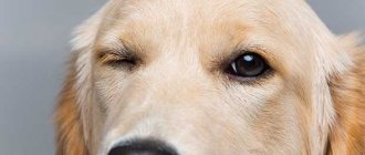
- the eye increases in size, bulging outward against the background of increased intraocular pressure;
- the animal suffers from pain in the eye area and the entire part of the muzzle as a result of a developed inflammation process.
In the most advanced cases, the dog loses the ability to navigate normally in space and photophobia (photophobia) is observed. The pet avoids bright light, trying to hide in a dark place. The retina and nerves of the eyeball atrophy. The dog loses the ability to see.
The primary type of pathology develops for a long time without characteristic symptoms.
The disease can only be diagnosed in a veterinary clinic. The owner may notice swelling in the area of the pet's cornea, excessive dilation of the pupil and slight redness of the mucous membrane.
Learn about other common eye diseases in dogs >>>
Predisposition
The main difficulty in diagnosing and visually determining abnormalities in the eyes lies in their slow manifestation, while pronounced severity can only be acquired in the later stages. Due to the long period of development, glaucoma is rarely diagnosed in the early stages, since it does not bother the animal at all, and a slight increase in pressure is not visually noticeable.
- It will be interesting about seborrhea on the fur of dogs
To prevent the disease, it is necessary to take into account the predisposition factors of the breed; if a dog has an increased risk of glaucoma, then it should undergo periodic examination. If at least one or more of the listed factors apply to your pet, you should especially carefully monitor the state of your eyesight and make an appointment with an ophthalmologist. Factors predisposing to glaucoma:
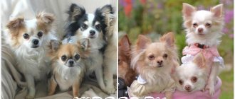
- Breed. Certain types of animals are more likely to encounter the disease than others; this must be taken into account when adopting Chihuahuas, beagles, divers, cocker spaniels, Dalmatians, huskies, poodles, etc.;
- Chronic eye diseases. If an animal has a seemingly harmless disease, perhaps the owner does not even know about it, the likelihood of glaucoma increases many times over. For example, conjunctivitis (inflammation of the mucous membrane) can provoke closure of the angle or thinning of the gap for fluid outflow. At the same time, improper treatment increases the risk of developing the disease; side effects from steroid drugs are especially dangerous. In this case, the disease progresses quite quickly and after 2 weeks the first manifestations may begin;
- Age. Age-related structural features of a dog’s body play a role, so animals over 6 years of age are much more likely to develop glaucoma;
- Hereditary predisposition. In addition to genetics associated with the breed, the disease can be passed on from parents. This factor can be determined by determining whether one of the parents had this disease. If the result is positive, you will have to more carefully monitor the pet’s health;
- Mechanical damage, eye injuries. This option plays a primary role; if the dog has received any damage, you should definitely contact a specialist. In the absence of timely assistance, there is a risk of developing a chronic, incurable disease;
- Other diseases. At risk are animals that have other types of illnesses: diabetes, cardiovascular diseases, they increase the risk of developing glaucoma.
Causes of glaucoma
From a medical point of view, there are some reasons that lead to the formation of diseases in dogs:
- Uveitis (inflammatory reaction in the inner lobe of the eye) - the cause of the disease is the penetration of infection into the intraocular fluid. In the presence of inflammation, the drainage channel is blocked, partially or completely, and leads to glaucoma;
- Subluxation in the lens of the eye is often a consequence of mechanical damage;
- The presence of benign or malignant eye tumors. This is how drainage is blocked due to an increase in the physical volume of the tissue;
- Intraocular bleeding - when blood clots, a clot is formed that can block the fluid outflow channel;
- Physical damage to the lens of the eye. Often this manifestation entails an inflammatory reaction, and, accordingly, swelling of the tissue and clogging of the canal;
- The occurrence of primary glaucoma is possible due to a defect associated with the anatomical structure of the iridocorneal angle. Moreover, it can be either narrow or closed due to the presence of parts of embryonic tissue.
Symptoms and treatment of glaucoma in cats and dogs
The eyeball is a ball filled with a special liquid - a modified blood transudate. The optimal pressure of this fluid in both humans and animals is 18-24 mm Hg. Art., its maintenance is regulated by the drainage system. When the balance is disturbed, when more fluid enters the eyeball than is removed, intraocular pressure (IOP) increases and glaucoma develops (from the Greek γλαύκωμα - “clouding of the eye”). This is one of the most severe and difficult to treat ophthalmological diseases in dogs and cats, which leads to gradual destruction of the retina and optic nerve, and ultimately to loss of vision.
Symptoms of glaucoma
Before moving on to treatment of the disease, you need to know for sure how cataracts manifest themselves and what are its main symptoms. If only a small part of the listed manifestations was identified, then it is likely that the disease is at an early stage or is not it at all. Nevertheless, this becomes a good reason to contact a specialist. Main symptoms of glaucoma:
- Increased tearing of the eyes;
- The eyeball increases in volume, but in the initial stages this is not very noticeable. The manifestation is popularly called “bull’s eye”;
- The vessels of the sclera protrude strongly, they become tortuous, with a pronounced red color;
- Pain in the eyes. This is evidenced by the dog’s behavior; it does not like it when the side of its head is touched, where painful sensations are observed;
- The pet eats much worse, the appetite disappears, or gets worse. This is accompanied by a depressed state and withdrawal from the society of animals or people;
- The dog has difficulty with orientation in space;
- Vulnerable to light, the pet tends to retreat to a dark corner, closes its eyes with the help of its paws or curls up into a ball, as with dropsy.
Glaucoma treatment
Treatment of glaucoma in a dog should be carried out upon examination by a doctor and determination of the cause or type of disease. So the information presented below should not replace a trip to a specialist, because self-medication can cause a lot of harm.
Treatment comes in many different forms and is highly dependent on the quality of the pet's vision when tested, the cost of the treatment needed, and the pet's temperament. If the dog can see, then it is necessary to reduce intraocular pressure, and it is important to do this as quickly as possible in order to preserve vision and also eliminate pain. So primary glaucoma in a dog should be treated in a timely manner. Emergency care may include:
- Osmotic diuretics:
- Glycerol. A fairly effective remedy, it should be administered intravenously in a volume of 1-2 ml per kilogram of animal weight at the beginning of 10-30 minutes and lasting 4-5 hours. The drug can be reused, but is contraindicated in cases of heart failure, renal failure, as well as systemic hypertension and dehydration. It is important that the animal does not inhale the substance;
- Mannitol. It is administered by intravenous injection at 1-2 g/kg body weight at regular intervals of 20 minutes. Initially, the cycle should be repeated every 30-60 minutes for 6 hours. The course can be reused. The drug is contraindicated in case of heart/renal failure and dehydration. After administration, you should refrain from drinking liquids, both orally and intravenously, for 2-4 hours.
1) Prostaglandins - they are great for monitoring the animal’s condition, but are expensive:
- The most common drugs in the group are Travoprost, Latanoprost, and Bimatoprost. Used topically, 1 drop every half day. Lead to the formation of strong mitosis.
2) Miotics - take special care in the presence of anterior uveitis, otherwise the formation of synechia, lens subluxation or exacerbation of glaucoma is possible:
- Pilocarpine – a 1-2% solution should be used 2-3 times a day;
- Dimecaine bromide – 0.125-0.25% solution instilled once a day.
3) Adrenergics:
- Levobunolol, Timolol (dosage 0.5%), Betaxolol - use 1 drop in the eye 2-3 times a day with a regularity of 8-12 hours. For mild severity, they are prescribed 2 times, in complex forms: three.
4) Neuroprotective agents:
- Amlodipine - taken orally in a volume of 0.625 mg once a day. It has a protective effect for ganglion cells.
5) Carbonic anhydrase inhibitors - effective for short-term or long-term blood pressure control. They also have a kaliuretic effect, so drugs containing potassium should be added to treatment if it is long-term:
- Dorzolamide, brinzolamide - used topically at 8-hour intervals;
- Acetazolamide - administered orally at 10 mg/kg twice a day;
- Methazolamide - dosage 5 mg/kg twice daily;
- Dichlorphenamide – dosage 2.5 mg/kg 2-3 times a day
Treatment without examination and diagnosis in a veterinary clinic is prohibited, as there is a high probability of side effects and worsening of the disease.
Currently reading:
- Diagnosis and treatment of liver cholestasis in dogs
- A warning sign for a dog with cloudy eyes
- Review for choosing carriers for small and large dogs
- Thyroid dysfunction in dogs (hypothyroidism)