Each breed has a specific feature associated with the physiological structure of the body. One of these pathologies is considered to be an anomaly in the development of decorative dogs, whose eyes fall out due to the structural features of the bone orbits. This condition is dangerous - in the absence of proper treatment, the pet may lose its sight.
How can you help your pet if he is a representative of a “special” breed of dog that has a physiological problem with his eyes?
Main symptoms
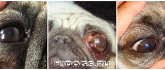
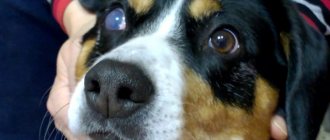
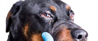
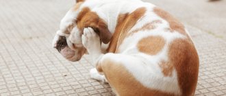
The prolapsed eye looks bulging, it extends beyond the eyelids, because of this, for a short period, the tissues around the eye become swollen, red, and very painful. In this case, the dog cannot blink, as a result of which the cornea (the outer transparent layer of the eye) develops. The dog becomes passive and often tries to scratch its eye with its paw.
If these symptoms are present, you should take your pet to a veterinarian as soon as possible, since hours and even minutes are counting, and there is a high risk of losing not only vision, but also the eye itself.
Causes and symptoms of eyeball prolapse
Changing teeth in a Yorkie: what time do they fall out?
Reasons for loss:
- skull injuries. This happens especially often during an accident, when the eyeballs can literally fly out of their sockets;
- failure to comply with hygiene rules;
- infections and inflammatory processes;
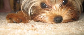
- mechanical damage: dog fights, etc. Small dogs can be injured even by grass, so the owner must walk them very carefully;
- collar too tight;
- the owner’s habit of carrying the animal by the withers;
- belonging to a breed with a predisposition to proptosis. In many animals, the eyeball is supported only by the muscles of the eyelids. Any damage can weaken them.
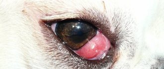
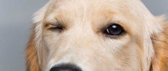
Important! A dog whose eyes fall out experiences severe pain and discomfort. The symptoms of the disease are difficult to miss, so you need to contact a veterinarian as soon as possible. This pathology is visible to the naked eye.
The veterinarian will conduct additional diagnostics and find out how advanced the disease is. The course of treatment depends on this.
Signs and general symptoms of hair loss:
- the eyeball is pinched by the eyelids;
- swelling of the conjunctiva;
- prolapse of the eyeball;
- bloody issues;
- strong pain;
- weakness, depressed state.
The operation must be done within the first day
First aid for proptosis in dogs
It is important that the dog does not try to scratch the eye with its paws and does not rub its head against surrounding objects, this can lead to even greater trauma and an increased risk of vision loss. To do this, you can wear a special protective (Elizabethan) collar. If there is no collar, you can carefully secure your pet’s paws or head with your hands or a towel.
If eye prolapse is detected and on the way to the veterinary clinic, you should rinse (and thereby moisten) the eye with sterile warm saline every 5-10 minutes. In the pharmacy it is called sodium chloride 0.9%. To rinse, you need to take a syringe with a volume of 5 or 10 ml, fill it with saline solution (or warm boiled water, if there is no sodium chloride), remove the needle and carefully water the eye. Antibacterial eye drops or moisturizing eye gels (Korneregel, Vidisik, Oftagel and others) are also suitable.
You cannot use solutions such as chlorhexidine, hydrogen peroxide, Miramistin and other antiseptics, as they can cause a burn to the cornea!
What absolutely cannot be done?
The use of standard antiseptics such as chlorhexidine is excluded. Even such a mild drug can cause irritation; moreover, it dries out greatly, and this is what is extremely dangerous for the affected organ. Owners of dogs prone to eye loss should always have hygiene lotions in their first aid kit to moisturize and relieve irritation. In this case, chamomile decoction is not suitable, as it can also cause dry mucous membranes.
Eyeball prolapse in dogs occurs as a result of injuries, certain diseases, and is even typical for certain breeds of dogs. So, Shih Tzus, pugs, bulldogs and other brachycephalics lose their eyes. This is not a reason to panic in advance. A bulging organ can be cared for, and a veterinary clinic can treat it. The main thing is to provide your pet with proper and regular care.
Diagnosis of eye loss
At the appointment, the doctor will examine the patient and announce prognoses regarding vision. An ophthalmologist will conduct tests and evaluate reflexes that will help understand the extent of damage:
- pupil reaction to light,
- blinding reflex,
- examination of the fundus using a special ophthalmoscope device,
- Ultrasound of the eye to evaluate internal damage or detachment of the retina (the inner lining of the eye that transmits visual information through the optic nerve to the brain).
The prognosis is considered good if:
- presence of vision,
- preservation of pupillary reflexes,
- reactions to blinding light,
- positive ophthalmoscopy data,
- rupture of less than two muscles that hold the eye in the orbit,
- lack of blood inside the eye.
Signs that give an unfavorable prognosis for the preservation of vision and the eye as an organ: no reaction of the pupil to light, retinal detachment is observed (according to the results of ophthalmoscopy and ultrasound), more than 2 muscles are damaged, there is a rupture of the structures of the eye or hemorrhage inside it.
Breeds Prone to Eye Loss
There are dog breeds in which eye loss is considered common. This occurs due to the anatomical features of the structure of the eyes.
Dog breeds with bulging eyes:
Pugs
Animals of this breed have charming and beautiful eyes, which are the main highlight of their appearance. But these organs are the most unprotected area. They do not have a protruding nose, which could protect the visual organs from negative factors. During a normal walk, the dog can injure the gas on the grass and various branches. Dust can cause severe inflammation and other unpleasant ophthalmic problems.
Pekingese
Dogs of this breed often experience eye loss. This may occur due to a deformed front part. They have wide eye sockets, and the eyes themselves are held in place only by the muscle tissue of the eyelids; all this can quickly lead to loss of the organs of vision. This can occur as a result of severe stress, a sharp jump, strong pressure on the neck from a leash, or problems with defecation.
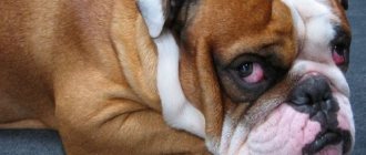
Shar Pei
Representatives of this breed are endowed with large sizes. Their eye problems are associated with the wrinkled structure of the skin. Quite often they experience entropion or entropy, which can lead to the development of serious eye diseases, including proptosis. This condition can be provoked by blows to the head or carrying the animal by the folds of its skin at the withers.
Shih Tzu
These are very beautiful dogs with long coats and friendly personalities. However, their eye orbits are not completely closed. For this reason, in the event of a blow or traumatic brain injury, the muscle fibers of the cheekbones simply cannot hold the eyeballs.
Treatment of proptosis in dogs
After determining the condition of the eye, surgery is performed to realign it. This procedure is performed under general anesthesia.
The eye is set by temporarily suturing the eyelids, which helps keep it in its normal position and speed up healing. Doctors try to save the eye even with poor prognosis. This is because signs such as lack of pupillary response to light may be temporary, and reduction is a less traumatic procedure than removal of the eye, which can be done electively at a later date. In the postoperative period, eye drops and tablets containing antibiotics, anti-inflammatory drugs to relieve swelling and pain, and a protective collar are used. After 14 days, the sutures from the eyelids are removed and the condition of the eye is re-evaluated.
Treatment for eye loss in dogs
After choosing a method to eliminate eye loss, the doctor will begin the operation. To install the organ of vision in place you will need:
- Apply general anesthesia to the dog.
- Gently adjust the eyeball. If necessary, an incision is made on the outer edge of the eyelid to provide more space for surgical manipulation.
- After the apple is set, the eyelids are sutured.
- While healing occurs, Atropine and antibiotics need to be instilled. This reduces pain and prevents inflammation.
- After 1–2 weeks, the doctor will remove the stitches, assess the condition of the organ of vision and give recommendations for further treatment.
When returning home from the hospital, your pet should wear an Elizabethan collar to prevent paws from rubbing its face. It is also recommended to limit the movement to the cage to reduce disturbance to the seams.
Did you know? A dog's eyes reflect the light of headlights or other light sources because they have a special mirror layer behind the retina.
Rules for treating the visual organs at home:
- Check your sutured eyelids daily. If swelling, bleeding, or yellow-green discharge appears, tell your doctor immediately.
- Administer the medications prescribed by your veterinarian exactly on schedule and in amounts. Typically, Atropine 1% is prescribed, which is instilled every 12 hours and a broad-spectrum antibiotic every 6 hours.
- Monitor for signs of infection such as lethargy, fever, or decreased appetite. If they are present, then this should also be reported to the doctor.
Premature removal of sutures may predispose the patient to recurrent eye loss. Therefore, the doctor determines and prescribes a schedule according to which he will examine the animal’s condition approximately once a week. If, when removing the stitches, the doctor sees swelling, he can re-stitch the stitches and continue treatment for another 2-3 weeks.
Consequences of eye loss
With a small amount of damage, preservation of reflexes and quick delivery of the pet to the veterinary clinic, such an eye will be able to continue to see (of course, after the stitches are removed).
If the eye has lost function due to loss, but there are no ruptures, internal bleeding, reflexes are preserved, and there is no dryness of the cornea (which can occur after damage to the glands that produce tears), then the doctor can leave such an eye under observation.
Removal is performed only with progressive deterioration of the eye condition.
If the eye is severely damaged due to proptosis (there are ruptures of many muscles and other structures, hemorrhages), then removal may be recommended immediately or a few days after the loss. The doctor makes a decision based on an assessment of the dog's condition. If your general health is poor or there are injuries to other organs, eye surgery may be postponed until the patient’s well-being has stabilized.
Why do some dogs' eyes fall out?
Prolapse of the eyeball (proptosis) is a disease that appears mainly due to traumatic exposure. The risk of its development increases with certain ophthalmological diseases that weaken muscles, prolonged constipation, an excessively tightened collar and breed structural features. The latter applies to brachycephals - dogs with a short muzzle and bulging eyes. The correct structure of the skull, deep eye sockets and developed brow ridges provide additional protection, but some breeds do not have it. However, proptosis can occur in any dog, only the strength of the traumatic physical impact differs.
With timely treatment, it is possible to restore vision to a companion in 90% of cases.