Pathologies of the cornea
The general health of the dog's cornea is determined primarily by the degree of its transparency. Therefore, as soon as you notice clouding of the dog’s cornea , this may already indicate the presence of some pathology. The following signs also indicate pathology of the cornea:
- hemorrhage in the eye;
- swelling;
- change in pupil color;
- calcium deposits (calcification);
- inflammatory cell infiltrates;
- destruction of enzymes by endogenous proteases of the body and, as a result, degradation and scarring of the cornea.
Such changes are a deviation from the norm and can be caused by various reasons. Pathogenetic reactions are often complex. Any pathology of the cornea of the eye is a consequence, and the causative factor always lies elsewhere. It is this root cause that should be sought, and once discovered, every effort should be made for proper and thoughtful treatment and restoration of the functions of the eye.
Superficial chronic keratitis in a dog
Superficial chronic keratitis (pannus) is an immune-mediated keratitis, the cause of which should be sought in genetics, sometimes in the environmental situation of a particular region. The most favorable areas for the development of such a disease are places with increased background radiation. If we talk about the influence of dog breed on the likelihood of chronic keratitis in a dog , then German shepherds and their crosses are the most susceptible. Greyhounds are also at risk. But most often dogs of all breeds suffer from chronic keratitis, and the statistical significance among shepherd dogs and greyhounds is visible only when a large number of sick individuals are studied. The disease begins with mild symmetrical redness of the cornea. Although it can begin in other quadrants of the cornea and be asymmetrical.
Histologically, the corneal infiltrate is determined by plasma cells, lymphocytes and blood vessels. As the process progresses, the entire cornea can be affected, leading to noticeable loss of vision and eventually blindness. Clouding of the cornea in a dog and the proliferation of fibrous tissue (fibrosis) are characteristic signs of a chronic course of the process. The disease occurs at the age of 3-5 years. Pannus is very difficult to treat in young animals. The diagnosis is made based on the clinical picture, the breed of the dog and cytology of the cornea or conjunctiva. Cytology usually shows increased white blood cells and plasma cells.
Like most immune-mediated pathologies, chronic keratitis in dogs is better prevented than treated. The complex of preventive measures includes the use of a number of corticosteroids, cyclosporines, Pimecrolimus (Elidel) and Tacrolimus (Protopic). The most preferred among steroids are drugs containing no more than 1% prednisolone and dexamethasone (0.1%). The required frequency of taking medications is determined by the complexity of the dog’s keratitis, the time of year and, on average, is about 2-4 times a day. Subconjunctival steroids may be administered as an adjunct to primary therapy or in particularly difficult cases. Such drugs may be Triamcinalone, Methylprednisolone or Betamethasone. They are all quite effective, however, for better removal of conjunctival formations, Betamethasone injections should be used. When treating keratitis in a dog, topical use of Cyclosporine at a concentration of 0.2%, 1%, 2% or Tacrolimus at a concentration of 0.02% or 0.03% is permissible. Sometimes superficial chronic keratitis in a dog can be treated only by using Cyclosporine or Tacrolimus separately. In some cases, the use of these drugs can reduce the use of steroids, which reduces side effects. The intensity of treatment for keratitis in a dog can be reduced in the winter months and increased in the summer. Beta irradiation and lamellar keratectomy may be additional treatment options, but these technologies are rarely used today. Plasma cells and lymphocytes are particularly sensitive to beta radiation, and this makes ionizing radiation the most effective treatment in severe cases. However, very strict licensing requirements for devices using strontium-90 have led to the fact that almost no one uses this treatment method.
How is keratitis treated in dogs?
Effective eye restoration with keratitis is possible if you consult a specialist in time. Therapeutic measures to heal the eyes can only be prescribed when the doctor identifies the cause of the inflammation. As a rule, treatment of keratitis in a dog begins with washing with antiseptic solutions (Furacilin, Rivanol, etc.). For keratitis, anti-inflammatory drugs - corticosteroids (Hydrocortisone, Dexamethasone) are also used. However, if there are ulcerations on the cornea, hormonal drugs are contraindicated.
If the disease is of an allergic nature, the administration of antihistamines (Tavegil, Diphenhydramine) will be required; if there is purulent discharge, medications with antibiotics will be needed (Levomycetin drops, Tobrex, Erythromycin ointment, etc.). For dry keratitis, dogs are prescribed drugs that stimulate the production of tear fluid (Cyclosporine, Tacrolimus) and antibiotics. Unfortunately, vascular and pigmentary keratitis cannot be cured without leaving a trace. But with the help of medical manipulations it will be possible to try to slow down the development of the disease.
Inflammation of the eyes in dogs (scleritis, episcleritis)
These are autoimmune and immune-mediated conditions that arise as a consequence of chronic infectious diseases, metabolic disorders or allergic reactions and are characterized by gradually spreading lesions of the sclera or episclera of the eye. Lesions may be unilateral or bilateral. Often only one quadrant is affected, and the resulting neoplasm is mistaken for scleritis. One way or another, scleral neoplasm differs from melanoma. There is a genetic predisposition to this disease in cocker spaniels and Airedale terriers. In Airedale Terriers, the condition can often be complicated by the simultaneous development of uvititis (sclerouvitis). A typical histological sign of scleritis and episcleritis in dogs may be the appearance of lymphocytes, plasma cells and histiocytes in the thickness of the sclera. The areas adjacent to the cornea of the eye, as a rule, suffer from numerous microinflammations of blood vessels and tissues, and lipid degeneration is also sometimes observed. Deep necrotizing inflammation of the eyes in dogs is quite rare, but can cause serious intraocular diseases (for example, retinal detachment). The diagnosis is made based on the clinical picture. A biopsy can be done, however, this is often not a necessary procedure. Immune system tests are usually not helpful and are not very informative. Treatment of eye inflammation in dogs consists of a combination of general and subconjunctival steroid medications and non-steroidal anti-inflammatory drugs. The latter include prednisolone, azathioprine and a combination of tetracycline and niacinamide. In some cases, the use of cyclosporine (both orally and locally) can be very effective. A long period of treatment is expected.
Pigmentary keratitis in dogs
Pigmentation of the corneal epithelium or stroma is called pigmentary keratitis (also sometimes called corneal melanosis or corneal pigmentation). contribute to the development of pigmentary keratitis in a dog , including developmental disturbances in the eye structure during extraembryonic (placental) formation, excessive muzzle folds, and dry eyes. Dry eyes are usually the most common cause of keratitis in most dog breeds (except pugs). Pigment growth (pigmentation) can appear after healing of a corneal ulcer (often post-traumatic) or in parallel with another disease, for example, pannus. A similar situation often occurs with pugs. In this breed, genetically predisposed exophthalmos and corneal exposure may be precipitating factors. In any case, the most important factor for the development of such a pathology remains the breed of the dog, since, despite the similar structure of the head, in dogs of such breeds as the bulldog, Pekingese and Shih Tzu, pigmentary keratitis is much less common. Canthoplasty and canthopexy (lower eyelid lift) are often used to slow the progression of the disease. The advantage of this procedure is to increase eye protection by reducing the palpebral fissure, eliminating trichiasis (abnormal growth) of nasal hairs and correcting the canthal ligaments of the inner and outer canthus, as well as nasal skin folds.
For pugs, an integrated approach is used to treat pigmentary keratitis , combining surgery and local treatment at the same time. Local treatment allows you to slow down painful inflammatory processes and consists of the use of cyclosporine, tacrolimus and corticosteroids. Cyclosporine and tacrolimus are approximately equal in their effectiveness and the final choice depends only on which drug is best suited for a particular patient. The use of steroids may only be beneficial in brachycephalic dog breeds due to their susceptibility to corneal ulcers. The use of beta irradiation is also sometimes acceptable, but it is justified only in cases where vision is significantly damaged and melanotic growths are significant; in other cases, the use of this type of treatment is inappropriate.
Causes of keratitis
Pigmentary keratitis, sometimes called color keratitis or corneal melanosis, is common in almost all brachycephalic breeds, but is most often diagnosed in pugs.
This serious ophthalmological disease can be triggered by several factors:
- injury or irritation of the cornea;
- inflammatory process that has become chronic;
- defects in eye development during extraembryonic (placental) formation;
- inversion of the medial corner of the eyelids (in brachycephalics);
- brachycephalic ocular syndrome;
- excessive folds on the muzzle;
- dry eye syndrome.
By the way, the last reason is to blame for keratitis in most dog breeds, with the exception of pugs.
The cornea is a convex-concave shell of the eyeball that refracts light rays. A healthy cornea free of pathologies is absolutely transparent . Keratitis of any etiology leads to a decrease in its shine and transparency. The inflamed epithelium of the cornea becomes cloudy or red, and microulcers appear on it.
With pigmentary keratitis, the clear epithelial cells are gradually filled with colored substances (biochromes), leading to darkening, clouding or blackening of the cornea.
Important! The determining factor for the development of pathology is (according to doctors) the breed of the dog: despite the similar structure of the head, pigmentary keratitis is much less common in bulldogs, shih tzus and Pekingese.
Pigmentation often appears after the healing of a corneal ulcer (usually post-traumatic) or in parallel with another eye disease, for example, pannus. This situation often occurs in pugs, when genetically caused exophthalmos and corneal exposure become catalysts for pigmentary keratitis.
Return to content
Corneal endothelial dystrophy in a dog
This pathology is primarily caused by a defect in the endothelium of the cornea, which leads to its swelling and, subsequently, gives the cornea a bluish-gray tint. The primary causes in the differential diagnosis of edema can be considered corneal ulcers, uvitae, glaucoma, which are easy to distinguish and recognize by their condition. endothelial dystrophy in dogs progresses slowly and usually begins in the lateral part of the cornea, but then spreads to its entire area. The risk of this disease is highest in breeds such as the Boston Terrier and Chihuahua, but dogs of all breeds are often affected. At an early stage, the disease, as a rule, does not cause painful conditions in the patient. Developing and progressive corneal endothelial dystrophy causes ulcerative keratitis and pain over time.
Therapy consists mainly of using 5% sodium chloride ointment or suspension (Muro-128) to minimize swelling. In any case, you cannot and should not expect a fairly quick cleansing of the dog’s cornea. Topical antibiotics or atropine are used only in cases of ulcers on the dog's cornea . Conjunctival hyperemia occurs in an already ill dog. If the eyes are particularly irritated and there are no ulcers, topical steroids may be used with caution. Local non-steroidal anti-inflammatory drugs (Flurbiprofan) are also successfully used in some cases. Thermal cauterization (thermokeratoplasty) is used in particularly severe cases and when ulcers open on the cornea. Although this procedure will not completely clean the cornea of the dog’s eye , it will prevent swelling and reduce the pain experienced during the opening of the ulcers. The essence of this technique is to use an ophthalmic laser to cauterize damaged areas of the cornea. It should be noted that the success of the operation depends on the skill of the surgeon, since careless movement or too long burning can lead to complete destruction of the dog’s cornea. Also, in some cases, it is advisable to use penetrating keratoplasty.
Pigmentary keratitis in pugs • Zoo-Vision™ Zoovision™
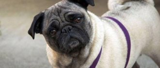
Pigmentary keratitis is the most common eye disease in pugs, in which the clear part of the eye becomes covered with an opaque pigment film. Pigmentary keratitis is the main cause of blindness or poor vision in pugs, Pekingese, Shih Tzu and some other dog breeds.
A dog's cornea must be perfectly transparent for normal vision. And not only dogs - all living creatures who look at the world through their eyes. Animals with an opaque cornea experience vision deficits. Some people just don’t see well, others don’t see at all. According to one study, about 80% of pugs suffer from pigmentary keratitis.
What is pigmentary keratitis in pugs?
Pigmentous keratitis (also known as corneal melanosis) is a disease in which brown pigment is deposited on the cornea. At first, these spots are pale, translucent, completely superficial, growing from the inner corner of the eye (from the medial corner of the eye).
As the spots grow, they acquire a more pronounced brown color, occupy a larger area, and grow deeper into the cornea. As pigment spots grow, dogs begin to experience vision deficits and may lose their vision. At first, do not notice small objects, then large ones. Then the dog may see worse at dusk, lose its owner, or crash into obstacles.
Typically the disease begins to progress by age 3. However, the disease can also occur in puppies up to six months of age.
Where does my dog get pigmentary keratitis?
The exact cause of pigmentary keratitis is not completely clear. A genetic basis is assumed, but so far this is at the stage of speculation without direct evidence.
It was previously assumed that pigmentary keratitis is a consequence of medial entropion of the eyelids in pugs and Pekingese.
However, studies have shown that in the presence of entropion of the eyelids in almost 100% of pugs, pigmentous keratitis develops in 80%.
What causes pigment growth on the cornea?
Any factors that cause the cornea to send an “SOS” signal. For example, eye injuries, entropion of the eyelids, improperly growing eyelashes, neoplasms on the eyelids, dry eyes. These factors accelerate pigment growth more than others.
How is pigmentary keratitis treated?
Treatment of this disease always begins with diagnosing the underlying cause. Or the main factor influencing pigment growth. Dogs with dry eye syndrome should first be treated for tear deficiency. Those who have suffered an eye injury should first treat the cornea.
If pathological eyelashes (for example, ectopic) are to blame, surgical treatment of these eyelashes is required. It also happens that the main cause is inversion of the eyelids. Almost 100% of Pugs have an inversion of the eyelids in the inner corner of the eye. This is where the pigment begins to grow.
In this case, the dog is indicated for eyelid surgery.
The pigment itself can also be removed. But this is carried out when the main factors of the disease have been eliminated. That is, a dog with dry eye syndrome should not have pigment removed until tear production is restored. Otherwise, at best, the pigment will grow back in 2 weeks. And in the worst case, you can get a corneal abscess.
Eyelid surgery in a pug. Whether it is necessary?
If this plastic surgery is performed according to indications, then it is definitely necessary. Sometimes this is the only thing that can protect a dog from blindness. But in order for this plastic surgery to be performed exactly according to the indications and have the desired effect, you should seek help from a highly specialized veterinarian, an ophthalmologist.
Green arrows point to the area where the eyelids turn in. Red – on the pigment growth zone.
Green arrows point to the area where the eyelids turn in. Red – on the pigment growth zone.
Pug 1 month after eyelid surgery. The palpebral fissure is of normal size, there is no longer any fur in the corner of the eye.
1 month after surgery. The eyelids are rounded and fit tightly to the eye. There is no more fur in the corner of the eye.
What about anesthesia? Pugs cannot be given anesthesia.
Pugs are the most common dogs that can do anything. And anesthesia as well. They once earned themselves a bad reputation because of their anatomical structure.
The long velum palatine (this is what causes them to grunt and snore) could be blocking the breathing of a sleeping, relaxed dog. Why the dog could have died. However, for more than 10 years this problem has not been relevant, because today all pugs are operated on with an oxygen-gas mixture.
And during operations they are intubated. That is, a tube is inserted into the trachea, thanks to which the dog breathes even easier than in normal life.
In addition, the dog will always undergo a heart and general medical examination before surgery. You will never go to the operating table without tests, will you? So your dog is not allowed. This way we get a patient about whom we know enough to decide whether he can be operated on.
What to do if your dog has pigmentary keratitis?
Regardless of whether you have a pug or another breed, pigmentary keratitis is a reason to contact a veterinary ophthalmologist. The illness is not an emergency and therefore does not require immediate attendance to a doctor. However, the visit should definitely not be delayed. Superficial pigment is much easier to remove without relapse. While total pigmentation of the cornea is very difficult to deal with.
Join us on social networks
Similar
Source: https://www.zoo-vision.com/thw_companion/pigmentnii-keratit/
Lipid or calcium keratopathy in dogs
Deposits of lipids and salts on the cornea of the dog's eye are quite similar to the diseases described above, but have completely different causes, and their clinical differences are sometimes difficult to distinguish. However, there are three main signs by which pathological deposits on the cornea can be diagnosed:
- corneal dystrophy in a dog;
- corneal degeneration;
- lipoid arc of the cornea (a grayish line on the periphery of the cornea, a kind of ring-shaped opacification of the periphery, separated from its other parts).
Corneal dystrophy in dogs can be: hereditary, bilateral, symmetrical. There are also cases when dystrophy begins to progress in one eye and then spreads to both. Lipid corneal dystrophy can occur in different breeds of dogs, but those most often at risk are: Siberian Huskies, Samoyeds, Cocker Spaniels and Beagles. Clinically, lipid deposits may produce a slight, almost imperceptible crystalline haze in the central cornea, or the affected portion of the cornea may appear completely opaque. Lipid deposits are usually subepithelial or stromal and consist of cholesterol, neutral fats and phospholipids. There is no systematicity in this disease and, as a rule, is not observed. The cornea is usually without ulcers, there is no inflammation. Very rarely, lipid deposits impair a dog’s vision , however, they also do not cause pain. For these reasons, dogs are not prescribed any specific treatment for this pathology. If treatment begins, then lamellar keratectomy is used, however, it does not guarantee recurrence of deposits, and the reason for this is persistent disorders of lipid metabolism in the parenchyma leading to new and new growths.
Corneal degeneration in a dog's eye can be caused by either lipid or salt deposits (and sometimes both). Initially, degeneration may be preceded by corneal ulcers of the dog's eye, uvitae and sometimes exophthalmos. Unlike corneal dystrophy , degeneration is often unilateral rather than symmetrical (bilateral). The affected part of the cornea is most often opaque, rough, with destroyed epithelium. And this already creates a certain discomfort for the animal. Inflammation, vascularization and pigmentation may also occur. The same lamellar keratectomy - it can reduce pain and restore vision, but at the same time, this treatment does not guarantee the avoidance of relapse. In some cases, the use of ointment in conjunction with keratectomy is useful. An alternative to surgery is the long-term use of abrasive and at the same time absorbable agents, such as powdered sugar, preparations based on natural honey and propolis, various combinations of bee bread, wax and bee pollen. It is imperative to use them simultaneously with ointments and drops without corticosteroids. However, their use (they are less traumatic) and effectiveness have not yet been sufficiently studied, and are only a weak alternative to modern methods and approaches for restoring a dog’s vision .
Lipid degeneration of the cornea can occur after a long period of treatment with corticosteroids, for example, cataract surgery, but such degeneration is not difficult to treat. The degeneration may regress after several treatment sessions.
Lipid deposition that occurs in the periphery of the cornea, in combination with persistent hyperlipidemia, is called a lipoid arc of the cornea (ring-shaped opacification of the periphery of the cornea). Clinically, the fat creates an opaque ring in the periphery of the cornea. The problem can occur in any breed of dog, but German Shepherds are especially susceptible to this. Ring-shaped opacification of the periphery of the cornea is a bilateral problem and is accompanied by mild inflammation and vascularization. Treatment is aimed solely at eliminating the underlying cause. Dogs suffering from lipid and salt deposits should first have their blood tested to check their cholesterol levels, triglycerides, and thyroid. If the test results are satisfactory, then the dog’s diet should be changed and this will partially solve the problem of lipid deposits.
Keratitis in dogs: how long does it last, treatment, eye restoration
Keratitis in dogs is a pathology that leads to loss of vision in the pet if treatment is not started. How long eye recovery takes depends on many factors. In some animals, due to the anatomical features of the structure of the skull, the disease requires lifelong care.
For what reasons do dogs develop keratitis and how can it be diagnosed? Treatment of this pathology, as well as restoration of the eyes after illness.
Description of the disease
Keratitis is an inflammatory disease of the cornea. The cornea is a clear, biconvex lens that sits at the front of the eye. Its main functions are light refraction and protection. It is thanks to the cornea that animals distinguish the brightness, shape and size of an object.
Most often, keratitis affects dogs of brachycephalic breeds (boxers, pugs, bulldogs, Pekingese, etc.). Due to the unusual structure of the skull, the eyes are not sufficiently moistened, resulting in inflammation.
Its varieties
Based on the degree of damage to the cornea, there are:
- Superficial keratitis in dogs - the process affects the external part.
- Deep – inflammation spreads to the inner layers. This type of pathology is accompanied by the formation of ulcers and scars, which remain even after treatment, worsening the quality of the animal’s vision.
Depending on the nature of the development of the pathology, the following types of keratitis are distinguished:
- Infectious. It develops when pathogenic microorganisms (fungi, bacteria) come into contact with the cornea. This inflammation is accompanied by profuse yellow-green discharge from the eyes.
- Vascular. Occurs when there is excessive growth of blood vessels in the tissue of the cornea. The front part of the eye becomes reddish and lumpy.
- Pigmentous keratitis occurs in brachycephalic dogs. With this type of pathology, brown pigment accumulates in the cells of the altered epithelium. This causes the cornea to become cloudy and then darken. There are local, subtotal and total.
- Dry keratitis develops, as a consequence, with pathologies of the lacrimal glands, with dry eye syndrome.
- Interstitial. A blue-white cloudy film appears on the cornea. The nature of this type of pathology is usually infectious (hepatitis, parvovirus, etc.)
- Phlyctenulous keratitis accompanies poisoning or chronic allergic reactions. The presence of irregularities is noted in the eyes, the third eyelid becomes inflamed and swollen.
Causes of disease development in animals
There are several causes of inflammation of the cornea of the eye, including:
- Breed predisposition in brachycephalic dogs. In animals, the eyelids do not close completely when blinking, which is why the central surface of the cornea is not sufficiently moistened. Also, in pets of these breeds, pathologies of the lacrimal duct are more often noted; as a result, the eyes do not receive the necessary amount of fluid for normal functioning.
- Chronic corneal injury. When the cornea is irritated, a kind of protective reaction is triggered in the body. In response to long-term damage, tissues become denser, but at the same time lose their main function - light transmission. This occurs in animals with abnormal eyelash growth, long-haired pets (especially if they have bangs), or if the dog is kept in a dusty room.
- Dry eye syndrome. For various reasons, little tear fluid is produced, nutrition and breathing of the corneal surface are disrupted. Keratitis in this case will be a secondary problem.
- After surgical interventions on the organ of vision.
- After viral keratoconjunctivitis (previous viral infections: canine distemper, hepatitis, parvovirus enteritis).
- Allergic reactions.
- Diabetes.
How long does keratitis last in dogs?
The duration of the disease depends on the degree of damage to the cornea and the timely and correct initiation of treatment. In the superficial form, the shell recovers quickly. Therapy will take 7-14 days.
With severe inflammation, the process can drag on for several months. However, it is not always possible to completely restore vision function.
The speed of recovery is influenced by many accompanying factors: nutrition, the age of the pet, the state of immunity, the presence of chronic diseases.
Symptoms of pathology
You can recognize the signs of keratitis in a dog at home and seek help from a veterinarian as soon as possible:
- Loss of vision. The pet moves uncertainly and bumps into surrounding objects.
- Fear of light. The dog hides in dark places and avoids daytime walks.
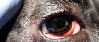
- Redness of the eyes and mucous membranes, blood vessels are dilated and filled with blood.
- Discharge from the eyes. Accumulations of pus, a large number of yellow-green crusts on the fur around the eyelids.
- Itching. The dog scratches its face with its paws or rubs against external objects.
- A cloudy white film appears before the eyes.
- General apathy. The animal stops playing and constantly sleeps.
It is more difficult to detect pigmentous keratitis, since the eyes of dogs are dark and against such a background it is difficult to see when the cornea becomes not transparent, but acquires a brownish color.
How to treat keratitis in a dog?
Treatment always begins with the use of local medications. Surgery is indicated for animals if there is no positive response to conservative therapy.
Medication
Therapy is aimed at:
- elimination of pathogenic microflora;
- reduction of toxic-allergic state;
- restoration of hydration of the ocular surface.
Groups of medications used to treat keratitis in dogs:
- Antibacterial drugs (Tsiprovet, Floxal, Normax, Tobrex).
- Lubricants (the action of this group of drugs is aimed at moisturizing the ocular surface). These can be drops, gels or ointments. The doctor must determine which form of release to choose during the examination. In each case the approach is individual. Examples of prescribed drugs: Hyphenlez, Hilabak, Systeine.
- Glucocorticosteroids (drops or ointments: Dexamethasone, Nevanak, Maxitrol) are prescribed with caution, as these drugs reduce the local immunity of the eye and worsen the process of regeneration of the cornea. Long-term use of the products can lead to corneal ulcers.
- Cytostatics (Optimmun ointment) are milder in their action, but a long course of administration also carries a number of side effects.
Before instilling the drops, remove all dirt from the eyes. A clean cotton pad soaked in freshly brewed tea or chamomile decoction is suitable for this. Rub the eye from the outer corner to the inner (towards the nose).
If it was not possible to remove all the discharge at one time, use a new cotton pad for each subsequent treatment.
To administer eye drops, the dog's head is fixed. The drug is applied by slightly pulling the lower eyelid, then make 3-4 blinking movements using your fingers. If several drugs are prescribed, the interval between treatments should be at least 15 minutes.
Application of eye gel is similar. Place a strip of gel under the lower eyelid and blink several times so that the medicine is distributed evenly.
If a dog intensively scratches its eyes, a special protective collar is worn throughout the course of treatment to avoid the development of secondary infection and self-injury.
By performing an operation
Surgical intervention is indicated in cases of:
- If keratitis progresses during therapeutic treatment.
- When allergic reactions to the drugs used occur.
- When a corneal ulcer occurs during treatment with drops.
- In case of total pigmentous form that does not respond to treatment.
The operation is performed in specialized clinics and consists of removing the pathological area of the cornea. The problem is solved surgically when keratitis occurs against the background of improper eyelash growth and other pathologies that are the root cause.
Eye restoration with keratitis in dogs
If you consult a veterinarian-ophthalmologist in a timely manner, keratitis can be completely cured. It is important to comply with the prescribed treatment and monitor the dynamics after the start of therapy.
If there is no effect after 10-14 days, you need to come back for a second appointment. The doctor will conduct an additional examination and change the treatment.
In some cases, a positive response occurs after a month or more.
In case of severe damage, complete restoration of the cornea is impossible. Scars and ulcers remain on the membrane, which significantly impair the pet’s vision.
If you do not consult a veterinarian, keratitis can be aggravated by the development of glaucoma, cataracts, and complete loss of vision.
Disease prevention
To protect your pet from keratitis, you need to follow a number of recommendations:
- Strict adherence to vaccination schedules according to the dog’s age.
- Brachycephalic dogs should be seen by an ophthalmologist annually.
- If pathology of the tear ducts is detected, the pet’s eyes need to be moisturized artificially with special means. For some animals, this procedure is lifelong.
- High-quality feeding, exclusion of harmful and allergenic foods or treats from the diet.
- Timely treatment of any eye pathology.
- In case of injuries to the organ of vision, it is necessary to undergo examination by an ophthalmologist, even if all visible causes (foreign bodies, dirt) have been eliminated.
- Control and treatment of chronic diseases.
Keratitis in dogs is a fairly common pathology of the visual organ. The process affects the cornea, but in severe cases it can also affect other structures of the eye. Inflammation leads to deterioration or complete loss of the animal’s vision, so it is important to recognize the problem in time and seek quality treatment. The sooner you start therapy, the easier your pet’s recovery will be.
Loading …
Source: https://dogworry.ru/veterinariya/keratit-u-sobak-skolko-dlitsya-lechenie.html
Punctate keratitis in dogs
Punctate keratitis is a fairly rare occurrence in dogs. Note that most often dachshunds suffer from it. Punctate keratitis is of immune-mediated origin and is a particular form of corneal ulceration. On the cornea affected by punctate keratitis, pinpoint opacities of the dog's cornea in the form of small fluorescent spots. Punctate keratitis affects either one or both eyes. Topical cyclosporine drops or ointment may help, but concurrent use of topical steroids may be more effective.
How to treat pigmentary keratitis in a pug
It is very difficult to treat pigmentary keratitis in pugs, especially if the cataract created by excess pigment has affected a large area of the cornea. The fight against creeping pigment must be waged decisively, otherwise the dog will simply go blind.
Causes of keratitis
Pigmentary keratitis, sometimes called color keratitis or corneal melanosis, is common in almost all brachycephalic breeds, but is most often diagnosed in pugs.
This serious ophthalmological disease can be triggered by several factors:
- injury or irritation of the cornea; inflammatory process that has become chronic; defects in eye development during extraembryonic (placental) formation; inversion of the medial corner of the eyelids (in brachycephalics); brachycephalic ocular syndrome; excessive folds on the muzzle; dry eye syndrome.
By the way, the last reason is to blame for keratitis in most dog breeds, with the exception of pugs.
The cornea is a convex-concave shell of the eyeball that refracts light rays. A healthy cornea free of pathologies is absolutely transparent . Keratitis of any etiology leads to a decrease in its shine and transparency. The inflamed epithelium of the cornea becomes cloudy or red, and microulcers appear on it.
With pigmentary keratitis, the clear epithelial cells are gradually filled with colored substances (biochromes), leading to darkening, clouding or blackening of the cornea.
Important! The determining factor for the development of pathology is (according to doctors) the breed of the dog: despite the similar structure of the head, pigmentary keratitis is much less common in bulldogs, shih tzus and Pekingese.
Pigmentation often appears after the healing of a corneal ulcer (usually post-traumatic) or in parallel with another eye disease, for example, pannus. This situation often occurs in pugs, when genetically caused exophthalmos and corneal exposure become catalysts for pigmentary keratitis.
Diagnostics
Pigmentation of the cornea resembles the gradual adhesion of dust and dirt on the windshield of a car . Individual splashes almost do not interfere with the view, but if the glass (corneal lens) is completely covered with dirt (excessive biochromes), complete blindness occurs.
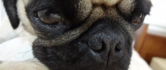
The clinical symptoms of pigmentary keratitis are quite specific: in pugs, it is usually noted closer to the nose, in the medial sector of the cornea. This is explained by permanent irritation of the eyeball by skin folds and hairs growing nearby, resulting in erosion and (if left untreated) corneal ulcer.
As the ulcer begins to heal, pigmented cells invade the damaged areas, contributing to the development of corneal melanosis. In complicated cases, biochromes take over a significant part of the corneal epithelium, which sharply reduces the visual function of the dog.
The diagnosis is made by a veterinary ophthalmologist who conducts a preliminary visual examination. Next, ophthalmoscopy is performed, as well as examination of the anterior segment of the eye using a slit lamp. To make corneal abnormalities visible during examination, it is stained with a fluorescein solution (1%).
Treatment of keratitis in pugs
It is believed that there are no ways to radically eliminate this disease. Overgrown pigmentation is difficult to respond to both conservative and surgical techniques.
In the treatment of pugs, both approaches are combined - a surgical scalpel and exposure to drugs . Local therapy is designed to stop the inflammatory process, often associated with pain. The four-legged patient is prescribed:
- cyclosporine; tacrolimus; corticosteroid drugs.
The first two drugs are equally effective, and the choice of one of them depends on the indications/contraindications of a particular dog. For pigmentary keratitis, the doctor usually prescribes corticosteroids, most often dexamethasone or prednisolone (in the form of direct injections into the conjunctiva of the eye).
But these medications have a significant disadvantage - they suppress the immune system, which begins to “miss” eye infections. Still, steroids are indispensable in the treatment of brachycephalic breeds prone to corneal ulcers.
Important! Occasionally, beta irradiation is used, which is justified in case of a large area of melanotic cataract and significant loss of vision. Removing pigmentation with a scalpel is considered ineffective, since after some time the spot appears again.
Eyelid surgery also helps to slow down the developing pigmentary keratitis, after which the inner corner of the lower eyelid is not able to cover the cornea. The doctor himself chooses the type of surgical intervention:
- Hotz-Celsus method; dissection of the medial canthal ligament; medial canthoplasty.
Pug owners pass on a recipe to each other, thanks to which the growth of pigment stops, and the thorn noticeably decreases in size.
First, the contents of the lidase ampoule are diluted with water for injection (5 ml). Next, dexamethasone (1 ml) and sea buckthorn oil (1 ml) are added to 1 ml of the resulting solution. Before instillation, done three times a day, the solution is shaken vigorously. Pugs tolerate the procedure calmly, as the liquid does not burn or sting.
After a course of instillation (about a month), take a week's break, since dexamethasone is a strong hormonal agent.
Disease prevention
The immunomodulators tacrolimus and cyclosporine A are recommended not only for treatment, but also for the prevention of pigmentary keratitis . The use (administration) of these medications continues for the rest of the pug’s life.
Immunosuppressive drugs become especially relevant when the dog reaches old age, often accompanied by keratoconjunctivitis. Cyclosporine A and tacrolimus also cope with this problem.
about pigmentary keratitis in pugs
Source: https://playdogs.ru/pigmentnyj-keratit-u-mopsa-chem-lechit/
Corneal dystrophy in Sheltie
A similar disease occurs in Shelties and sometimes in Collies, but the cause has not yet been thoroughly elucidated. Many dogs have multifocal corneal opacities with persistent fluorescent spots. Secondary corneal degeneration may occur. The disease is very similar to punctate keratitis and is treated using the same method. However, topical use of steroids must be done very carefully, since the animal's reaction to the drug is sometimes difficult to predict. In the affected eye, with dystrophy of this kind, a small amount of tears is produced, which leads to an aggravation of the process (“vicious circle”).
Neoplasms of the cornea in dogs
Tumors of the cornea in dogs are quite rare. The most famous are dermoid and lumbar melanomas. Dermoids are benign congenital neoplasms that are most often observed on the temporary cornea. They are quite easily removed using lamellar keratectomy. Lumbar melanomas are histologically malignant tumors, but they develop as benign tumors. Their growth is very slow, however, if ignored for a long time, they will fill all the space available to them. From their classification name it is clear that they are formed in the corneoscleral junctions (limbs). Surgical treatment significantly slows the progression of the disease. However, the surgery can cause too much damage to the eye, making this treatment questionable. Surgical intervention refers to either complete excision followed by grafting, or partial removal followed by laser correction.
Spontaneous chronic defects of the corneal epithelium in dogs
This is a particularly unpleasant disease (in terms of diagnosis and treatment) for veterinary medicine specialists, representing specific ulcerative processes. Typically, these processes are chronic, superficial, non-infectious (with the exception of feline herpes) and practically painless. In most cases, there is excessive layering of the corneal epithelium and variable vascularization. Ulcers form abnormalities of the corneal epithelium and basement membrane. All breeds of dogs are susceptible to the disease. Middle-aged and elderly dogs are most often affected. Appropriate local treatment should include antibiotics and atropine 1% once or twice daily. To reduce the likelihood of relapse, sodium chloride ointment or drops are applied topically. Also, topical use of cyclosporine reduces the likelihood of scarring. Surgical debridement and grid keratotomy are often recommended for complete cure. The goal of the procedures is to restore the basement membrane and improve the connection between the epithelium and stroma.
Reticulate keratotomy
This procedure is only intended to treat superficial, non-infected ulcers and should never be used on deep corneal ulcers. Reticulate keratotomy is performed after preliminary surgical debridement as described above. General anesthesia is recommended for restless dogs or when surgery is being performed for the first time. In other cases, mild sedatives are sufficient. Needles, 22 or 25 gauge, are used for superficial mesh cuts. The mesh is created by “drawing” the needle across the cornea, with its inclination of 30-45°. Deep penetration into the cornea should be avoided. Otherwise, continue the prescribed treatment. After the procedure, anti-inflammatory analgesics (for example, non-steroidal analgesics and tramadol) are used. Immediately after the operation, it is recommended to put a protective Elizabethan collar on the dog. Most ulcers heal within two weeks after the reticular keratotomy procedure.
Diagnostics
When diagnosing your pet’s condition, the doctor begins with an external assessment of the condition and a conversation with you. In other words, from the medical history, you will greatly help your pet if you can give the veterinarian as much information as possible.
- What does the dog eat?
- How often does he walk, is there any contact with other animals?
- When did the first signs of the disease appear?
- Was the animal sick before?
And most importantly, never hide from your doctor if you have already tried to treat the disease with something; these details are very important for making the correct diagnosis, and therefore for effective treatment.
Testing the dog's vision and a thorough examination of the condition of the eye must be carried out under very good lighting.
A keratoscopy is performed to determine the condition of the cornea, and an ophthalmoscope is performed to study the permeability of light. If necessary, a swab is taken from the cornea to detect infections or fungi.