A smooth and silky coat is a standard sign of a dog’s health. But the owners are not always able to enjoy such a wonderful spectacle. And the cause of the unsightly condition of the coat can be adenitis of the sebaceous glands in dogs. This disease in itself does not pose a mortal danger, but it certainly will not improve your pet’s health.
Let us immediately note that the exact causes of this pathology have not yet been properly studied. It is assumed that the disease is either caused by some genetic defects, or represents some special form of hormonal dysfunction. Some veterinarians suggest that the “trigger” for the disease is ordinary otitis media. How these two diseases are connected is not known for certain, but the fact is that after ear inflammation in the same poodles, adenitis often follows... In a word, the owners have every reason to be wary.
Definition, clinical picture
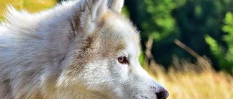
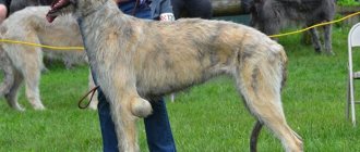
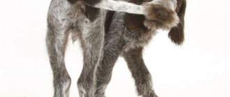
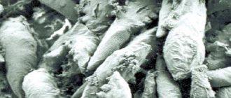
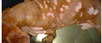
Sebaceous adenitis is a rare type of inflammatory skin disease that affects the sebaceous glands of young and middle-aged dogs. Most often, the pathology manifests itself in poodles and Japanese Akita Inu dogs (for the latter this is generally a disaster). There are two main types of sebaceous adenitis. One is typical for short-haired breeds, and the other for long-haired breeds. The clinical picture in long-haired dogs is as follows:
- Baldness.
- Plaque along the hairline.
- Small clumps of matted hair.
- “Cylinders” of sebaceous plaque that form around the hair shaft.
- The wool quickly becomes matted, greasy, and looks untidy.
- Intense itching along the hairline; the dog scratches frequently, sometimes scratching the skin until it bleeds.
- Hair follicles may become inflamed.
- Silvery-white spots on the skin.
- Especially many affected areas are observed in the head and, especially, ears.
Sweat glands
These glands look like a long tube, the end of which is often curled into a ball, and the rest is usually twisted in the shape of a corkscrew. The walls of the sweat gland consist of single-layer cuboidal epithelium, covered on the outside with a layer of smooth muscle fibers. The sweat gland develops in the form of a dense outgrowth of the main layer of the epidermis, growing deep into the dermis. Later, a channel appears in this outgrowth. Usually the sweat gland is formed along with the hair. Sweat glands most often open into the hair follicle. The secretion of the sweat glands is a product of the secretion of glandular cells that make up their walls. In addition, sweat glands filter intercellular fluid and are able to extract water with substances dissolved in it from the blood and lymph, participating in water-salt metabolism. Sweat contains sodium, potassium and chlorine ions, urea and other metabolic products. Thus, the sweat glands play the role of additional excretory organs.
There are two types of sweat glands, differing in their structure and functions. Human skin is dominated by small eccrine glands that secrete watery sweat, the evaporation of which from the surface of the skin causes cooling and plays an important role in thermoregulation. The intensity of sweating greatly depends on the ambient temperature, but can also occur under the influence of other factors, including emotional ones. Sweating is regulated by the endocrine system and nerve centers located in the brain and spinal cord. A dog has glands of this type on the soft parts of its paws. Since dogs do not produce liquid sweat, it is widely believed that they do not have sweat glands at all. However, this is fundamentally wrong.
Mammals covered with thick hair, including dogs, and also in small quantities in humans, have larger apocrine sweat glands, usually connected to hair follicles. Their secretion has a high content of organic substances, it is thicker and more fragrant. Mixing with the secretion of the sebaceous glands, it forms a natural fatty lubricant for the skin and hair. Unlike eccrine glands, apocrine glands are located in specific areas of the body and often begin to function only after puberty. A variety of apocrine glands are the glands of the eyelids and the glands that secrete earwax.
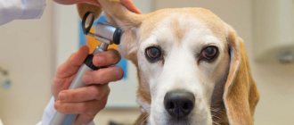
Diagnostic measures
It is very important to understand that all of the above signs may indicate not only adenitis of the sebaceous glands, and therefore it is extremely important to make the correct differential diagnosis. Here are the main diseases, the symptoms of which are similar to the pathology we describe:
- Classic seborrhea. A rather nasty skin disease, which is also known under the term “keratinization.” It is quite simple to distinguish it from adenitis, since with seborrhea the skin becomes hard, scales appear on it, and the upper layer of the epidermis may peel off.
- Demodecosis. Colonies of mites of the genus Demodex “live” in the thickness of the skin, which can cause itching, hair loss and inflammation. Since many of the signs of this pathology are similar to adenitis, the final diagnosis is made only based on the results of a microscopic examination of a sample of pathological material.
- Dermatomycosis. Fungal skin disease. Poorly diagnosed and not very readily treatable.
- Endocrine skin disease of unknown etiology. It's difficult to say anything here. A complete blood test can help make a diagnosis; sometimes the type of pathology is determined by the animal’s response to treatment.
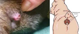
“Fat (greasy) tail” (Hyperplasia of the tail glands, “Tail of breeding cats, males”)
Why is the tail fat?
Tail gland hyperplasia is a seborrheic condition that is associated with hyperplasia of the sebaceous glands in the tail gland region (dogs and cats) or perianal region (dogs). In cats, breeding cat tail occurs as a localized idiopathic condition. In dogs, this pathology may be localized or may be associated with generalized primary or secondary seborrheic disease. Tail gland hyperplasia is common in dogs, and unneutered males are predisposed. This condition is uncommon in cats, with the highest incidence seen in cage-based catteries or in cats that do not properly groom their coats. Unneutered males may be predisposed.
In dogs, the lesion appears as a slowly enlarging, asymptomatic, oval, raised area of hair loss on the dorsal portion of the tail approximately 2.5 to 5 cm below the base of the tail. Damaged skin may be oily, scaly and... Pustules may be observed due to secondary bacterial infection. Dogs with primary or secondary seborrhea may have other skin lesions.
In cats, it appears as a strip of matte fur or a collection of waxy, seborrheic discharge along the dorsal part of the tail. Damaged skin from breeding cat tail may become hyperpigmented or partially bald. The lesions are asymptomatic and no other symptoms of skin involvement are noted.
In dogs, if there is generalized skin disease, the cause of seborrhea should be identified and monitored. Appropriate systemic antibiotic therapy should be given for 3-4 weeks if the dog's lesions become secondarily infected.
Clinical improvement in dogs and cats may be observed with localized antiseborrheic therapy.
What to do with the tail?
In cats, grooming should be improved. Regular grooming and brushing by owners may be necessary in cats that do not groom themselves well.
In unspayed male dogs, castration may induce partial or complete regression, or may prevent further expansion of the lesion. Improvement should be observed within 2 months after castration. In unspayed male cats with breeding cat tail, neutering may not cause the lesion to disappear, but may help prevent further progression.
For cosmetically unacceptable lesions in dogs, excess gland tissue can be surgically removed. Without concurrent castration, however, the lesion is likely to recur within 1 to 3 years. It can be very difficult to achieve closure of the wound.
The prognosis is good. This is a cosmetic disease that does not affect the animal's quality of life.
In our Center you can rid your animal of a “fat tail” using special cosmetic and therapeutic drugs and techniques.
Therapeutic measures
Treatment will depend on the stage of the disease, as well as the breed of the animal. It is important to take into account that the duration of therapy can and should be quite long, since “unfinished” pathology can easily turn into a complicated chronic form and appear within a few years, when it will be much more difficult to treat. So the final decision to discontinue therapeutic measures should only be made by an experienced veterinarian.
Some dogs are more sensitive to treatment than others. Veterinary practice shows that the most unpleasant in this regard are those same Japanese Akitu Inu. They respond poorly to therapy. All this suggests that these “Japanese women” have some kind of original genetic defect. However, let's not talk about sad things. How to cope with this pathology?
Typically, the following methods are used in practice:
- The dog's coat should be brushed more often to prevent the fur from permanently sticking together and forming tangles.
- Multivitamin complexes and corticosteroids are prescribed internally.
- The animal must be washed. It is better to ask your veterinarian about the type of shampoo, since conventional detergent compositions are contraindicated.
- If the affected skin has been contaminated with pathogenic microflora and there are signs of purulent inflammation, special antiseptic shampoos are used for washing. In most cases, antibiotic therapy cannot be avoided, since without antibiotics the risk of general sepsis is too great.
- If you are not squeamish, then, wearing surgical gloves, regularly pick out pieces of skin from your pet’s fur and cut out tangles of matted fur. By the way, it is generally advisable to cut dogs of long-haired breeds during the treatment period, since in this case it is possible to achieve better results.
Sebaceous glands
Alveolar, i.e. pouch-shaped, holocrine-functioning sebaceous glands are found on the follicles of primary and secondary hair in all areas of the body. With the secretion of this type, the epithelial cells of the gland undergo fatty degeneration, and more and more fat droplets accumulate in their cytoplasm. Eventually, the cells are destroyed and form a sebaceous secretion, which accumulates in the cavity of the gland. The sebaceous glands appear during fetal development along with the formation of hair roots, but completely complete their development only with the onset of puberty. The size and shape of the sebaceous glands vary depending on the type of animal, breed, body area and type of hair follicle. Especially large and lobular glands are located on the dorsal side of the neck, on the back and on the tail, smaller and simpler ones are on the stomach. All sebaceous glands belonging to the same hair group are located approximately at the same level, in the reticular layer of the dermis.
The secretion of the sebaceous glands is discharged into the hair canal, where it is mixed with the secretion of the apocrine tubular glands. Glands of both types function, as a rule, simultaneously, having common external excretory ducts. This mixed secretion forms a sticky film on the surface of the skin. It protects against ultraviolet radiation, maintains elasticity of skin and hair and has a strong water repellent property, especially in animals with thick fur.
Signs
The course of the disease occurs in several stages:
- first, the outflow of secretions stops, the glandular sacs become overfilled, the internal contents thicken;
- inflammation of the glands develops, causing a painful itching under the dog’s tail, and if left untreated, the animal injures the skin with its teeth, provoking infection with secondary microflora;
- internal inflammatory processes are intensified by external ones, which ends in suppuration with the formation of an abscess.
Over time, the abscess breaks through and a hole (fistula) forms in this place in the gland. All these processes are accompanied by an increase in body temperature.
Attention! Inflammation of the gland under a dog’s tail to the point of an abscess can only be caused by a complete lack of attention to the pet’s condition or by improper treatment. Therefore, it is extremely important not only to diagnose the disease in a timely manner, but also to treat it with a competent veterinarian.
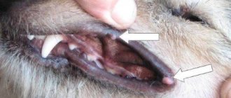
Typical symptoms of inflammation of the glands in a dog are the following:
- the area under the tail near the anus swells, turns red and becomes covered with a rash, the hair around the anus becomes wet and falls out;
- the animal becomes restless, often crawls with its butt on the floor, jumps up sharply, “chases” its tail, and chews on itchy areas;
- The pet emits an unpleasant odor, the source of which is the area under the tail.
The symptoms of inflammation of the glands are in many ways similar to a perineal hernia, helminthic infestation or the manifestation of allergic reactions. Therefore, self-medication is unacceptable in any case. In addition, in Moscow, all veterinary clinics provide professional treatment for inflammation of the glands in dogs. Lack of treatment or delayed contact with a specialist can result in death. If inflammation of the glands in a dog recurs repeatedly, it is recommended to remove them.
How to clean a dog's glands at home?
There are two methods of sanitation. Algorithm for an option that is easy to do yourself:
- If the process takes place along with bathing, the pet is left in the bathroom. Raise the tail with your hand and move it up towards your back. This will cause the muscles to relax and the ducts to open.
- Place a paper handkerchief on the anus, gently press on all sides of the anal sphincter. The secret will be on the napkin. Then you can wash your pet, then apply Vaseline to the anus area.
- If during the process there is restless behavior, pain or cracks and redness of the sphincter, it should be treated with ointment (synthomycin liniment). For the next three to four days, you will need to use rectal ichthyol suppositories.
The second method is usually used by veterinarians, since it will be possible to immediately palpate the paraanal glands for diagnostic purposes. It is important to provide protection by wearing a rubber glove.
- Apply Vaseline oil to the anus and fingers.
- Send your index finger into the rectum and grab the skin near the sphincter with your thumb. Using gentle massage movements, press down on both sides in turn.
- Wipe the affected area with a paper handkerchief previously dipped in chlorhexidine. Use rectal ichthyol suppositories for three to four days. It is better to start the procedure after the pet has finished defecating.
- After defecation, the dog's anus should be treated with a chlorhexidine wipe. This is quite simple and prevents possible inflammation.
Video: inflammation of the paraanal glands in dogs: symptoms and treatment
How to treat inflammation?
Diagnosis of the disease is based on the history reported by the dog owner, visual examination, and rectal examination. If the case is advanced, other tests will be required, such as:
- Blood test (biochemical and general).
- Examination of skin scrapings using a microscope.
- Blood chromatography.
Differential diagnosis is necessary in order not to confuse the pathology with an allergic reaction, hernia, or helminth infection.
Drugs are prescribed during therapy depending on the severity of the disease. Suppositories for the inflammatory process are necessary in any case. Typically used are methyluracil, Proctosedyl, and ichthyol. The duration of therapy depends on the size of the tumor. The following drugs are often prescribed:
- "Methyluracil". They are suppositories that promote rapid wound healing and stimulate protection. During administration, a burning sensation is noticeable. In humans, they often cause an allergic reaction, headache and dizziness. Cannot be used for malignant diseases of lymphoma and bone marrow, leukemia.
- "Cefotaxime". Novocaine blockades are used to eliminate pain. In case of abscesses and the appearance of fistulas, antibiotics are prescribed without fail. Broad-spectrum agents that eliminate gram-negative and gram-positive bacteria help best. The injections are intended for intramuscular administration (0.5 g of medication is mixed with 2 ml of clean water). Side effects: diarrhea and vomiting. Cannot be used for pathologies of the kidneys and liver. Inflammation leads to itching in the anal area, which causes restless behavior, fidgeting on the surface, and biting the affected area. The product will help eliminate itching.
- "Dexafort". It is a glucocorticosteroid hormone. It is used for a single subcutaneous or intramuscular injection in a volume of 0.5-1 ml. After seven days, you are allowed to repeat the procedure. If used for a long time, the pet may lose weight and experience muscle weakness. During pregnancy it should be used with caution.
- "Prednisolone." Usually an injection is given at the withers. Dosage – 0.5 ml once a day. With long-term use, excess weight and ulcers in the digestive tract may appear.
- "Fluconazole". Used for antifungal therapy. Available in solutions, syrups, capsules. Dosage – 10-20 mg/kg 2 times a day. Side effect: stomach disorders.
The pet will need vitamins and minerals to normalize the gastrointestinal tract and proper bowel movements, which is necessary for treatment. A balanced diet and vitamin complexes are also important.
The diet should be therapeutic, made from easily digestible food, enriched with fiber. Dietary fiber naturally clears accumulated secretions from the anal sacs, preventing it from stagnating.
To avoid problems with stool, it is important to provide your pet with high-quality and fresh products. You should follow the regime and avoid overeating the animal. The menu should not include pickles, smoked foods, preserves, fatty or spicy foods. Giving your pet pieces from the table is prohibited. Sometimes the veterinarian recommends adding bran, complexes of vitamins and minerals to the diet.
Physiotherapeutic methods include massage and warm compresses. Often, owners want to use traditional methods in order not to suffer from the aggressive chemical effects of medications:
- plants that eliminate inflammation: parsley, cumin, anise (fruits);
- antifungal: St. John's wort, calendula, wormwood, tansy;
- promoting wound healing (basil, plantain);
- anthelmintics (garlic, tansy).
This therapy includes internal use of herbs and external use in the form of treating the area where the glands are clogged. If the dog resists and does not want to drink them, you can add them to the food.
Homeopathy uses toxic substances. The method is based on eliminating the harm and toxicity of a substance by increasing its biological activity. Then dilutions will be effective. The proportions of the substance to the solution should be 1 to 10. For pets, it should be diluted more strongly.