Pathogenesis of urinary tract infection
It is very difficult for healthy animals to get UTIs due to the normal functioning of the urinary tract's defense mechanisms.
With the exception of the distal urethra, the urinary tract of healthy dogs remains sterile. Microorganisms that inhabit the lower genital tract and distal urethra prevent UTIs by inhibiting the attachment and growth of pathogenic bacteria. Frequent and complete urination physically removes bacteria from the urinary tract. Anatomical factors that cause unilateral movement of urine and prevent penetration of UTIs are ureteral peristalsis, vesicoureteral valves, prostatic secretions, urothelial surface properties, urethral length, urethral peristalsis, and urethral sphincter contraction.
The properties of the mucous membrane, which produces antibodies and has its own antibacterial properties, and the surface layer of glycosaminoglycans also prevent the proliferation of bacteria in the urinary tract. Urine has its own antibacterial properties - very acidic or alkaline urine pH, hyperosmolality and high urea concentration. Finally, systemic humoral and cellular immunity also protect healthy animals from UTIs.
Most UTIs result from bacteria entering the distal genitourinary tract and establishing themselves in the urethra or bladder, and possibly also in the ureters and kidneys. The bacteria that cause UTIs are the same ones that colonize the distal genitourinary tract and perineum in healthy dogs. Any disturbance that interferes with the normal functioning of the defense mechanisms and causes dysfunction of the urinary tract (production of low-density urine or the presence of stones) predisposes the animal to UTI. Female dogs are more likely to get UTIs, possibly because their urethra is shorter and they lack prostate secretions.
Several mechanisms appear to predispose dogs with DM and HAC to UTI. Both endocrine disorders cause polyuria and decreased urine osmolality, which may increase the likelihood of a UTI. Excessive cortisol production in dogs with HAC may cause immunosuppression or a decrease in the normal inflammatory response to infection.
UTIs in dogs with diabetes and OAB are caused by the same microorganisms as in healthy dogs. Escherichia coli was isolated from 65% of dogs, other organisms isolated were Klebsiella spp. (15%), Streptococcus spp. (7%), Enterobacter spp. (7%), Staphylococcus spp. (7%), Enterococcus spp. (7%) and Proteus spp. (7%). Approximately 80% of dogs with UTI, DM, and HAC are infected with one microorganism, and 20% are infected with two or more microorganisms.
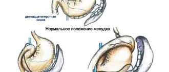
Urolithiasis disease
Urolithiasis is the formation of stones or sand in the kidneys or bladder, which prevents the normal flow of urine.
It is more common in cats than in dogs. However, some dog breeds are more prone to this disease. This is associated with a genetic disorder of phosphorus-calcium metabolism. In addition, urolithiasis can be caused by a genitourinary tract infection.
Improper feeding - the predominance of proteins over carbohydrates, excess fish and dairy products.
The pathology is characterized by the accumulation of calculi or stones in the bladder and renal pelvis. Essentially, stones are calcium or phosphorus salts that accumulate and prevent urine from being excreted normally. If too many of them form, blockage of the urinary ducts can occur. This entails the death of the animal.
The disease manifests itself symptomatically:
- pain when urinating;
- lethargy;
- refusal to eat;
- frequent or difficult urination.
The dog should be taken to the veterinarian as soon as possible. He will prescribe antispasmodics, special nutrition that excludes large amounts of calcium and phosphorus salts. Diet is an important component of therapy. Therapeutic nutrition can dissolve stones and sand in the kidneys and bladder.
For prevention, you need to create the correct diet for feeding your dog, appropriate for its breed. Also avoid infections of the genitourinary system.
Causes
- entry into the urinary system from the lymph or blood of various pathogenic bacteria, viruses and fungi (Escherichia coli, strepto- and staphylococci, pasteurella, chlamydia, candida, trichomonas, etc.);
- complication of kidney diseases (urolithiasis, nephritis or pyelonephritis);
- hypothermia (the most common cause) due to the animal’s habit of sleeping in cold places and drafts, excessively long walks in cold and wet weather, owners pampering themselves with swimming in cold ponds, etc.;
- the appearance of inflammation in bitches against the background of endometritis, vaginitis, improper catheterization in violation of antiseptic rules;
- in exceptional cases – parasites (worms), an allergic reaction, taking toxic medications or tumors;
- abdominal injuries in the perineal area:
- poor quality drinking water (saturated with metal ions or harmful salts);
- problems in the blood supply to the bladder or urine flow;
- allergy to food (very rare cause).
Cystitis is an inflammatory process localized in the genitourinary system. This pathology is inherent in dogs of both sexes and all ages.
In more than half of the cases, the disease is bacterial in nature. In this case, pathological microorganisms enter the mucous membrane of the dog’s bladder in two ways:
- the descending route is the transfer of bacteria from neighboring organs using blood or lymph. The source of infection can be any organ - a tooth with caries, a sore throat, etc.;
- ascending tract - an infection that penetrates the bladder directly from the urethra.
Normally, bacteria are constantly present in the dog's urethra, but they are washed out of the body when urinating. If the dog rarely urinates or his immunity drops, then pathogenic microorganisms begin to multiply quickly and rush into the bladder.
There are a number of other reasons why dogs develop cystitis:
- infection with endoparasites;
- food allergies;
- neoplasms of the genitourinary system;
- toxic drugs;
- urolithiasis disease;
- inflammatory kidney diseases.
There have been cases of idiopathic cystitis in dogs, in which the exact cause has not been determined, although all symptoms point to this pathology.
Bitches more often suffer from this disease due to the anatomical features of the genitourinary system. Their urethra is shortened and located closer to the anus, making it easier for bacteria to enter the bladder. In addition, girls are characterized by purely “female” diseases that cause disruption of the microflora.
Most often, a dog gets sick after severe hypothermia. A walk in prolonged rain or a long swim in a cold river contributes to a sharp decrease in the body's resistance.
At this time, the microflora living in the genitourinary tract (streptococci, chlamydia, staphylococci, mycoplasma, pasteurella, as well as microscopic fungi) is activated, its virulence increases, and it begins to develop rapidly, causing an inflammatory reaction.
Cystitis
Cystitis can be either acute or chronic, lasting long and indolently. And the nature of the inflammation can be purulent, catarrhal, fibrinous.
Cystitis can also be caused by tumors or allergies to certain drugs and food.
Cystitis in dogs: features of the disease
The disease occurs in both elderly (geriatric) patients and young animals, including puppies. However, older animals (7 - 8 years +) are most prone to developing this disease. Due to the natural processes of aging, combined with concomitant systemic diseases, errors in diet, stress, the immune system is not able to perform its functions at the original level, which allows bacterial agents to freely penetrate into the bladder cavity and multiply there.
If you notice the symptoms of this disease in time and consult a doctor, you can avoid sad consequences.
Note! Cystitis in dogs occurs in chronic or acute form.
According to the nature of the manifestation of the inflammatory process, cystitis can be classified into:
- Catarrhal.
The inflammatory process occurs within the mucous membrane, without going beyond it. In this case, a general clinical urine test reveals protein, while the urine is cloudy. - Hemorrhagic.
The inflammatory process is accompanied by the release of blood in the urine. In this case, blood can be observed both at the end of urination and color the entire portion of urine. - Dystrophic.
This type is characteristic of the chronic course of the disease. The presence of epithelial cells is noted in the urine. - Purulent.
It is observed in advanced cases, in the absence of proper treatment. In this case, the urine acquires an unpleasant odor + the release of purulent masses in the urine.
In most cases, the cause of cystitis is bacteria, viruses, fungi, protozoa, which enter the bladder in the following ways:
Ascending path.
In this case, pathogenic agents penetrate and “rise” into the bladder “from outside,” from the urethra. In the lower (distal) parts of the urethra, microorganisms are always present, and this is considered the norm, but with the flow of urine during urination, they are “washed away,” which does not allow them to actively increase their numbers. However, for various reasons, the number of bacteria may increase, and the bacteria "climb" up the urethra into the bladder cavity. These reasons include the following factors:
- Age of the animal;
- Gender of the animal: females, due to the fact that their urethra is shorter and wider compared to the urethra of males, are more prone to developing cystitis. Also, in females, the urethra is located close to the anus, which also contributes to the development of cystitis due to the penetration of bacteria from the surface of the anus, while the pathology of the reproductive system (endometritis, vaginitis) + complications during childbirth also makes females more susceptible to this disease;
- Rare walks: bacteria are not removed by urine current and, when actively multiplying, make it possible to penetrate the urethra;
- Inactivity of the animal;
- Hypothermia: drafts in the room where the animal is kept, lack of bedding, bathing the animal in cold water, bathing the animal in open water at low ambient temperatures;
- Stress;
- Unbalanced diet.
All these factors weaken the body's defenses and give rise to the inflammatory process.
Descending path.
In this case, the infection is initially localized in another organ or organ system. The infection can enter the bladder through the lymphatic vessels from the pelvic organs; through blood vessels from any infectious focus localized in the animal’s body.
Cystitis can also develop due to parasitic infestation, against the background of an allergic reaction, when the tumor is localized in the organs of the genitourinary system. In this case, cystitis can manifest itself as a symptom of another disease of the genitourinary system (secondary):
- urolithiasis disease;
- pyelonephritis;
- jade and so on.
Therefore, when observing symptoms of this disease in an animal, it is necessary to contact a veterinarian and not neglect additional diagnostic measures, since in order to effectively treat cystitis, like any disease, it is necessary to eliminate the very cause that provoked the disease.
ICD in dogs - symptoms and course of treatment
Dogs, like humans, can develop a variety of bladder diseases. Urolithiasis, or bladder stones, are more common in dogs than kidney stones. These stones can vary in size, although in some cases one large stone may form in the bladder.
One of the most common crystals in canine urolithiasis are calcium oxalate (CaOx) crystals.
The exact causes of bladder stones are poorly understood. In a healthy dog, the mineral salts in the urine remain dissolved as long as the pH level remains within a narrow range or until the urine becomes too concentrated.
Modern research shows that urine high in calcium, citrate, oxalate and acid predisposes to the formation of calcium oxalate crystals and stones. Large studies show that a diet that causes urine to become more acidic (ph level less than 6.5) may also increase a dog's risk of developing bladder stones.
Symptoms of urolithiasis are very similar to those of bladder infections or cystitis. The most common signs of stones are hematuria (blood in the urine) and dysuria (difficulty urinating).
Hematuria occurs due to stones that rub against the walls of the bladder and cause irritation and damage to its tissue, as well as bleeding.
Dysuria may result from inflammation and swelling of the walls of the bladder or urethra.
Small stones can be passed into the urethra through urine, where they can become lodged and cause an obstruction. In this case, the dog may completely lose the ability to urinate, which sometimes leads to rupture of the bladder.
Complete obstruction can be life-threatening for your pet and requires immediate emergency care.
In some cases, the veterinarian will be able to detect stones through palpation if the dog is calm enough and the bladder is not too painful.
However, if the stones are too small, palpation will not help determine their presence. Bladder stones are often diagnosed using an x-ray or ultrasound.
DETAILS: If I have chronic kidney disease and hepatitis C
Typically, your veterinarian will be able to determine the type of stones based on palpation, x-rays, and urine test results.
Treatment of urolithiasis in dogs
There are two ways to treat bladder stones in dogs:
- without surgical intervention;
- using surgical removal.
Small stones can be removed without surgery. However, this is only possible when the diameter of the stones is very small.
In some cases, this procedure is performed under heavy sedation, although general anesthesia is often used.
Surgical removal is usually recommended in cases where the bladder stones are too large or in very large numbers, i.e. when there is a high risk of developing complete urinary tract obstruction. Surgery is also used if the problem needs to be resolved as quickly as possible.
Male dogs have a significantly higher risk of developing urinary obstructions, so when stones are found in a male dog's bladder, the veterinarian will usually immediately recommend surgical removal. Surgery is also used in cases where diet treatment has not been successful.
Your veterinarian will discuss with you the most appropriate treatment strategy for your dog based on your specific situation.
Recently, the ultrasonic stone dissolving method has been increasingly used, which uses high-frequency ultrasonic waves to break up the stones and remove them from the bladder naturally. This method has numerous advantages, the most important of which is that there is no need for surgery.
It is advisable to feed dogs that have already experienced urolithiasis on a special therapeutic diet. A diet that will help reduce acidity and reduce urine concentration is recommended.
Most dogs are recommended to be fed canned or wet food and encouraged to drink plenty of water. In some cases, drugs are used to lower pH, such as potassium citrate.
If the dog is fed homemade food, vitamin B6 supplements are often added.
If your dog exhibits any clinical signs such as frequent urination, urinating in unusual places, painful urination, or blood in the urine, he should be seen by a veterinarian immediately.
Unfortunately, bladder stones have a fairly high recurrence rate, despite careful attention to your dog's diet.
Anatomical features of the bladder in dogs
The bladder is a storage device, but the muscular system of the bladder, sphincter and urinary canal are involved in the excretion of urine.
Therefore, when considering the problem of the inability to urinate, we cannot talk exclusively about atony (loss of tone in the muscle walls of the bladder).
The dog's genitourinary system.
If the sphincter or urinary canal is blocked, then urine will also not be excreted , and the symptoms will be similar, but we cannot talk about loss of muscle tone.
Blockage can occur from stones, tumor tissue, and inflammatory products such as fibrin strands.
Causes of atony
Very often, bladder atony is a consequence of injury. In this case, both the spinal cord and the brain can be damaged.
Even if there is visually no violation of the integrity of the skeleton, then infringement of the nerve ending may be present, but this cannot be diagnosed by palpation.
Nervous regulation of urination in dogs.
Pathogenic microflora
Atony may also be observed as a consequence of the inflammatory process caused by pathogenic microflora. In this case, antibiotic therapy quickly helps to cope with the disease.
Taking antibiotics will help cope with the disease.
Age
Age may also be a cause of atony. Atony tends to be more common in older dogs.
In this case, a complex of changes occurs, both in the mucous membrane and in the muscle membrane. As a rule, animals are difficult to treat due to destructive age-related changes, and catheterization is performed on a permanent basis.
Old dogs are difficult to treat.
The cause may be neurogenic disorders, mainly after injuries. The muscular system and sphincters function normally, but the brain does not transmit the signal that the bladder is full. In this case, there will be involuntary release of urine and it is not correct to talk about atony.
Clinical picture of the disease and diagnosis
Symptoms depend on the type of disease. When the disease proceeds according to an acute scenario, the dog becomes lethargic, it is depressed, a febrile state may develop, the act of urination becomes painful, and blood often appears in the urine. An experienced specialist, upon palpation, can feel an enlargement of the bladder, as well as thickening of the walls of the urethra, which is caused by the infiltration of lymphocytes into the thickness of their tissues.
It can be useful to collect all the urine excreted by the animal during the day: if its amount is reduced, this indicates serious kidney damage. In all cases, a general urine test is performed. If inflammation is present, under a microscope you can easily see a huge number of “fallen off” epithelial cells, as well as tiny crystals of uroliths, “casts” of the renal tubules.
The worst thing is when the urine gives off a pronounced cadaverous or putrid odor, since these signs indicate the gangrenous nature of the inflammation. Note that in the chronic course of the disease, the clinical picture is very blurred; some animals do not show any signs of anything wrong at all.
Diagnosis is quite simple: palpation and urine analysis play the main role; in more complex and doubtful cases, ultrasound examination of the bladder and urethra, as well as contrast radiography, are used.
How to recognize the disease
Depending on the course and manifestations of the disease, acute and chronic cystitis are distinguished. The symptoms of the disease are similar in both cases, but with acute cystitis they will be more pronounced:
- The dog begins to urinate at home, and can not only make puddles, but also damage the furniture.
- The dog whines and worries when urinating, as well as immediately after the process.
- After urinating, the dog finds it difficult to move its hind legs; it moves them very carefully.
- The urine becomes cloudy and has a bad odor. It may contain mucous veins and drops of blood.
- The abdomen is tight and painful.
- The dog pees often and in small portions. At the same time, the male, who previously raised his paw when urinating, begins to sit down or raise his paw even higher. Bitches seem to pull their pelvis forward when urinating.
- If the process has gone far and the animal has developed hemorrhagic cystitis, then purulent discharge appears in the urine.
- With severe inflammation and blockage of the urinary canal, urine comes out drop by drop or its release stops completely.
- The general condition of the dog changes - it becomes lethargic or, on the contrary, too aggressive.
- The inflammatory process causes hyperthermia.
- The animal loses its appetite, and with severe pain the dog may vomit.
- Signs of cystitis in dogs are quite typical, so any manifestation of the disease should worry the dog’s owner and cause a visit to the veterinary clinic.
Clinical symptoms
- Increased urination. You can only notice it in pets who live indoors - he will constantly ask to go outside or make small puddles right in the room. The volume of urine produced per day does not increase: the dog goes to the toilet more often, but in small portions.
- Impurities appear in the urine: mucus, blood or pus. It becomes cloudy and acquires a sharp and unpleasant odor that is not typical for its normal state.
- At the time of urination or at the end of the process, the dog may whine in pain. Males stop raising their paws and urinate while crouching on both paws (a clear sign of cystitis). It is clearly evident that the procedure causes pain and discomfort for the pet.
- The dog does not allow you to touch the belly in the perineum - it twitches its paws, squats, whines and tries in every possible way to avoid inspection. The abdominal wall is tense, like the membrane of a drum.
- With a protracted process, general malaise, lethargy develops, appetite decreases, and body temperature rises. Signs of intoxication may appear at the time of development of the purulent process, because a sick animal will increasingly try to hold in urine in order not to feel pain.
- Cystitis in a dog has symptoms similar to urolithiasis - a more serious pathology that is excluded in the first place.
- The general condition of the animal initially does not cause concern, but an increase in temperature can be detected by a simple touch. In this case, the animal will experience increased thirst.
- Observing the behavior, the reason becomes clear. An inflamed bladder increases neuroreflex excitability, its muscle walls contract more often than necessary, and this causes the urge to urinate. At the same time, the bladder itself may be empty, which causes even more anxiety in the dog.
- The dog crouches, or the male raises his paw unnecessarily, and the puddle is of absolutely insignificant size. Sometimes a male dog may involuntarily sit down due to a painful reaction during the passage of urine. The animal does not choose a place to urinate, and may make whining sounds. Urination may be accompanied by whining both at the end and at the beginning.
- If no help is provided and the inflammation continues to increase, the dog becomes apathetic and sometimes aggressive. Urinary discharge acquires an unpleasant, pungent, unusual odor due to the developing microflora. Upon careful examination of the discharge in the urine, you can see blood, fibrin threads, and turbidity.
- If you try to palpate the abdominal area, it may be problematic due to the pain reaction. Place the dog's head away from you and slowly, with stroking movements, try to run your hand over the abdomen; it will be tight and painful.
- Due to poor urine flow, waste products are absorbed and this can cause the dog to vomit or gag.
Diagnosis
Ultrasound examination does not make it difficult to detect inflammation of the bladder. This may reveal sand and rocks.
DETAILS: Is it possible to eat dairy products if you have chronic kidney disease?
Most dogs with UTI, DM, or HAC are older animals, with an average age of 9 years. Miniature Schnauzers, Cocker Spaniels, and Poodles are predisposed to UTIs, while Golden Retrievers, Labrador Retrievers, and mixed breeds are less susceptible to UTIs.
Clinical signs of UTI are stranguria, dysuria, hematuria and pollakiuria, and are observed in less than 10% of dogs with DM and HAC. This may be due to the anti-inflammatory effects of excess cortisol in dogs with HAC. This can also be explained by the fact that owners are more likely to notice polyuria, which is common in dogs with diabetes and HAC.
The absence of stranguria, dysuria, and pollakiuria in dogs with diabetes and OAB is an indicator of kidney and ureteral infection, which may not cause symptoms of a urinary tract infection. General examination findings are typical for dogs with DM and HAC—cataracts, skin lesions (pyoderma, skin thinning, alopecia, cutaneous calcification), hepatomegaly, and abdominal enlargement.
Clinical picture of the disease and diagnosis
Before starting treatment for cystitis in dogs, it is necessary to conduct a thorough diagnosis. This is done to ensure that the diagnosis is correct, because some pathologies of the genitourinary system have similar symptoms.
For a veterinarian, signs such as frequent urination in small portions and the presence of blood in the urine already confirm the diagnosis, but the doctor prescribes additional studies to clarify it:
- The bladder is palpated to determine its fullness. Fullness of the urinary tract and difficulty in excreting urine are characteristic symptoms of cystitis.
- A bacteriological analysis of urine is carried out to identify the presence of pathogenic microorganisms in it and determine their sensitivity to antibiotics.
- The urine is examined under a microscope to look for pus, blood and sand. Such impurities indicate stagnation and development of the inflammatory process in the bladder and that the dog has urolithiasis.
- A swab is taken from the dog's genitals to check for infectious diseases.
The diagnosis is confirmed if the study shows the presence of pathogenic microorganisms and sediments of inorganic substances in the urine.
Further diagnosis is carried out only after urine is removed by catheterization. In the emptied bladder, the doctor can use palpation to detect stones that have blocked the urinary canal and caused cystitis.
Using X-rays and ultrasound of the dog's pelvic organs, the size of the stones and the degree of inflammation in the bladder are assessed, and the presence of tumors in it is determined.
Cystitis in dogs is curable, so after diagnosis, the animal owner will need to strictly adhere to the doctors’ recommendations so that treatment takes a minimum of time.
Cystitis in dogs: diagnosis
It is determined by a veterinary specialist based on medical history, laboratory and special studies, which include:
- general urine analysis;
- blood analysis;
- biochemical blood test;
- Ultrasound of the abdominal cavity;
- X-ray of the pelvic cavity.
It is important to differentiate this disease from other diseases of the excretory system, where cystitis can be observed as a secondary manifestation (urolithiasis, pyelonephritis, nephritis, neoplasms of the genitourinary system, and so on).
The diagnosis is made by a veterinarian not only based on the owner’s description of the pet’s condition and clinical examination, but also on the results of ultrasound and blood and urine tests.
ICD in dogs - symptoms and course of treatment
Urolithiasis is a disease in dogs in which stones (calculi or uroliths) form in the urinary tract. Stones can be located anywhere: in the urinary tract, kidneys, ureter or bladder, but most often in the bladder. Stones are formed due to the oversaturation of urine with certain minerals!
A number of factors can contribute to mineral overload in urine: increased concentrations of individual minerals, changes in pH (acidity or alkalinity), high concentration of urine, use or absence of stimulants and stone inhibitors.
When your dog has difficulty peeing, you assume that it is urolithiasis, but you do not know what to do and are looking for advice on the Internet on forums, we recommend that you do not self-medicate and experiment on your beloved dog, because the consequences of your experiment may disappoint you and your family.
A number of factors can contribute to the development of urolithiasis in dogs. They include:
- Genetic predisposition;
- Incomplete diet and lack of water;
- Basic metabolic diseases, such as high concentration of calcium in the blood, consequences of overactivity of the parathyroid glands;
- Congenital problems such as abnormal blood vessels around the liver as they contribute to the formation of uric acid stones;
- Bacterial urinary tract infections (stone formation).
In many cases, the cause of stone formation is unknown!
According to the predominant mineral composition, different types of stones are divided. In dogs, the most common stones are those that consist of the minerals ammonium, magnesium phosphate, and calcium oxalate.
Urate stones are typically less common in most breeds, but are very common in Dalmatians or English Bulldogs. Cystine and silica stones are relatively rare.
Different types of stones in dogs are treated differently. Therefore, it is important for the veterinarian to obtain stones for chemical analysis.
- Oxalate stones in Lhasa Apso;
- Urate stones are common in Dalmatians and English Bulldogs;
- Cistine stones in Newfoundlands;
- Struvite and oxalate stones in miniature schnauzers and bichon frizes.
The risk of recurrence for urolithiasis is high and ranges from 20 to 50 percent. Your pet's symptoms depend on the number of stones, their location in the urinary tract, the physical characteristics of the stones (smooth or jagged), and the presence of a bacterial urinary tract infection.
- Difficulty or frequent urination;
- Urinating in inappropriate places;
- Blood in urine.
Symptoms caused by kidney stones include back or stomach pain, and sometimes an unusual odor in the urine if a bacterial infection is present. Surprisingly, many animals with kidney stones may have no symptoms at all.
To identify urolithiasis in dogs, it is necessary to conduct diagnostic studies that will identify both the causes of symptoms and other disease processes. Your veterinarian may recommend:
- A complete medical history and physical examination, including palpation of the abdomen. Bladder stones are difficult to palpate due to the tendency of many pets to tense their abdomen when the veterinarian tries to palpate the stones. The medical history includes questions about the pet's urine stream pressure, frequency of urination, presence of blood in the urine, changes in water intake, changes in appetite, weight loss, and history of previous illnesses or infections;
- Urinalysis to assess concentration, acidity or alkalinity (pH), the presence of red blood cells, white blood cells, bacteria;
- An x-ray of the abdomen to look for stones, which are usually hard enough to be seen on an x-ray.
Other diagnostic tests that may be performed:
- Culture and sensitivity to detect bacterial urinary tract infection;
- Serum biochemistry to assess renal function;
- Complete blood count to evaluate for infections;
- Abdominal ultrasound to evaluate urinary tract stone obstruction;
- X-ray contrast dye studies to visualize some stones that do not show up on X-rays;
- Stone analysis to determine mineral composition and guide treatment for urolithiasis.
Treatment methods
Treatment for urolithiasis in dogs may include one or more of the following:
- Treating a bacterial urinary tract infection with antibiotics. Removing stones surgically or through dietary intervention. There are advantages and disadvantages to both methods. Surgery usually removes all stones and allows the mineral composition of the stones to be assessed.
- Dissolving stones using dietary methods is not invasive, but does not provide the mineral content of the stones. It also requires the veterinarian to make an educated guess as to the type of stone present. Some stones can be broken down only by changing the diet, which cannot be achieved by other means.
DETAILS: Urological collection will ensure kidney health
Whether or not to use a stone-dissolving diet will depend on your pet's overall health, the type of stone, the location and number of stones, and other factors that your veterinarian will discuss with you.
What treatment is prescribed for urolithiasis in dogs at home? If your dog is being treated at home, make sure you use the medications prescribed by your veterinarian. Give antibiotics as scheduled. It is important that your pet has free access to fresh, clean water.
Work with your veterinarian to perform regular physical examinations and urine tests to monitor your dog's progress. Urine culture should be repeated 5-7 days after the end of antibiotic treatment to check for infection. If your pet does not tolerate the prescribed treatment, further testing may be required to look for underlying disease processes.
Analysis of the stones will help you draw up an accurate treatment plan for your veterinarian:
- If struvite stones (magnesium ammonium phosphate) are found, antibiotics are prescribed to treat bacterial infections;
- Oxalate stones: thiazide diuretics and potassium citrates;
- Urate stones: allopurinol;
- Cystine stones: penicillamine or 2-mercapto propionyl glycine (2-MPG or Thiola).
conclusions
In most cases, it is difficult to prevent stones from forming in the body in dogs. Preventive care for urolithiasis in dogs is to provide the pet with the opportunity to urinate frequently and an adequate supply of fresh, clean water.
Prompt treatment of urinary tract infections can help reduce the potential for a certain stone to form.
Treatment medications are prescribed, dosed and combined strictly by a veterinarian! The disease is not cured by eliminating symptoms alone.
- Bacterial infection. Treatment of cystitis in dogs with antibiotics is used only after urine culture and determination of the sensitivity of the microflora to one or another appropriate drug. If there is no time to wait, the veterinarian usually prescribes broad-spectrum drugs or nitrofurans (for example, furadonin for cystitis).
- Hemorrhagic factor. Dogs are required to use hemostatic agents when blood is detected in the urine.
- Secondary diseases. If, in addition to cystitis, pathologies suspected of causing it are found in a dog, then the underlying disease is first eliminated, and then the inflammatory process is eliminated. Or treatment is carried out in parallel.
- Rinsing the bladder using a catheter is carried out with antiseptic liquids and only by a specialist! A good way to cleanse the bladder of waste products of microorganisms and reduce the risk of intoxication. It must be done after the bladder has been emptied. In rare cases, antibiotic solutions are used.
- Allergic cystitis is diagnosed very, very rarely. In addition to antihistamine therapy, the diet must be changed, excluding potential allergens.
- During treatment, a special drinking regime is observed - plenty of it and only clean drinking water.
- The pet should be fed either with special food intended for diseases of the urinary system, or the diet should be formed independently. The most famous foods for cystitis and diseases of the genitourinary system (including urinary stones): Royal canin urinary s/o and Royal canin urinary;
- Hills Prescription Diet canine u/d;
- Farmina Vet Life for dogs;
- Purina urinary.
It often happens that cystitis in a dog can be completely cured only in combination with the treatment of another disease that has become the cause or consequence of cystitis. This situation is caused by delaying contacting a doctor at the first signs of illness or the presence of primary diseases, for example, diabetes.
The owner needs to reconsider the diet of his pet and switch him to dietary food. If the dog is accustomed to industrial food, then it is necessary to buy it medicinal food intended for animals with problems of the genitourinary system. Feeding with natural products is also carried out taking into account the dog’s disease - everything spicy, salty, fried, and fatty is excluded.
Preference is given to porridge with meat broth, fermented milk products, and boiled vegetables. Meat in its pure form is not recommended for feeding a dog in the acute period of the disease. The animal must always have access to clean drinking water, to which you can add ½ teaspoon of soda per 1 liter of water.
To treat cystitis, antibiotics are prescribed in 90% of cases, since the disease is most often caused by a bacterial infection of the bladder. The most commonly used medications are:
- "Ceftriaxone".
- "Baytril."
- "Amoxicillin."
- To relieve spasmodic contractions, no-spa, cystone, and analgin are prescribed.
- The use of antibiotic therapy is mandatory. If possible, it is necessary to conduct research to identify the most sensitive microflora; if this is not possible, broad-spectrum antibiotics (baytril, ciftriaxone, cephalotoxime) are prescribed.
- It is necessary to use one of the sulfonamide drugs (furodonin, urolex, furogin) in combination.
- After conducting a urine test, pH is an important indicator for bladder inflammation. If the reaction is alkaline, it is necessary to use salol, and if it is acidic, hexamethylenetetramine is prescribed.
- To quickly release inflammatory products, drink plenty of herbal diuretics; for this purpose, bearberry leaves, dill or fennel seeds and oregano flowers are brewed. Among veterinary drugs, the drug “Stop Cystitis” has a good effect. A herbal suspension can be given to a dog, either with food or separately, for both medicinal and preventive purposes.
- If cystitis is detected quickly and comprehensive, correct treatment is prescribed, then the symptoms disappear after 1–2 days. The course of treatment lasts up to 7 days, during which time the inflammatory reaction on the mucous membrane completely disappears.
- If the animal has already developed cystitis, the owner should be careful and understand that this is a “weak” point in his dog. For prevention purposes, hypothermia of the body and the occurrence of gynecological complications should not be allowed.