The development of inflammatory processes in a dog’s brain very often begins so rapidly that the animal’s external manifestations resemble nothing more than the last dying hours. Unfortunately, in some cases this is true, but at the same time, depending on the form of meningoencephalitis, emergency veterinary care can save the dog.
What it is?
Encephalitis is inflammation of the brain. The most common pathology of the central nervous system in dogs. They are divided into subtypes depending on the location and extent of the lesion. With meningoencephalitis, both the membrane of the cerebral cortex and the medulla are affected.
By origin they are distinguished as primary and secondary, by the nature of the course they are divided into acute and chronic, and depending on the nature of the lesions they are differentiated into bacterial and non-bacterial.
Inflammatory diseases of the nervous system in dogs
Inflammatory diseases of the nervous system in dogs
Inflammatory diseases of the nervous system of dogs cover a fairly large group of diseases - meningoencephalitis/meningomyeloencephalitis of various etiologies (causality).
Meninitis is inflammation of the meningeal membranes of the central nervous system, myelitis is inflammation of the spinal cord, encephalitis is inflammation of the parenchyma (tissue itself) of the brain. Meningitis is characterized by inflammation involving the subarachnoid space, that is, inflammation of non-neuronal (containing nerve cells) tissue.
As a rule, one cannot talk about specific meningitis or encephalitis, because both processes occur simultaneously, because inside the skull, the tissues are anatomically located very close to each other, so it is more legitimate to use the term meningoencephalitis.
Meningoencephalitis, regardless of the cause, is not a widespread disease, but in general it makes up a significant percentage of the total number of neurological diseases.
Inflammatory diseases of the central nervous system (meningoencephalomyelitis) according to etiology are divided into infectious and non-infectious.
Infectious
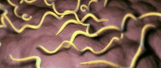
can be caused by bacteria (Staphylococcus spp., Pasteurella multocida, Actinomyces spp., Nocardia spp., Listeria monocytgenes), fungi (Cryptococcus neoformans, Asppergillus spp.), protozoa (Toxoplasma gondii, Neosppora caninum, Babesia spp.), parasites (Dirofilaria immitis, Cuterba spp., Toxascaris spp., Ancilostoma spp., Taenia spp., Angiobylus spp.), rickettsia (Ehrlichia spp., Rickettsia ricketsii, Neorickettsia helminthoeca, Borrelia burgdorferi - Lyme disease) and viruses (rabies, canine distemper, Aujeszky, parvovirus and herpesvirus in puppies).
With viral, protozoal (caused by protozoa) and parasitic infections, signs of damage to the brain parenchyma (encephalitis) are more pronounced; with bacterial infections, signs of damage to the meningeal membranes (meningitis) are more pronounced.
When infected by fungi or rickettsia, signs of damage to both those and other formations may be observed (diffuse symptoms).
To non-infectious
include steroid-dependent meningitis, granulomatous meningoencephalitis, and several breed-specific meningoencephalitis.
It is assumed that the pathology is based on immunological disorders, since almost all animals respond to treatment with immunosuppressive doses of glucocorticoids.
Granulomatous meningoencephalitis
(GME) is a non-purulent inflammatory disease in which limited (focal) or diffuse (multifocal) damage to the central nervous system occurs.
There are 3 forms: limited GME involving the brain stem; disseminated GME, which damages the cerebrum, lower brainstem, cerebellum and cervical spinal cord; Visual GME is characterized by damage to the eyes and optic nerves.
The cause of GME is unknown, but the disease is believed to be immune in nature. Treatment includes administration of glucocorticoids; the prognosis is uncertain, especially in the long term. With rapid progression of the disease, it is always unfavorable.
Beagle pain syndrome -
a severe form of steroid-dependent meningitis with polyarthritis causing pain in the cervical spine.
It is assumed that the disease is caused by immune disorders, since steroid therapy leads to complete remission.
Bernese Mountain Dog meningitis
- this breed is susceptible to necrotizing vasculitis and polyarteritis (aseptic meningitis). The cause of the disease has not been established, but clinical manifestations disappear in almost all animals when treated with steroids.
Pug meningoencephalitis
– a disease of young and middle-aged dogs (9 months – 4 years), characterized, as a rule, by a rapid course and a poor prognosis.
At the onset of the disease, convulsions and a picture of diffuse damage to the central nervous system occur. Characteristic features include circling, ataxia (shaky, uncoordinated gait), “head resting” against the wall, blindness, and pain in the cervical spine.
Therapy with steroids and anticonvulsants does not give good results; animals usually die within a few weeks after the onset of symptoms.
Clinical manifestations of inflammatory diseases of the central nervous system can be varied, depending on which area is affected and how severely; disorders can be limited (focal), diffuse (spread), and quickly develop from limited to diffuse.
Classic signs of meningitis are pain (usually in the neck) and fever. Animals resist being taken on a leash, they exhibit hyperesthesia (increased sensitivity to touch and influence) and rigidity (tone) of the neck muscles. In severe cases, lateral positioning, opisthotonus and hyperextension of the front legs occur.
The nature of the clinical manifestations of encephalitis is due to damage to the brain parenchyma. Violations are usually asymmetrical. The severity of symptoms can increase gradually - impaired consciousness (depression) up to stupor, coma; changes in behavior; visual impairment (while maintaining the normal reaction of the pupils to light - so-called central blindness); impaired coordination of movements and voluntary motor functions; convulsions.
In the presence of encephalomyelitis, sensory ataxia (impaired gait and body position in space, impaired posing reactions), motor dysfunction and dysfunction of the cranial nerves are detected.
A diagnostic method for identifying meningoencephalitis and its cause is cerebrospinal fluid (CSF) analysis. The collection of cerebrospinal fluid requires general anesthesia, and is associated with a certain risk (both anesthetic and surgical, since a puncture of the occipital cistern is performed). Non-invasive methods (however, also performed under general anesthesia) are CT (computed tomography) and MRI (magnetic resonance imaging), however, on the basis of these studies it is not always possible to accurately make this diagnosis, because changes may be specific to pathologies of different causes (for example, autoimmune, fungal, bacterial cannot be verified).
Causes
Causes of bacterial meningoencephalitis:
- severe damage to the liver and kidneys;
- oncology;
- sepsis;
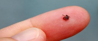
- encephalitis tick bite;
- chronic rhinitis, frontal sinusitis, nasopharyngeal polyps;
- lymphadenitis of the submandibular and retropharyngeal nodes;
- undergone chemotherapy;
- long-term treatment with corticosteroids;
- odontogenic abscesses.
Reference. At particular risk are dogs that have had one of the diseases: distemper, leptospirosis, adenovirus, herpes virus or viral hepatitis.
What causes nonbacterial meningoencephalitis:
- helminthic infestations;
- autoimmune pathologies;
- hereditary predisposition;
- traumatic brain injury.
Carrying out an examination of the animal
If the doctor suspects the appearance of encephalopathy, he should prescribe an additional minimum of tests. Testing for the presence of bile acid must be performed twice - the first sample is taken before meals, the second two hours after meals (in this case, the test is taken from a vein).
Encephalopathy is characterized by a standard or slightly higher than normal presence of bile acids in a sample taken in the morning before meals and up to 10 times higher than the established measure of their indicator after feeding.
A laboratory diagnostic method, as a result of which conclusions can be drawn about the functioning of the liver, often shows a low albumin level. Using biochemical analysis, a high degree of urea is detected with normal or reduced creatinine levels. Occasionally, low potassium and low cholesterol levels in the blood are observed.
In dogs with liver shunts, tissues and organs experience oxygen starvation (microcytic anemia). Ammonium biurate is present in the urine, and large stones may form. Using additional analysis techniques, characteristic changes can be determined:
- X-rays of pets with cirrhosis reveal a significant decrease in liver volume;
- Application of ultrasound. In this manner, a decrease in the size of an internal organ is detected. In some cases, it is possible to identify vascular pathology. Dopplerography detects another vessel, but this technology is not considered as an expert method;
- Thanks to portography, abnormal vessels are identified;
- Portosystemic shunts can be detected by MRI of the liver;
- Biopsy;
- Manifestations characteristic of hepatic encephalopathy are caused by various pathologies, which must be excluded by a veterinarian.
Kinds
All meningoencephalitis in veterinary medicine is divided into 2 large categories: necrotizing and granulomatous. Each of them has its own subspecies depending on its narrow specificity.
Necrotizing (NE)
Characterized by multiple necrosis in brain tissue. Pugs, Papillons, Chihuahuas, and Maltese dogs are more susceptible than others. As a rule, it affects the bark, the trunk - with rare exceptions.
Lesions form at the boundaries of gray and white matter. The disease begins extremely acutely and progresses rapidly, leading to death in the first few days. The prognosis is poor, with a high mortality rate within the next few weeks due to lack of response to treatment.
Granulomatous (GME)
Occurs for unknown reasons. Most often it affects young dogs under 5 years of age of small and medium breeds. It occurs in three forms: focal (1 lesion), multifocal (multiple lesions) and visual with damage to the optic nerve and the structure of the eyeball. It is a direct threat of blindness as a provoking factor of uveitis and retinal detachment.
In the case of a multifocal form and proper adequate treatment, life expectancy is on average 1.5 years. Timely treatment of the focal form can result in long-term remission for many years. The ocular form is characterized by a chronic course and favorable survival prognosis.
Despite all the clear theoretical differentiation, in most cases, in most cases, after the fact, a dog that has already died is given a conclusion about inflammation of the brain of unknown etiology.
Types of disease and routes of infection
Meningitis in dogs is classified according to several criteria.
Depending on the type of pathogen (etiology), there are several types of infectious forms of the disease. Most often they are purulent lesions of the brain. In this case, neutrophils (leukocyte cells) predominate in the cerebrospinal fluid (CSF):
- bacterial meningitis (causative agents - pneumococci, meningococci, mycobacterium tuberculosis);
- viral (enteroviruses);
- fungal (candida, cryptococcus);
- protozoal (consequence of toxoplasmosis).
In addition, there are aseptic forms of the disease, most often representing serous (non-purulent) forms (in this case, lymphocytes will predominate in the cerebrospinal fluid):
- meningitis caused by helminths;
- various toxins (chemical and organic);
- autoimmune reactions.
With the disease, there are several ways of infection of the meninges:
- contact route (consequence of purulent infection);
- sinusogenic (complication of nasal infection - sinusitis, sinusitis);
- otogenic (complication of ear infections);
- ondogenic (complication of dental infections);
- infections can affect the brain by lymphogenous, hematogenous, placental routes;
- meningitis can develop after traumatic injury to the skull.
Forms of the disease differ in the nature of the lesion and involvement:
- generalized (cerebral) meningitis;
- limited (focal form).
Symptoms
In each individual case, the dog’s symptoms will manifest itself very differently, since everything depends on the scale and location of the lesion.
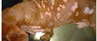
- General weakness, extremely apathetic state with threat of coma.
- Skin hyperesthesia (twitching).
- In severe forms, dramatic changes in behavior occur: the dog becomes either unusually aggressive, or overly affectionate, or does not recognize the owner at all.
- While walking, he staggers and loses his balance.
- Shows unreasonable obsessive anxiety.
- Appetite is noticeably reduced or completely absent.
- There is almost complete disorientation in space.
- Seizures or convulsions occur, similar to epileptic seizures.
- Vomiting, bloody diarrhea.
- The pupils are constricted.
- Muscle tremors.
- While eating, he experiences significant difficulties with swallowing reflexes and chewing.
- With purulent inflammation - increased temperature.
Bacterial meningoencephalitis is characterized by an acute onset and rapid progressive development over several hours. Over the course of 1-2 days, the dog’s condition and behavior may change beyond recognition.
Diagnostic methods
If meningitis is suspected, the animal should be referred to a well-equipped veterinary clinic as soon as possible for diagnosis and treatment. After an external examination, the doctor must take diagnostic measures.
The most informative way to diagnose meningitis is to take cerebrospinal fluid (CSF) for examination using a lumbar puncture. The procedure is performed under general anesthesia. After taking a sample of the liquid, the state of the cerebrospinal fluid, general appearance, protein content are described, and a cytological examination is performed. The presence of the disease is indicated by the following data:
- when a puncture is taken, the cerebrospinal fluid flows out under high pressure;
- with serous meningitis - the presence of lymphocytes, transparent consistency;
- with purulent meningitis - the presence of neutrophils, cloudy consistency, brown color, glucose content reduced to zero;
- with tuberculous or fungal meningitis - reduced glucose levels.
In addition, to clarify the diagnosis, the following diagnostic methods are prescribed:
- Craniography (radiography of the skull without the use of contrast agents);
- magnetic resonance imaging or computer diagnostics;
- electroencephalography;
- clinical analysis of blood and urine.
Differential diagnosis must be carried out to exclude the following diseases:
- hydrocephalus;
- renal failure;
- rabies;
- leptospirosis;
- plague.
Treatment
The purpose of treatment directly depends on the reason for which meningoencephalitis developed. But in most cases, this reason can be identified after autopsy. Therefore, corticosteroids are prescribed as first aid.
As a rule, in the near future the dog's condition is assessed as significantly better, however, such drugs are notorious for their severe side effects. The key factor in the success of treatment will be the correctly calculated dosage.
Some veterinarians immediately use cytarabine injections to avoid complicating risks. The entire course takes 4 injections over 48 hours.
Acute meningoencephalitis often responds well to antibiotics in loading doses. The drug must penetrate the blood-brain barrier (Enrofloxacin, Ampicillin), therapy should not be stopped earlier than after 14 days.
In the complex, immunomodulators and vitamins will be important, and in order to minimize the risk of epileptic seizures, it is important to take anticonvulsants.
For the entire treatment period, it is recommended to provide the dog with a secluded place without bright light or loud sound sources. Feed your pet small fractional portions of liquid cereals and soups with boiled vegetables and a small amount of beef. The diet must be enriched with vitamin complexes.
Attention. Walking the dog during treatment is strongly discouraged.
Diagnosis
Thus, meningitis is dangerous because the clinical manifestations when it appears are not strictly specific. They can equally well be attributed to many other diseases. Therefore, successful and accurate diagnosis is only possible if there is an experienced veterinarian and a well-equipped clinic that has all the necessary equipment.
Very few tests can specifically determine the presence of meningitis. The most effective are computed tomography, magnetic resonance imaging (MRI), and analysis of spinal fluid (CSF). Blood tests are also important, because they help identify the causative agent of the primary infection. Please note that inflamed meninges can only be seen using computed tomography, which is not available in all clinics. So, in most cases, veterinarians have to be content with only indirect signs that allow them to judge the presence of meningitis.
Forecast and consequences
Despite all the success of treatment, one way or another, inflammatory processes in the brain are often irreversible. The most common complications after encephalitis are:
- cerebral hemorrhages;
- necrosis in tissues;
- formation of purulent infiltrates;
- degeneration of the nervous system.
After illness, the dog’s immune system level is at zero, and individual organs function only partially. High risk of relapse of encephalitis or development of any inflammatory pathologies. To improve the general condition of the dog, it is advisable to carry out blood purification: plasmapheresis and hemosorption.
What should you do if you notice the above signs in your pet?
First of all, it is very urgent to take the animal to an appointment with a veterinary neurologist, who, after examining the animal, collecting anamnesis and comparing clinical signs, will determine a list of differential diagnoses.
Next, you should be prepared for the fact that in order to establish a clear diagnosis, the doctor will prescribe additional tests, which may include: blood tests, tests for infections, computed tomography or magnetic resonance imaging, and analysis of cerebrospinal fluid (CSF). All this is necessary to identify the cause and prescribe the right treatment.
Meningomyelitis in a small breed dog. MRI results.
Remember, under no circumstances should you self-medicate, this will not only not help, but can only make things worse for your pet.
About diagnostic problems
Routine blood tests (including white blood cell count) are very often useless. The only case when the tests can be used to understand something is the bacterial origin of the disease. In this case, the tests are characterized by a significant increase in the number of leukocytes. In general, only a brain biopsy will help determine the type of pathology with 100% reliability. Alas, the procedure is not only very expensive, but also quite dangerous; it can only be carried out by an experienced and competent specialist.
Large veterinary institutes can perform a brain biopsy in our conditions. Much safer and easier is a puncture to obtain cerebrospinal fluid. The method is much cheaper. If inflammation has affected the brain and spinal cord, leukocytes will definitely be found in the fluid (which normally should not be there at all). The disadvantage of this method is that one can only guess about the true root cause of the disease.
However, blood, stool and urine tests are extremely important for this very reason - they make it possible to determine what caused the inflammation of the nervous tissue. If all checks have not revealed any signs of infection, a diagnosis of “autoimmune encephalomyelitis” is made - in cats, as we said, this happens much less often than in the case of dogs. Alas, there are currently no other ways to identify autoimmune pathologies of the nervous system. They are determined solely by the method of exclusion.
Prevention of encephalitis
Since encephalitis is most often caused by ticks, it is against them that you should fight first. Unfortunately, today there is simply no effective preventive vaccine for dogs, but a wide range of measures that can prevent the development of this disease can save the situation. Thus, special collars, drops on the withers and sprays are widely used to prevent encephalitis.
Preparations (oils and ointments, sprays) contain substances of different concentrations. In collars, these protective enzymes are located on the surface (synthetic tape). When the skin and these fillers come into contact, the constituent elements begin to spread, as they are able to dissolve in the fatty layer of the skin and accumulate there. Then the substances can then be released in those areas where it is needed. Animals treated with such preparations can take water treatments within a couple of days after application.
Since ticks can even cling to a dog’s hair, treatment is recommended when walking in forests and parks. The production of enzymes from the drug can protect the dog, even if the parasite is not on the skin.
However, it is also worth keeping in mind that only one or several areas of the animal’s body receive the maximum or required dose of the active substance (sprays are distributed pointwise). They will protect against the most common parasites, such as fleas or lice. However, to protect your dog from ticks, which are much more resistant to medications, you should additionally examine the dog after walks, use the above-mentioned sprays every day, spraying them all over the animal’s hair.
Perhaps in veterinary practice there are rarely cases more severe than diseases of the nervous system in animals. They are extremely difficult to identify and even more difficult to treat. The prognosis is also often quite vague. A classic example is encephalomyelitis of domestic animals.
Diagnosis of cats and dogs
The symptoms of meningitis are not specific; the clinical picture is also typical for other infectious diseases. Making the correct diagnosis largely depends on the professionalism of the veterinarian.
Blood is taken from a sick animal for general and biochemical analysis.
. If myelitis is suspected, a painful procedure – a spinal puncture – is required. To determine the source of infection, a urine test is taken.
The most accurate diagnosis can be made after performing a computed tomography scan of the animal’s brain. In the absence of special expensive equipment, veterinarians diagnose the disease based on indirect signs and test data.
Once a diagnosis is made, veterinarians do not guarantee the animal’s recovery; their prognosis is cautious. The outcome of treatment depends on many factors: timeliness of treatment, age of the pet, general condition, etc.
Treatment with cyclosporine in combination with prednisone for autoimmune meningoencephalitis in three dogs
Journal of Veterinary Medical Science Vol. 75 (2020) No. 12 December
Most small breed dogs may be predisposed to GME. NME and NLE occur primarily in Pugs, Maltese, Pomeranians, Chihuahuas, Yorkshire Terriers, Shih Tzus, Pekingese, West Highland White Terriers, Boston Terriers, and Miniature Pinschers. Currently, the main treatment for autoimmune CNS inflammation in dogs is immunosuppressive doses of glucocorticoids. However, the response to treatment with glucocorticoids alone varies and clinical symptoms often return during dose reduction of glucocorticosteroids. There are limited reports of the previous use of various immunosuppressive drugs in autoimmune inflammation of the CNS. Several recent reports suggest that treatment with cyclosporine and prednisolone in combination reduces the dose of prednisolone and demonstrates synergistic effects in autoimmune CNS inflammation. One study demonstrated that the median survival time after treatment with a combination of cyclosporine and prednisone for CNS inflammation was 930 days. Another study reported that the median survival time of 15 dogs with GME with prednisone alone was 41 days. In one previous report, however, the mean survival time of dogs with histologically confirmed NME treated with cyclosporine and prednisone was 305.7 ± 94.7 days, and the mean survival time of dogs with histologically confirmed NME treated with prednisone alone was 58.3 ± 30.5 days. Further, one study described survival of 1096 days after treatment with cyclosporine and prednisolone in a dog with a histopathologically confirmed case of NME.
Several reports (albeit without histopathological confirmation) describe cyclosporine therapy as showing beneficial effects in dogs with suspected autoimmune CNS inflammation. In this study, we administered cyclosporine plus prednisolone to three dogs with autoimmune CNS inflammation. However, the survival time of the three dogs was relatively shorter than in previous reports. One dog lived 170 days and two others less than 100 days after treatment with cyclosporine plus prednisone in this study.
There are several possible reasons for the shorter survival time in this study. (1) In case 1, clients refused a brain CT scan at the initial appointment and we simply prescribed prednisolone plus furosemide for 70 days. In our experience, the timing of administration of immunosuppressants (as early as possible) is very important to increase survival time in dogs with autoimmune CNS inflammation. Prednisolone can cause improvement in inflammatory and edematous changes in almost all cases of autoimmune CNS inflammation, but it cannot delay the progression of autoimmune disease at a lower dose. Side effects such as polyuria/polydipsia/polyphagia, hepatotoxicity, weight gain, iatrogenic Cushing's disease and depression are frequently observed during long-term prednisone therapy. According to a previous report, immunosuppressants such as cyclosporine may reduce the rate of disease progression when used in the appropriate manner and at the appropriate time. We hypothesized that in case 1, cyclosporine was not prescribed at the appropriate time. Therefore, late administration of cyclosporine may not show the significant beneficial effect described in previous reports. Although case 1 showed a relatively short survival time, we should focus on the improvement of clinical symptoms after cyclosporine administration compared with the clinical status of prednisolone alone.
(2) Because cyclosporine is in the form of an emulsion in capsules, it may be difficult to administer it in the appropriate manner. Further, one previous study described that administration of cyclosporine with food may reduce bioavailability by 22% and increase interindividual variability in drug absorption. Owners in Cases 2 and 3 were seriously concerned about prescribing cyclosporine at home and sometimes did not administer the medications. Cyclosporine was prescribed less than 3 times per week in both cases. Although we used cyclosporine plus prednisolone initially in cases 2 and 3, in both cases it was an acute form and inconsistent use of cyclosporine may not have delayed disease progression. The acute form of autoimmune inflammation of the CNS progresses rapidly and shows high mortality even in the early stage. According to a previous report, cyclosporine cannot stop disease progression, but may improve inflammatory and edematous changes initially and then reduce the rate of disease progression. (3) We did not measure cyclosporine concentrations in these 3 cases, therefore, we could not show the exact therapeutic concentrations after treatment.
Several reports (albeit without histopathological confirmation) describe cyclosporine therapy as showing beneficial effects in dogs with suspected autoimmune CNS inflammation. In this study, we administered cyclosporine plus prednisolone to three dogs with autoimmune CNS inflammation. However, the survival time of the three dogs was relatively shorter than in previous reports. One dog lived 170 days and two others less than 100 days after treatment with cyclosporine plus prednisone in this study.