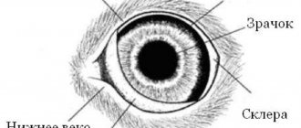
A dog's third eyelid is a separate eye organ that is needed to protect the eyes from dirt and dust. It also performs the function of wetting the cornea of the eyeball. People who decide to get a dog at home should become familiar with the features of the third eyelid and the diseases that are associated with it.
The third eyelid is an organ that all dogs have.
What is it: main functions and how it works
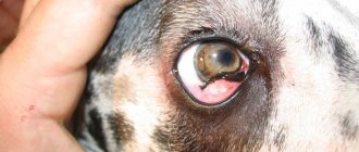
The third eyelid is a popular name often used by dog breeders. Veterinarians call this a conjunctival fold. It is located in the corner of the eyeball.
The main function of the conjunctival fold is to protect the eyes from dust and mechanical damage. Even if you press lightly with your finger on a dog's eye, an eyelid will immediately appear and completely cover the surface of the cornea.
Inside the third eyelid there is an additional gland that produces about 30% of tears. As the eyelid moves, it distributes fluid evenly over the surface of the cornea. This ensures that it is cleaned from bacteria and foreign particles.
Additional Information! There are cases when there is no pigmentation on the edge of the eyelid. This is not a pathology, but a feature of some animals.
What is a dog's third eyelid?
The third eyelid is a semilunar leathery fold of the conjunctiva in an animal, located in the inner corner. The main purpose of this supplement is to protect the corneal layer from injury, which can be seen by lightly pressing the surface of the eye (the eyelid will quickly cover the surface of the eye, often even with a slight tilt of the head to the side). In addition, an additional gland is hidden in its thickness, producing about 30% of tears, which, when it moves, cover the entire cornea in doses. This helps to moisturize the eyeballs, wash away foreign particles and bacteria, thereby improving the general condition of the animal’s visual organs.
Adenoma: main causes
Adenoma or prolapse of the third eyelid is a common condition that often develops in domestic puppies and adult dogs. Before treating such a disease, it is necessary to understand why it appeared.
There are several reasons why this disease may occur:
- Age-related changes. Some people believe that adenoma appears only in adult dogs, but in fact this is not the case. Most often, young dogs under two years of age encounter this disease. It often appears in cocker spaniels, poodles and pugs.
- Ligament weakness. In some dogs, the ligaments that support the lacrimal gland begin to weaken. This leads to the appearance of third eyelid adenoma in dogs.
- Position of cartilage. Sometimes the eyelid can become inflamed due to the incorrect position of the cartilage. When left in a deformed state for a long time, it begins to lose its frame properties.
- Heredity. There are cases when eye adenoma in a dog appears due to heredity. Perhaps one of the dogs' parents has suffered from this disease before.
- Damage. If you accidentally damage the 3rd eyelid in dogs, an adenoma may appear. Therefore, you need to carefully monitor your pet's eyes.
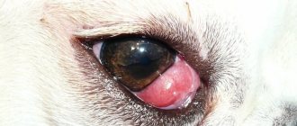
Enlarged gland is one of the signs of ocular adenoma
Disease prevention
Carrying out preventive measures will protect the animal from possible disease. Diseases of the third eyelid most often occur as a result of mechanical damage. Which means all kinds of animal injury prevention. This is especially true for hunting and guard dogs.
For owners when buying a dog, the main thing is to learn about all sorts of breed characteristics. Some dogs are more genetically predisposed to third eyelid disease. If various pathologies and symptoms occur, you need to contact a specialist. Doctors warn about the possible consequences of home treatment. Disease of the third eyelid in a dog requires attention and treatment in the clinic. During treatment, the owner must remain close to the pet at all times to prevent the dog from scratching the eye and getting an infection into the wound. During treatment, you should also reduce or eliminate walks outside (especially in the cold season).
During treatment, you also need to monitor your diet. Doctors recommend using dietary food to avoid allergic reactions and complications. Additionally, after treatment, immunostimulants and vitamin supplements should be used, which should be aimed at improving the microflora after taking antibiotics. Therefore, avoiding walks and proper treatment and care can speed up the animal’s recovery process. Every owner should remember that eye diseases in dogs can lead to blindness or death if not treated promptly.
Currently reading:
- Neoplasms and infections in diseases of the eyes of dogs
- Thyroid dysfunction in dogs (hypothyroidism)
- Blindness is a faithful companion of entropion in a dog
- Fear of blindness due to Blepharitis in a dog
Symptoms
Inflammation of the third eyelid in dogs, like most other diseases, is accompanied by certain symptoms. The development of the disease is indicated by the following symptoms:
- Enlarged gland. First, due to the development of the disease, the pet's eyes become red. Then the size of the gland begins to increase, which leads to increased tear production. This is why many dogs with adenoma have watery eyes.
- Deterioration of vision. Since sick animals develop a reddish formation in the corner of the eye, dogs begin to see worse.
- Itching and damage to the cornea. Due to the appearance of an adenoma, dogs' eyes begin to itch very much. Many dogs cannot tolerate severe itching that does not go away, and therefore constantly try to scratch with their paws. Frequent scratching damages the cornea.
- Lacrimation and purulence. It is no secret that inflammation of the third eyelid in dogs is accompanied by increased lacrimation. However, if nothing is done or treated, prolapse leads to the release of a large amount of pus.
Important! Over time, if left untreated, it only increases in size, which also negatively affects vision.
Lacrimal gland prolapse in dogs
The structure of the eye in the process of evolution has led to the formation of various means of protection - barriers - one of which is the tear (tear fluid).
The tear washes the outer surface of the anterior part of the eye - the cornea and conjunctiva of the eyeball and eyelids. Tear is the environment in which certain metabolic processes and a number of immunological reactions occur.
The third eyelid complements the protective mechanisms of the eye in animals. On the inner surface of the third eyelid, at its base, is the lacrimal gland of the third eyelid.
Lacrimal gland prolapse in dogs is quite common in many dogs, regardless of breed.
Visually, the owner of the animal can see the so-called “cherry eye” - a pinkish-red formation of a round shape. Of course, such an formation causes pain and inconvenience for the dog. That is why it is necessary to deliver the animal to a specialist – a veterinary ophthalmologist – as quickly as possible.
Most often, according to statistics, lacrimal gland prolapse occurs in breeds such as French and English bulldogs, Cane Corso, Toy Terrier, Pug, and Pekingese.
Why is this happening? This is a congenital pathology - genetically determined. The gland is initially increased in size. Due to the increased size, the gland moves outward from its bed and is infringed by the third eyelid. This can lead to eyelid deformation. It is also believed that prolapse (loss) of the gland occurs due to rupture of the fragile ligament that attaches the gland to the periosteum of the orbit. Prolapse of the lacrimal gland in dogs can be caused by injury to the inner corner of the eye.
For an animal, prolapse of the lacrimal gland is incredibly worrying - therefore, it is necessary to protect the eye from scratching with paws - for example, put a special veterinary protective collar on the head - to prevent injury to the prolapsed gland, eye (cornea, eyelids, etc.) with paws, because this can lead to This leads to necrosis of the lacrimal gland, corneal ulcers and other complications.
Treatment of lacrimal gland prolapse in dogs is most often surgical, performed by a veterinary ophthalmologist under general anesthesia. The purpose of surgical treatment is to secure the lacrimal gland in such a position that it does not fall out again. There are several techniques for this operation; each ophthalmologist uses the one that is most convenient for him.
In any case, if you notice that your animal suddenly has swelling in the eye area, you should immediately see a specialist.
The article was prepared by Radevich M.A.,
veterinary ophthalmologist at MEDVET © 2021 SEC MEDVET
Types of adenoma
Cystic adenoma is the most common type of tumor.
There are several types of adenomas, which are most often found in dogs:
- Cystic
This is most often a benign tumor that forms in the third eyelid of the dog. This tumor grows quite slowly. However, despite this, its size can reach several centimeters.
You should not self-medicate a cystic neoplasm with medications. It is better to seek help from a specialized veterinarian for surgery to remove the tumor.
- Papillary
This is a cystic type of neoplasm of the papillary type. Inside such a tumor there is a dark liquid.
Unlike the previous type of adenoma, this one grows faster. If such a tumor is detected, you should contact your veterinarian and have a biopsy performed to ensure that it is benign.
- Polypoid
This is a benign neoplasm that can appear in the third eyelid of the domestic dog. Even though the tumor is not malignant, it still needs to be gotten rid of. The most effective method of treatment is surgery.
Attention! The cost of such surgery largely depends on the size of the tumor.
Breeds characterized by adenoma
The pug is a dog breed that often develops adenoma.
There are several dog breeds that most often develop diseases of the third eyelid. These include the following dogs:
- Pug. These are fairly small and calm dogs that are popular among dog breeders. On average, such dogs live about 10-12 years. However, due to weak immunity, not everyone can live to that age.
- Shar Pei. Most often, the disease appears in Shar Peis under the age of three months. Small puppies have a very weak immune system, which strengthens only 2-3 months after birth.
- Cane Corso. Despite good immunity, representatives of this breed may develop adenoma of the third eyelid. Most often, the disease appears in dogs under six months of age.
- Bulldogs. Many believe that these dogs rarely get sick due to their strong immunity. However, this is not always the case. If you do not properly care for your eyes, your English and French bulldog will develop an adenoma in the third eyelid.
- Doberman. Representatives of this breed have a greatly reduced immune system and because of this they often get sick.
- Dog. Quite large dogs, which are often bred to protect territories. They have good health, but some dogs may still develop an adenoma.
- Sheepdog. Outwardly, shepherds seem to be healthy dogs who rarely get sick. However, some representatives of this breed suffer from inflammation of the third eyelid.
Infectious diseases
Treatment of Cane Corso dogs is always a costly and expensive matter, especially if the disease is severe. Therefore, it is worth more closely monitoring the health of your dog and preventing disease by paying due attention to preventive measures.
A real test for both the dog and all family members is the pet’s infectious diseases. These include the following: plague, enteritis, ringworm and leptospirosis. Treatment for the dog will take quite a long time and it will need constant care. For example, trichophytosis is difficult to treat.
To cure a dog of ringworm, more than just medications are needed. It is necessary to carry out a number of comprehensive measures for the pet’s recovery:
- regular disinfection of toys, beds, kennels and other “personal belongings” of the dog, as well as the room where he is constantly located, in order to destroy the fungus;
- it is necessary to completely isolate the dog from all family members and other animals;
- organize proper nutrition and proper care.
Most infectious diseases are transmitted through contact with sick animals. This includes not only other dogs, but also cats and wild animals. This is how, for example, lichen and plague are transmitted.
Preventative measures to protect your dog:
- It is necessary to minimize your dog's contact with other dogs you don't know, especially strays. Don't let your dog off the leash, so you can control his behavior and communication with other dogs. To become infected with distemper, a healthy dog only needs to touch the nose of a sick animal. In most cases, this disease is transmitted during sniffing. And for infection with trichophytosis, just one touch with a sick dog affected by lichen is enough. Of course, it is extremely difficult and unnecessary to completely exclude your dog from communicating with others. The dog must be socialized! Here are two such contradictory points that you will encounter. Just choose dog lovers as friends who take care of their dog. If you adhere to this, you will eliminate the risk of pathogen transmission by 80%.
- Do not walk your Cane Corso dog in dump sites. It is garbage that attracts most rodents, in particular rats - carriers of various infections. Gray rats are carriers of a serious disease - leptospirosis, which affects both dogs and people.
- Don't let your dog sniff other people's feces. This is a source of helminths.
- Monitor your dog's well-being closely. Pay attention to even the most minor changes. Perhaps the dog became lethargic, his appetite disappeared, and his nose became dry. Or you began to notice that the dog sheds profusely, and this has nothing to do with seasonal shedding. Excessive hair loss is the first sign that your dog has health problems. All of the above may be symptoms of serious illnesses. And only you can prevent them. If you detect and take your pet to the veterinary clinic on time, the easier it will be to defeat the disease. You should not ignore the symptoms, otherwise it can lead to serious consequences. Advanced infections are much more difficult to treat, and sometimes even impossible. The dog either dies or suffers serious complications such as loss of vision, hearing, paralysis of limbs, and so on.
- Keep all necessary vaccinations for your Cane Corso dog on schedule. This is one of the most effective methods of prevention. But before vaccinations, it is imperative to treat helminthiases.
Treatment of adenoma
Adenoma should be treated by a professional doctor.
Many people are interested in what treatment methods exist and how long it will take to eliminate the disease.
At an early stage, you can get rid of the tumor using a special eyelid massage technique. You need to massage the closed eye diagonally towards the muzzle. A veterinarian will demonstrate the technique.
Most often, surgical treatment is necessary to eliminate the adenoma. To do this, you need to go to a veterinary clinic and find out how much such an operation will cost.
Important! You should not self-medicate or give your pet pills without a veterinarian’s prescription.
Rehabilitation of the animal after treatment
During the rehabilitation period, a person will have to do the following:
- regularly treat your eyes;
- give your pet more vitamins;
- make sure that the dog moves less;
- Examine your eyes daily and clean them to ensure they are normal.
The third eyelid is an organ that is responsible for protecting the eyes. There are times when it becomes inflamed and an adenoma appears. Before getting rid of it, you need to familiarize yourself with the reasons for its appearance and the main methods of treatment.
