Mammary tumors in cats and dogs are a fairly common problem. We have been specializing in the treatment of animals with cancer for a long time and today we are opinion leaders in this field of veterinary medicine in Belarus.
In our clinic we have all the necessary equipment and, most importantly, competent specialists to competently approach the diagnosis, complete preoperative examination, safe anesthesia using narcotic drugs that provide complete pain relief with a minimum number of side effects, complete control of the patient and experienced surgeons , correctly and quickly performing the required operation.
Often, having discovered any lump on the mammary gland of a cat or dog, the owners themselves try to figure out this problem by studying gigabytes of information on the Internet. Below is part of the speech of the oncologist of our clinic, Alexey Sas, at the Belarusian Veterinary Oncology Conference.
We hope that this information will be useful to you and you will seek help from a competent specialist in time.
Mammary tumor in dogs
Mammary gland tumors in dogs are the most common oncological pathology and account for 70%, including benign and malignant neoplasms. There are several studies on the ratio of benign and malignant breast cancer and they vary from 2.5% to 53% of malignant neoplasms of all breast tumors. This significant difference is explained by the regions where the studies were conducted. Thus, AMF is less common in regions where it is customary to sterilize dogs at an early age, but the percentage of malignant neoplasms from these tumors is much higher. Also, a significant percentage of malignancy was observed in regions where animals are not sterilized, and progestins are used to suppress sexual cyclicity, which increase the risk of developing benign and malignant neoplasms. The percentage of malignant neoplasms also increases in the case of late surgical treatment of breast tumors, which is associated with malignancy (malignancy) of neoplasms. From our practice, malignant neoplasms (including in situ cancer) in dogs account for about 45-50%.
The key point in preventive veterinary medicine is sterilization. Conducted before the first estrus, before the second estrus and before the third estrus reduces the risk of developing breast cancer to 0.5, 8, 26%. Further, sterilization does not affect the development of malignant neoplasms (25-30%), but only reduces the risk of developing benign ones. By the way, false pregnancies do not increase the risk of primary malignant neoplasms.
WE RECOMMEND STERILIZING DOGS BETWEEN THE FIRST AND SECOND HEAT , when the hormones produced during the first heat will trigger the closure of bone growth zones, which will reduce the risk of developing orthopedic problems, and the bladder sphincter, which will prevent the development of urinary incontinence in the dog.
Mammary tumor in a dog development symptoms
In dogs, it is customary to distinguish between such forms of mammary gland cancer as nodular and diffuse. At the same time, the second type does not have a clear neoplasm limited in shape and is essentially a diffuse form. In turn, the nodular form can be expressed in a single or multiple tumor.
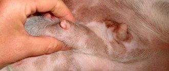
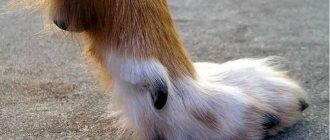
The most commonly identified signs of tumor development include the presence of a solid mass in the subcutaneous space of the mammary gland in a dog. It can be either one tumor of significant size, or many small inclusions that resemble peas. Such tumors remain mobile for a long time and remain small in size. And the diffuse form affects the entire breast tissue and does not always have a clear tumor node. The skin of the affected part of the bitch's udder becomes dense and inflamed.
Most often, these inclusions cannot be moved under the skin due to their fusion with nearby tissues. Often there is a very rapid increase in size of these tumors, when they become twice as large in a month.
Another symptom of the development of mammary gland tumors in dogs is the localization of the oncology. They most often begin to develop in the last two pairs of glands, but these are not the only places that are most often affected by cancer. Unlike the malignant form of tumors, fibroids do not grow as quickly and are distinguished by their smooth structure, which is felt by palpation.
For early detection of the onset of cancer development in a dog, it is imperative to independently check the mammary glands at least once every six months. Upon palpation, various types of compactions and nodules in the gland are easily detected. We advise you to pay attention to their elasticity and the condition of the surface of the neoplasm.
Non-cancerous tumors are small in size and have a smooth surface. But malignant tumors are distinguished by the following symptoms:
- Heterogeneous and bumpy surface.
- The tumor growth rate is too rapid.
- Immobility of relatively nearby gland tissues.
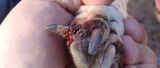
- Bleeding ulcerations of the skin over the tumors cannot be ruled out.
- The neoplasm is not limited to clear boundaries.
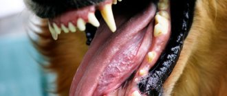
- Local signs of inflammation: redness and fever.
- Bloody and purulent discharge from the nipple of the affected lobe of the gland.
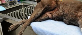
- The general depressed state of the dog and its noticeable lethargy.
If carcinoma symptoms are detected early, the dog's chance of recovery and survival is greatly increased.
Determining the type of lesion and making a clinically significant diagnosis is possible only taking into account numerous analyzes and studies. All these studies, as well as veterinary care in severe cases, are provided by veterinarians at the Center for Emergency Veterinary Care for Animals. If a tumor is detected in a dog, you cannot hesitate and you must immediately seek help from specialists.
The role of hormones in the development of breast cancer
As already mentioned, sterilization significantly reduces the risk of developing breast cancer. At the same time, exogenous administration of estrogen, which stimulates the development of ducts, does not affect the risk of developing breast cancer. And exogenous use of synthetic progestins dose-dependently increases the risk of developing benign neoplasms. At the same time, the combined use of progestins and estrogens significantly increases the growth of malignant neoplasms. The use of progestins in non-sterilized bitches to suppress sexual cyclicity, when it accumulates with the natural titer of progesterone, stimulates the activation of growth hormone in the mammary epithelium, which can play a significant role in the development of malignant neoplasms, which in the process of malignancy lose growth hormone receptors. The influence of prolactin still requires research: in some studies there were significantly more receptors for prolactin in malignant neoplasms, relative to benign ones, in others such a pattern was not noted.
Receptors for estrogen and progesterone. Previously, it was believed that 70% of breast tumors (benign and malignant) have estrogen receptors. Progesterone has not been considered previously.
Normal breast tissue contains receptors for estrogen and progesterone. In benign neoplasms, such receptors are isolated in 90%. In malignant neoplasms, receptors are isolated much less frequently, and in the case of metastasis to a regional lymph node, receptors for estrogen and progesterone are not isolated either in the metastatic tissue or in the primary tumor. There are also no receptors identified in the tissue of inflammatory carcinoma (the most malignant AMG). Thus, it is believed that the moment a tumor loses receptors is associated with the process of malignancy.
Progress of mammary gland removal surgery in dogs
Mastectomy
(from ancient Greek mastòs “breast” and ek tome “remove”) - a surgical operation to remove breast tissue. The essence of the operation is to remove the mammary gland, fatty tissue that contains lymph nodes (probable sites of metastasis), and also, depending on the stage of the oncological process, removal of the superficial pectoral muscle, possibly part of the abdominal wall.
Indications for surgery
• Breast tumors
(the most common reason for mastectomy in dogs and cats) • Suppurative process, severe mastitis with gangrene of most of the mammary gland, or serious wounds of the mammary gland (although this is quite rare).
There are several options for this operation:
• Unilateral mastectomy
- includes the removal of the entire ridge of five mammary glands on one side, subcutaneous tissue and regional lymph nodes (axillary and inguinal).
Always used in cats
and most often in
dogs
.
• Bilateral mastectomy
- characterized by an increase in the volume of surgical intervention: with this method, two ridges of mammary glands are removed from both sides along with regional lymph nodes (axillary and inguinal). This operation is very traumatic, difficult to tolerate and should be avoided.
• Regional mastectomy
It is performed exclusively in dogs and will be discussed in this article.
It is used only in dogs, since this operation is not practical in cats. In order to understand what a regional mastectomy is and why it is needed, you need to know some basics of oncological surgery
and animal anatomy, in particular the structure of the lymphatic system of the mammary gland.
Histology
On the mammary gland there is a fairly wide variety of neoplasms with adenomas and their variations in the top among benign ones and carcinomas among malignant ones. It is important to note the extent of the tumor: intraductal, infiltrative and invasive (degree of invasion into blood vessels). Basically, all tumors are of epithelial origin, but sarcomas are extremely rare. Usually these are simple (consisting of one type of cell) - carcinomas; or complex neoplasms containing epithelial and myoepithelial cells; mixed - with areas of cartilage and bone (metaplastic carcinoma) having a pluripotent stem cell in its origin.
Sarcomas occur, but are rare; they are usually rapidly developing tumors with central necrosis and infiltrative growth.
Inflammatory carcinoma is considered separately - it is a rapidly developing tumor with massive swelling, erythema and pain, without clear boundaries. It is often confused with mastitis. It can affect either one milk carton or several in a row, or both rows of milk cartons. Cytology can differentiate mastitis from tumors with reasonable accuracy (70%). From a histological point of view, it can be any malignant tumor of the breast (tubulopapillary carcinoma, or solid carcinoma) with characteristic invasion into the dermal lymphatic vessels.
Usually, due to the sudden onset, owners quickly contact a veterinary clinic, but metastases are detected at the initial visit in 80 to 100 percent of cases. Because of the rapid progression and early metastasis, surgery is usually not advisable, and the median survival time without treatment is 30 days.
Presentation
Usually these are single neoplasms of one centimeter or more, sometimes multiple (60%). Benign OIs are well limited from surrounding tissues and are not fixed to the skin or underlying tissues. In some cases, during estrus or in the first month after it, or against the background of the use of progestins, small 1-2 mm multiple NOs are detected, which are more correctly classified as hyperplasia, and which should disappear with a drop in titers of steroid hormones in the blood. If the tumors do not disappear or their size is more than 2-3 mm, surgery should be planned.
Malignant neoplasms usually grow more quickly, have inflammation of the soft tissues around them, infiltrative growth, fixation to the underlying tissues or growth into the skin, erosion (opening).
One animal and one mammary gland may have several histological types of tumor.
Prognostic factors
- Age. In young animals, benign ones are more common, but vice versa.
- Reproductive status. Pathologies of the reproductive cycle (short, long, painful estrus, false pregnancy) do not in any way affect the prognosis. Whereas sterilization done 2 years before the presentation is a bad sign.
- Breed. Yorkies, poodles, spaniels and German shepherds have some predisposition to AMF.
- Tumor size. Up to 3 cm, 3-5 cm and more than 5 cm. The prognosis worsens as the tumor increases from 3 cm.
- Metastases in the lymph nodes. The presence of pure lu does not guarantee the absence of metastases 100%, since carcinomas metastasize, both hematogenously and lymphogenously. Naturally, the more lymph nodes are affected by metastases, the worse the prognosis.
Stage of the disease: staged according to the TNM system according to WHO. The higher the stage, the worse the prognosis. It worsens significantly with ulceration of the tumor, fixation to the skin and underlying tissues, local relapse, and distant metastases at the time of examination. Staging.
Staging is carried out according to the WHO TNM scale. For complete staging and, consequently, the adoption of the correct course strategy, a mandatory study of the tumors is required if they are detected. Distant metastases can be found in parenchymal organs, bones, and lungs. When the nodule is located in the caudal abdominal and inguinal breast, additional ultrasound of the caudal abdominal region is required to check for enlarged iliac lymph nodes.
On average, metastasis to regional lymph nodes at the time of the first examination is detected in 20% of cases. Metastases in the lungs in 5%.
Cytology and biopsy
Malignant neoplasms of the breast usually have insufficient signs of malignancy during cytological examination, which determines the accuracy of fine-needle examination at 80-85%. In any case, preoperative examination is usually not performed since benign and malignant neoplasms require the same treatment. Preoperative examination is only necessary in doubtful cases (usually suspected inflammatory carcinoma, when surgery is not entirely advisable).
POLICY - LET'S WAIT, WATCH UNTIL CLEAR SIGNS OF MALIGNANCITY APPEAR - USUALLY MAKES THE TUMOR INOPERABLE OR WHEN SURGERY FAILS TO RESULT IN A CURE.
Surgical treatment of mammary tumors in dogs
This is the main method for effectively treating mammary tumors in dogs and cats.
A correct and timely operation will ensure that the animal is completely free of cancer.
There are several surgical strategies:
According to some sources, it is possible to remove small ones, but with a margin of 1 cm in healthy tissue, but I do not perform this operation due to the rather poor prognosis and the high risk of local relapse.
A simple mastectomy is the removal of one mastectomy. This method of surgery is considered for benign or without signs of malignancy neoplasms 0.5-2 cm. But in some locations, tumors of 5 or 1 are more difficult to perform than performing a regional sectional mastectomy.
Unilateral sectional mastectomy. 1,2,3 are removed when the tumor is localized in 1,2 packages, and 3,4,5 are removed when the tumor is localized in 4,5 packages. During this surgery, the regional lymph node is removed and submitted for histological examination. In case of tumor invasion into the underlying tissue, it is recommended to remove one muscle layer in depth. In this case, the axillary lobe is removed only if it is enlarged.
Unilateral mastectomy - removal of the entire mammary ridge. When localizing but in a 3-pack or multiple but throughout the row.
Bilateral mastectomy is not tolerated by dogs in most cases. But if we have tumors in two packages, it is always worth planning the most extensive operation possible. This is because we allow the primary tumors that we leave intact to continue to grow and metastasize. And during the first operation, we will injure the adjacent mammary gland, which will disrupt its single lymphatic block and increase the risk of dissemination of metastases.
I usually do a unilateral mastectomy on one side and a 3,4,5 sectional mastectomy on the other side at the same time, and if there is a large skin deficit, the flap can be transferred from the knee to the groin area.
According to studies, the risk of re-formation of the primary breast tumor in the remaining part is 48% in the case of removal of the initially malignant tumor and 22% in the case of removal of the initially benign tumor.
Diagnosis and treatment of mammary cancer in dogs
The most informative research method is a biopsy of tumor tissue, but removal of carcinoma followed by histological examination of the tissues that form it is also used. A chest x-ray and a detailed examination of the lymph nodes help confirm or refute the terrible diagnosis.
The most reliable way to diagnose breast cancer is considered to be histological examination of the tumor. With this method, several cells are collected intravitally and the type of neoplasm is determined based on their structure.
Carcinoma is characterized by the formation of metastases. With mammary carcinoma, the oncological neoplasm affects the regional lymph nodes and lungs of the animal. For example, with the development of a malignant process in the first two pairs of glands, metastasis affects the lymph nodes that are located near the forelimbs. But the defeat of the last two pairs of glands already gives metastases to the lymph nodes of the hind limbs and pelvis.
Given the wide coverage of organs with metastases of a cancerous tumor, ultrasound of internal organs (liver, pancreas) and x-rays of the hind limbs are performed to confirm or exclude the presence of metastases in the bone tissue of the dog’s limbs. Differential diagnosis is also carried out with mastopathy in diffuse forms of breast cancer.
General laboratory tests of peripheral blood and urine and a biochemical blood test are required. And only based on the results of all the tests performed, a clinically substantiated diagnosis can be made.
Mammary tumor in a dog surgery
Treatment of a mammary gland tumor in a dog is carried out in a variety of ways available to modern veterinary medicine. Ultimately, the effectiveness of the treatment is always directly dependent on a wide variety of factors. This:
- Type of neoplasm.
- What stage is the tumor at the time of diagnosis?
- Age of the sick dog.
- Physiological state of the animal.
Treatment of benign tumors is most effective. The tumor is resected under anesthesia, and the animal becomes practically healthy. But it is much more difficult to treat malignant tumors.
Due to the spread of metastases, not only the affected mammary gland is removed, but also nearby lymph nodes. If there is significant damage to organs by metastases, mastectomy and chemotherapy may be prescribed. Malignant tumors are also subjected to local irradiation with ionizing rays.
It is very important for the owner to understand that the last two listed methods of treatment have a very detrimental effect on the dog’s condition and if the animal’s body is weakened, it can be fatal.
Veterinary surgeons at the Center for Emergency Veterinary Care for Animals perform mastectomy with maximum organ-saving and minimal damage to surrounding tissues.
Forecasts
The main prognostic factor is tumor differentiation, type of tumor and its type of growth (invasion). Thus, sarcomas are extremely metastatic and the prognosis is cautious, closer to unfavorable, even in the absence of metastases in the lymph nodes and lungs at the time of surgery.
19% of in situ carcinomas and 22% of solid carcinomas develop metastases in the next 2 years after radical surgery in the absence of detectable metastases at the time of surgery.
In the case of detectable lymph node metastases, mortality after surgery is 86% within 2 years, compared to 8% in the absence of detectable metastases. In the case of detectable distant metastases, survival is about 5 months.
If you step back from mathematical terms such as median, life expectancy, and think about the patient and client who will look and care for their pet, evaluating your work by comparison with their expectations about the effect of your actions. That is, the owner will not notice a slight increase in the median life expectancy of his pet if you perform a super wide operation against the background of detectable metastases in the lymph nodes, when the operation will not be curable, but will only reduce the primary focus of metastasis, without noticeably affecting the course of the oncological process itself.
In my opinion, animals with signs of absence of metastases should be taken for surgery, when surgery can lead to a complete cure.
Adjuvant therapy
Radiotherapy, which is widely used for breast cancer in humans, has not been sufficiently studied in dogs. Estrogen blockers (tamoxifen), widely used in human medicine, cause significant complications in 60% of cases in dogs. In addition, in malignant breast cancer, estrogen receptors are not detected in most cases, and ovario/hysterectomy does not affect the course of the oncological process itself.
Chemotherapy for a mammary tumor in a dog
The combination of 5 fluorouracil and cyclophosphamide is considered effective, which increases the median duration from 6 to 24 months, compared with animals receiving only surgery.
Doxorubicin, which has long been considered effective in dogs, does not produce clinical results, which we could observe for a long time in clinical practice before reading the corresponding article at the end of 2016.
Treatment of breast tumors
Surgery
Surgery is the main and most effective treatment for mammary tumors in dogs. In dogs with mammary tumors, this operation is called a mastectomy (from the Greek words mastos - breast and ectome - excision) - a surgical operation to remove mammary tissue (photo 3).
- Lumpectomy (photo 4) - sectoral resection of the breast;
- Simple mastectomy - removal of one breast;
- Regional mastectomy (photo 5) - removal of several mammary glands;
- Unilateral mastectomy (photo 6) - removal of the right or left ridge of the mammary glands;
- Total mastectomy - removal of all mammary glands.
The scope of the operation is chosen individually in each specific case. Many factors are taken into account: the age of the animal, concomitant diseases, previous treatment, stage of the disease, size and number of tumors, and a number of others.
The decision-making algorithm can be seen in Diagram 1.
General recommendations: lumpectomy is performed if the tumor is less than 5 mm in diameter; simple mastectomy - if the tumor occupies the central region of the gland; regional mastectomy - if the animal has several tumors or it is located between the glands; unilateral mastectomy - if the dog has several tumors on one ridge; total mastectomy (carried out in two stages: first, one ridge is removed and after a few weeks the second) - with multiple lesions of the mammary glands (MacEwen EG, Harvey HJ, Patnaik AK, et al.)(Chang SC, Chang CC, Chang TJ, et al. .).
Removal of ovaries (castration) for breast tumors
There are conflicting opinions regarding spaying dogs with mammary tumors. Thus, initial studies did not reveal positive effects from sterilization of dogs with AMF (Yamagami T, Kobayashi T, Takahashi K, et al.). However, recent studies have noted a longer life expectancy in dogs that were neutered before or during surgery (Sorenmo KU, Shofer FS, Goldschmidt MH.)(Chang SC, Chang CC, Chang TJ, et al.).
Chemotherapy
Chemotherapy is recommended for animals with stage III and IV breast cancer as additional (adjuvant) therapy. It can also be used in cases of incomplete surgical removal of the tumor or in case of aggressive histological characteristics of the tumor (Simon D, Schoenrock D, Baumgartner W, Nolte I.).
Radiation therapy
Although radiation therapy is often used in humane medicine for people with mammary tumors, there is little veterinary literature on radiation therapy for dogs with mammary glands.
Hormone therapy
Hormone therapy is rarely used in dogs with mammary tumors:
- Due to the high cost of analysis (immunohistochemistry) to determine the presence of estrogen and progesterone receptors in tumor cells.
- Because dogs are often spayed at the same time as a mastectomy. Removing the ovaries stops the body's production of estrogen and progesterone, thereby making additional hormonal therapy unnecessary.
Immunotherapy
Various nonspecific immunotherapies have been tried in dogs with AMF, but none have been shown to be effective (Parodi AL, Misdorp W, Mialot JP, et al.)(Rutten VPMG, Misdorp W, Gauthier A, et al.)(Teske E, Rutteman G, Ingh T, et al.).
Mastectomy
(from ancient Greek mastòs “breast” and ek tome “remove”) - a surgical operation to remove breast tissue. The essence of the operation is to remove the mammary gland, fatty tissue that contains lymph nodes (probable sites of metastasis), and also, depending on the stage of the oncological process, removal of the superficial pectoral muscle, possibly part of the abdominal wall.
Indications for surgery
• Breast tumors
(the most common reason for mastectomy in dogs and cats) • Suppurative process, severe mastitis with gangrene of most of the mammary gland, or serious wounds of the mammary gland (although this is quite rare).
There are several options for this operation:
• Unilateral mastectomy
- includes the removal of the entire ridge of five mammary glands on one side, subcutaneous tissue and regional lymph nodes (axillary and inguinal).
Always used in cats
and most often in
dogs
.
• Bilateral mastectomy
- characterized by an increase in the volume of surgical intervention: with this method, two ridges of mammary glands are removed from both sides along with regional lymph nodes (axillary and inguinal). This operation is very traumatic, difficult to tolerate and should be avoided.
• Regional mastectomy
It is performed exclusively in dogs and will be discussed in this article.
It is used only in dogs, since this operation is not practical in cats. In order to understand what a regional mastectomy is and why it is needed, you need to know some basics of oncological surgery
and animal anatomy, in particular the structure of the lymphatic system of the mammary gland.
Lymphatic system of the mammary gland
Dogs and cats normally have five packets of mammary glands on both sides. The first and second pairs of mammary glands have regional lymph drainage to the accessory axillary and axillary lymph nodes. The fourth and fifth pairs of mammary glands have lymphatic drainage to the inguinal lymph nodes, and the third pair has mixed lymphatic drainage (both axillary and inguinal lymph nodes).
Basic concepts of oncological surgery:
• Zoning:
involves excision of areas along the path of regional lymphatic drainage with the tumor and surrounding healthy tissue (this is subcutaneous tissue containing lymphatic vessels and lymph nodes of the first and second order).
• Blocking:
involves excision of the tumor and the area of potential invasion of its cells as a single block. For example, in case of a breast tumor, it is necessary to remove the additional axillary lymph nodes and the tissue containing them as a single block.
That is why, if a tumor appears in the first or second pair of mammary glands, the first, second and third pair with axillary tissue and lymph nodes must be removed. If a tumor appears in the fourth or fifth pair, the third, fourth and fifth pairs of mammary glands with inguinal tissue and lymph nodes are removed. And in case of a tumor in the third pair, all mammary glands and both groups of lymph nodes are removed, as this ensures the radicality of the operation.
A regional mastectomy in dogs involves the removal of at least three mammary glands on one side. And in most cases, this operation is needed for cancerous lesions of the breast. Failure to comply with these simple principles will lead to the development of tumor relapses at the site of its removal.
Main stages of the operation
1. A bordering skin incision and removal of breast tissue with subcutaneous tissue. 2. Axillary lymphadenectomy - removal of tissue containing the axillary lymph node located in the axillary region (there is no need to look for the inguinal lymph node, since it is removed along with the mammary gland). 3. Suturing the wound.
Complications after regional mastectomy
• Bleeding in the early postoperative period. It occurs rarely; when using an electrocoagulator, this problem can be solved (most often observed in the presence of blood clotting disorders).
• Suppuration of the postoperative wound. Maintaining asepsis - antiseptics and the use of antibiotics solve this problem.
• Seroma (lymphorrhea) is the release of fluid (lymph and a small amount of blood) under postoperative sutures. Post-mastectomy seroma occurs to varying degrees in many animals. The accumulation of seroma is due to serious surgical trauma, as well as the fact that lymph does not coagulate, unlike blood, and can continue to ooze under the sutures for up to 2-3 weeks. The amount of lymphorrhea may vary. Most often, this problem bothers obese animals and is eliminated by installing drains (plastic or rubber tubes through which lymph flows out) under the seams or by performing evacuation using a puncture (performed with a regular syringe).
2-3 days of inpatient treatment
, especially considering that such an operation is often needed in older animals. Monitoring of tests and condition is desirable, 2-3 days - intravenous infusion, antibiotics, treatment of sutures and use of a protective blanket. On the second day, the animal can get up, move independently, and walk. Full activity is restored on days 7-10, although this is all individual. Sutures are removed on days 12-14 depending on healing. For the first few days, painkillers are usually prescribed.
So, regional mastectomy is most often used for the radical removal of tumors in the first, second, fourth or fifth packages of the mammary glands in dogs, includes the removal of at least three packages, is rarely used for the radical treatment of purulent mastitis and almost never as a palliative operation for mammary tumors glands in cats. The duration of this operation is less than an hour and its complexity is low, unlike, for example, unilateral mastectomy, where the surgical trauma is much higher and the surgical intervention is more invasive. The postoperative period most often passes without complications if the veterinarian’s prescriptions are followed correctly and accurately.
Removal of a breast tumor (mastectomy) in our clinic
The veterinary clinic “Sas Animal Service” is a team of true professionals in their field. Our veterinary surgeons and oncologists have extensive experience and constantly maintain the level of their education and professional skills. You can make an appointment with us for a consultation or operation by leaving an online application on the website or by calling +375-29-685-37-01, +375-17-235-05-81. Clinic opening hours: from 9:00 to 21:00 all days of the week, except Wednesday - on this day we are waiting for you at the clinic from 11:00 to 21:00.