Causes
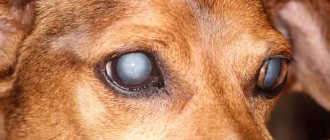
If you notice a light spot in the pupil area on your pet, then this alarm signal indicates the beginning of the development of cataracts. The lens of the eye is responsible for the refraction of light, and its clouding leads to the formation of a light spot. When clouding occurs, the light, penetrating deep into the eyeball, is not refracted and the desired image is not sent to the retina. From this we conclude that the development of cataracts leads to deterioration of vision, and sometimes to complete blindness.
Scientists from all over the world argue about the causes of the disease. What kind of shock provokes the formation of cataracts is not determined. One thing is clear: cataracts in dogs are a disease with a genetic predisposition. The development of this disease is also caused by:
- diabetes;
- long-term treatment of cataracts with medications;
- disturbances in the functioning of the endocrine glands;
- age (in dogs over 8 years old);
- glaucoma;
- contusions, clinical death;
- a sharp decrease in the body's immunity;
- eye injuries;
- chronic infections;
- acute poisoning, intoxication.
Breed Predisposition
According to statistics, the following breeds are most susceptible to this disease: golden retrievers, poodles, terriers, including Yorkshire, cocker spaniels, schnauzers. The reason why representatives of these particular breeds are more likely to develop cataracts is not clear.
Symptoms of cataracts in dogs
Just like in humans, cataracts in dogs develop over the years, regardless of breed. In most cases, age-related cataracts affect animals aged 8 years and older, and the leading positions are occupied by animals genetically predisposed to cataracts - these are miniature poodles, Yorkshire terriers, cocker spaniels, and retrievers. Signs of cataracts are varied, but the main thing that pet owners can identify is a visual disturbance in their pet. With the development of cataracts, partial and then complete clouding of the lens occurs.
A cloudy lens becomes a natural barrier to light rays, and the visible image deteriorates. With cataracts, the clouding of the pupil occurs unevenly; if the process of clouding of the lens begins in the peripheral parts, then the cataract can be asymptomatic for a long time, but if the central part is affected, then vision deterioration occurs quite quickly.
Kinds
By type, cataracts are divided into congenital and acquired.
- Congenital lens opacification is a rare phenomenon and is associated with concomitant chronic diseases such as diabetes mellitus. It also occurs with intrauterine injury to the eyeball, underdevelopment.
- The acquired form is the most common type and occurs at a certain age due to genetic inheritance. It occurs more often in dogs over eight years of age, because by this age the condition is aggravated by chronic illnesses.
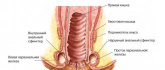
Entropion or eversion of eyelids in dogs
Entropion in dogs is an incorrect position of the eyelids relative to the eyeball, in which the plane of the free edge of the eyelids is turned inward all or over some extent. Normally, the eyelid, resting on the eyeball, copies its contour, i.e. located along the hemisphere of the cornea. Due to eyelashes rubbing the cornea of the eye, constant irritation occurs, which can lead to corneal ulceration, perforation, pigmentary keratitis and even loss of vision.
Causes of entropion in dogs
- breed predisposition (English bulldog, English mastiff, Dogo Argentino, Dogue de Bordeaux, bullmastiff, ca-de-bo, Mastino Napoletano, Tosa Inu, Fila Brasileiro, Shar-Pei, pug, French bulldog, chow-chow, spaniels.);
- age of the animal: bloat can occur both in a young animal (due to the active growth and development of tissue structures of the animal’s body) and in an adult animal (this is associated with atrophy of muscles, subcutaneous fat, changes in the properties of the skin);
- various pathologies, such as chronic conjunctivitis, keratitis, ulcers and erosions of the cornea, foreign body, eyelash defects can also cause entropion.
Eversion of eyelids in dogs
Eversion of the eyelids in dogs is a position of the eyelid in which part of it or it is completely turned out. In this case, the conjunctiva, according to the magnitude of the inversion, is exposed, exposed to external influences and contamination. Eversions can be either primary - congenital, or secondary - developing against the background of the underlying pathology (wounds, burns, dermatitis, neoplasm, also observed with facial paralysis and Horner's syndrome).
Treatment
For entropion/eversion of the eyelids, surgical treatment is a more reliable method of treatment, and depending on the condition of the animal’s eyes, immediate surgery may be required. Thanks to modern research and diagnostic methods (blood tests, ecg, cardiac echo), which can be used to judge the animal’s health status, preoperative preparation of the animal, surgical intervention at home, as well as postoperative outpatient treatment become possible. There are several methods for correcting volvulus/eversion; in each specific case, the doctor determines the appropriate method. Along with surgery, local use of medications is recommended.
The article was prepared by doctors of the ophthalmology department "MEDVET" © 2013 SEC "MEDVET"
Stages and symptoms of disease development
Signs of cataracts often do not appear in the early stages of its development. Symptoms make themselves felt too late, when visual acuity has noticeably dropped. It is even more difficult to distinguish this disease from other eye diseases in dogs of advanced age, because clouding of the eyes in this situation is a normal physiological process and it signals the development of nuclear sclerosis, and it has absolutely no effect on the quality of vision.
Clouding of the lens, a decrease in its transparency, is the main sign of cataracts, noticeable in the pupil area. But in some cases, white spots with a blue tint form on peripheral tissues, which significantly complicates early diagnosis. You can notice a decrease in vision by the dog’s everyday habits. If an animal does not fit into the doorway, does not notice its own toys, does not find the bowl the first time, or sometimes by smell, this is a cause for concern.
In an animal with a cataract in the central part of the lens, located directly behind the pupil, twilight vision is better than in daylight. This occurs due to the constriction of the pupil in bright light, which reduces the field of view. Double image phenomena are possible.
The development of the disease is divided into four stages:
- Initial stage. A long period, it lasts more than one month or even a year. If a doctor identifies a disease during a preventative eye examination, this will help significantly reduce the risks. Timely therapy without surgery will prevent complications and prevent the disease from moving to the next stage of development. Vision is practically not affected.
- Immature. At this stage, a decrease in visual acuity begins. The area of turbidity increases noticeably. Interventions during this period are not recommended.
- Mature. An easy stage to diagnose and treat. The clouding is clearly visible, vision is greatly reduced, the animal does not orient itself in space, relying only on the senses. At this stage, surgical intervention is prescribed.
- Overripe. Condition of critically poor vision. Without urgent medical surgery, the dog may completely lose vision or develop glaucoma, a complication of cataracts.
Note! It is also possible that the disease may re-develop some time after complete recovery during therapy or surgery.
It occurs against the background of a sharp decrease in the immune properties of the body, during exacerbation of chronic diseases, after an acquired injury. There are not many cases of recurrent illness, but this situation is no exception. Treatment is difficult.
Entropion of the third century
A disease that most often occurs in young dogs. This occurs due to the rapid enlargement of the eyeball. The ligamentous apparatus does not support the elongated cartilage stalk. In this case, it is not possible to insert the eyelid back, since the cartilage seems to break. During such a disease, the dog feels discomfort in the eye, the visual organ becomes vulnerable to various infectious diseases, and hyperemia is very often observed. Entropion of the third eyelid is typical for: Great Danes, Shepherds, Newfoundlands.
Treatment of the disease
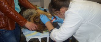
When visiting a veterinary clinic after the first signs of illness appear, the animal is prescribed a series of diagnostic tests. In addition to a general eye examination, they check the condition of the heart, do an ultrasound, and take a blood test to determine insulin and glucose levels. Anamnesis is collected and owners must indicate the animal’s chronic diseases, temperament, and activity. The veterinary passport is checked for vaccinations.
If all the signs of cataract are present, then the animal is prescribed an electroretinogram (ERG), which allows you to accurately determine the presence or absence of the disease in a matter of hours. The essence of the method is to check the retina, or, more precisely, its reaction in the form of impulses to an incoming beam of light. The study is carried out in a dark room and in the light using radio wave fluxes. Before the examination, an anesthetic is injected into the mucous membrane of the eye. This method allows you to diagnose cataracts in the early stages of development.
Drug treatment
Treatment of the disease in dogs will directly depend on the degree of development of the disease. So, at the initial stage, surgical intervention is not required. It is enough to carry out therapy. It consists of prescribing vitamin complexes, medications, enzyme agents, and special drops.
Remember! There is no special medicine to treat cataracts!
Removal operation
Just a few years ago, it was believed that in order to remove cataracts from dogs, they waited until they were “ripe.” But many veterinarians recommend getting rid of the disease in the early stages. The operation is performed using ultrasound. A special device directs ultrasonic waves to the lens and crushes it into a pulp. Then, using a puncture method, the split lens is removed from the eyeball. It is replaced by a special lens or an artificial lens.
No sutures are required for this type of surgery; healing occurs 10 days after surgery. This procedure is prescribed to dogs from six months of age to old age. In cases where both eyes are affected, the ophthalmologist performs the operation alternately on two lenses.
After surgery, owners must ensure that the pet does not injure the operated eye.
Cataract surgery for dogs in Rostov
Cataract is the loss of transparency of the eye lens, partially or completely.
In this condition, the lens becomes cloudy and prevents light from reaching the retina, which can lead to vision loss. The lens is an optical lens that, using the cornea of the eye, focuses light rays onto the retina. In a healthy eye, the lens is a transparent formation that appears as a result of the special structure of specific proteins. With age or under the influence of negative factors (metabolic disorders, diabetes, radiation, trauma), the unique structure of the lens proteins may be lost, which impairs its transparency. As practice shows, the first symptoms of cataracts most often appear in animals over the age of 4 years. The complexity of this problem and its huge prevalence among dogs and cats is due to several factors. Firstly, the type of inheritance of some types of cataracts is still unknown, and not every breeder will agree to exclude all relatives of a sick animal from breeding. Secondly, hereditary cataracts may appear in old age, or not appear at all if the animal died relatively young. Third, owners rarely have their pets diagnosed by an ophthalmologist throughout their lives, and the animal may be mistakenly classified as free. This makes research into this disease even more difficult. Cataracts can be not only congenital, but also acquired. In the latter case, the disease usually occurs as a result of injury or acute ophthalmological disease and is localized in one eye. Hereditary cataracts are always bilateral, although the simultaneous development of pathology in both lenses is possible. Cataracts are diagnosed through an ophthalmological examination. The only treatment method is surgery (replacing the affected lens with an artificial one). Cataracts are perhaps the most common genetic eye disease in both dogs and cats of all breeds.
The main method of treating cataracts
The most modern, painless and effective method of treating cataracts and restoring vision in our time is considered to be phacoemulsification in combination with lens implantation.
To perform cataract surgery, you do not need to wait for it to “mature” and, as a result, experience a constant decrease in vision. Now cataracts can be eliminated even at the earliest stage of its development:
1. Using a special instrument, the surgeon makes a micro-incision about 3 mm in size on the cornea. All further manipulations are carried out through it. This incision is made in such a way that after the operation it self-seals without consequences, that is, it disappears.
2. A special tip is inserted through a micro-incision, which, using ultrasound, turns the animal’s native lens into an emulsion. It is removed from the eye, after which the probe is also removed.
3. Through the same micro-incision, a flexible lens is inserted into the capsule (that is, in place of the removed lens), which will subsequently perform all the functions of the removed lens.
4. After the lens independently unfolds inside the eye, it is securely fixed using special flexible “antennae”. This lens does not require replacement; it lasts the animal’s entire life. Lenses are made from materials that are biocompatible with eye tissue. Our complex uses lenses from the world's leading manufacturers.
5. Upon completion of all surgical actions, the micro-incision, as already mentioned, self-seals. As a result, the patient’s vision regains the clarity lost due to cataracts.
It is worth noting that in the vast majority of cases, cataract surgery is performed through a micro-incision, that is, it does not require sutures. Within a few hours after the operation, the animal will be able to see quite well, and after two weeks, visual acuity will be completely restored. By decision of the World Health Organization, cataract surgery is recognized as the only completely rehabilitative operation among all, and not only eye, surgical interventions. Since the main feature of cataracts is their gradual development, some believe that there is no need to rush into surgery. This is a dangerous misconception, since cataracts are often accompanied by other diseases of the eye tissue, and they themselves can cause vision impairment.
For questions regarding the provision of specialized ophthalmological care, you can contact the ophthalmological complex of microsurgical profile, created on the basis of the Center for Veterinary Medicine.
All questions by phone 89885798289 or by e-mail: [email protected]
The data is not supported by your browser.
source
Prevention
No preventive measures have been developed to completely prevent a dog from developing cataracts. Owners are advised to monitor their pet’s diet and daily routine, take frequent walks, and enjoy active rest. All this will help reduce the risk of developing chronic diseases, and they serve as an impetus for clouding of the lens. Since genetic predisposition plays a big role, when purchasing a puppy, do not be lazy to ask the breeders about the presence of this disease in the parents or members of his family.
Visit your veterinarian regularly for preventive examinations, because early detection of the disease makes the treatment process easier. Add vitamin complexes to your pet's diet. Pay attention to changes in your dog's habits. Examine the condition of your eyes yourself periodically. If you notice a bluish tint, seek medical help.
Cataracts are an insidious disease, the development of which is difficult to predict. Do not be indifferent to your pet, regularly monitor its condition and do not delay visiting the veterinary clinic. Remember that we are responsible for those we have tamed.
Symptoms of the disease
In the initial stages, the symptoms of cataracts in dogs are practically not noticeable, so treatment begins quite late. Cataracts are manifested by signs typical of eye diseases:
- decreased visual acuity - the dog is poorly oriented in space, bumps into objects;
- the animal's eyes become cloudy and become covered with an opaque film.
The disease can have 4 stages:
- Clouding of the lens occurs along the perimeter, as a result of which vision is slightly reduced, and the dog cannot see only certain parts of objects.
- When pathological changes occur in the central part of the eye lens, the dog’s vision decreases significantly, and it can only see the outlines of objects - this is an immature cataract.
- In the stage of the disease, which is called mature cataract, the animal ceases to see the outlines of things and distinguishes only light. In this case, the clouding affects the entire lens.
- With overripe cataracts, the dog's eyes are completely covered with a thick, milky-blue film, which causes the dog's ability to perceive light to be lost. The fibers in the lens liquefy, which causes its disintegration.
If time is not lost and the diagnosis is made in the early stages, then you can cure your dog of cataracts and give him the opportunity to look at the world with healthy eyes.