Etiology
Oral neoplasms are common in dogs and cats
. The most common soft tissue masses in the oral cavity are neoplasms, and most are malignant (ie, melanoma, squamous cell carcinoma, fibrosarcoma). However, acanthomatous ameloblastoma (formerly called epulis), fibromatous epulis (classically in boxers), as well as oral papillomatosis and eosinophilic granuloma (eg, in Siberian huskies and Cavalier King Charles spaniels) also occur.
Causes of the disease
If rough growths are found on the dog’s body, one can judge the presence of viral papillomatosis. The disease affects the mucous membranes and the upper part of the skin - the epidermis.
Growths are diagnosed on the face, in the eye or mouth area, and can also occur on the limbs (between the pads of the fingers), and in the genital area. Infectious lesions of the skin and mucous membranes are caused by a viral nature. The virus is transmitted from one dog to another, but does not pose a danger to humans.
The main way of transmitting viral agents from one pet to another is through close contact. Infection occurs through coitus (sexual intercourse), eating food from the same container, or using ammunition (in particular, a collar). Small growths begin to form on the dog's body several weeks after infection. Typically, the incubation period is 3 to 5 weeks.
Papillomatosis is divided into types depending on the location of the viral agent. The following types of disease are distinguished:
Viral papillomatosis is not a fatal disease. In this case, warts formed on the skin are fed by small vessels. The danger lies in mild trauma to the tumors and the opening of bleeding. Infection in the wound surface can lead to sepsis (blood poisoning).
Any dog can become infected with human papillomavirus. But veterinary experts note that there is a risk group. The most commonly affected dog breeds are pugs, terriers and giant schnauzers. The virus is also dangerous for small puppies up to 6 months, adult dogs with reduced body defenses and elderly animals.
Diagnostics
A thorough examination (which may require sedation) will usually reveal masses in the gum area, although the tonsil area, hard palate and tongue may also be affected. Diagnosis requires cytological and histological examination, although papillomatosis or melanoma can be suspected based on their appearance and volume.
The preferred diagnostic approach for a dog with an oral mass is an incisional biopsy and a chest and skull radiograph or CT scan of the affected area. If malignancy is suspected, chest x-rays are needed to identify metastases (rare, but the prognosis is very poor if they are present), and x-rays of the maxilla and mandible to determine bone involvement. A fine-needle biopsy of regional lymph nodes is recommended; even if they appear normal, metastases can often be detected. Melanoma may be apigmented and may be cytologically similar to fibrosarcoma, carcinoma, or undifferentiated round cell sarcoma. A biopsy and subsequent histology are required to establish a definitive diagnosis.
How to detect papillomas on an animal’s body and how they can be dangerous
If you have a smooth-haired pet, then detecting papilloma in a dog is quite simple. When carrying out hygiene procedures, you will immediately be able to notice foreign growths on the body of the four-legged animal. But there are cases when sometimes even experienced dog breeders may not notice this disease for years.
It is especially difficult to detect the virus in long-haired dogs. Lesions on the genitals are also not noticed by all owners, except perhaps professional breeders who regularly conduct preventive examinations of their animals. It is also difficult to detect warts in the oral cavity and in the ears.
Only when the disease becomes significantly more complicated do multiple growths appear, causing pain when chewing food. Fearing pain, the dog may refuse to eat, and when it eats food, the bitten growths may bleed. Increased salivation and an unpleasant odor from the mouth appear.
Papilloma on the paws, between the toes, can cause big trouble. In this case, the pet will just as quickly make it clear about the danger: it begins to limp, tuck its paw, and constantly lick and chew its fingers. Fresh wounds that appear can easily become infected or fungus.
Very often, papillomas may not bother your pet for years. And in the case of spontaneous remission, you can guess that the dog once had a wart by the remaining scars. In some animals, warts that appear may spontaneously disappear, while in others they persist for life.
In order not to bring the development of papillomatosis to a critical situation, you need to regularly examine the animal. And not only open, but also “secret” areas of the body.
What areas of the body should you pay attention to? As a rule, warts appear around the eyes, on the lips, in the mouth, and near the ears. They often cover the limbs. Please note that hair does not grow on the growths. If the papillomas are of the “cauliflower” type or resemble a honey mushroom colony, then you will immediately identify them.
But they can often look like a simple pinkish pimple. In this case (and in any other case, if you discover any neoplasm of unknown origin), you should not try to remove it yourself. After all, as we mentioned earlier, this could be an ordinary papilla.
Papilloma in dogs is a benign neoplasm. But some varieties can degenerate into malignant ones. This happens quite rarely. But such cases have happened in world veterinary practice.
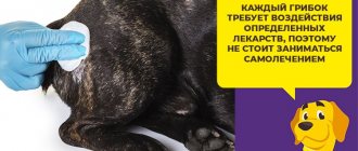
Treatment/Prognosis
The preferred treatment approach for dogs with confirmed oral malignancies and no clinically detected metastases is aggressive surgical excision of the mass and surrounding tissue (eg, marginal resection, maxillectomy). Enlarged regional lymph nodes should be removed and sent for histology, even if cytologically there are no signs of neoplasia. Early and complete removal of squamous cell carcinoma, fibrosarcoma, acanthomatous epulis, and (rarely) melanoma of the gingiva or hard palate may be effective. Acanthomatous epulis and ameloblastoma may respond to radiation therapy without surgery (but complete surgical treatment is preferable), squamous cell carcinoma and fibrosarcoma may respond well to adjuvant radiation therapy in the postoperative period. Squamous cell carcinoma of the tongue affecting the base of the tongue and tonsil cancer have a very poor prognosis; complete removal and irradiation still leads to the development of a serious disease. Melanoma metastasizes early and the prognosis is guarded. Chemotherapy is usually unsuccessful in dogs with squamous cell carcinoma, acanthomatous epulis, and melanoma. Piroxicam has been effective in some patients with squamous cell carcinoma. Combination chemotherapy has been successful in several cases of fibrosarcomas. Radiation therapy has been successful in some dogs with oral fibrosarcoma.
Papillomatosis usually goes away on its own, although in some cases resection of the masses is required if they interfere with eating.
The main reasons for the appearance of growths on a dog’s tongue
Oddly enough, growths in the oral cavity are often much safer than “harmless” spots.
The point is the reasons for their appearance, which are not always limited only to oncological pathologies:
Unfortunately, in some cases, the formation of growths may indeed indicate oncological pathologies.
Table - Some characteristics of oral tumors in dogs
| Tumor | Typical appearance/location | Biological properties | Therapy |
| Squamous cell carcinoma Gums Tonsil Edge of tongue (dogs) | Induration or ulceration/rostral gingiva Induration or ulceration/one or less often 2 tonsils Ulceration/tongue edge | Malignant, locally invasive Malignant, spreads to regional lymph nodes Malignant, locally invasive | Surgical removal ± irradiation; Piroxicam Special no treatment, Piroxicam Tongue removal, irradiation; piroxicam |
Oral cancer
Diagnosed in 30-40% of oral tumors in dogs, while in cats it is by far the most common type of oral tumor (70%). The most common location is the gums.
Elderly animals are primarily at risk, but there are no gender or breed patterns. Very young dogs (often less than 6 months old) develop a specific type called papillary. The tumor is most often localized in the area of the premolars/molars of the upper jaw, premolars of the lower jaw and the tongue.
The defeat of the latter can manifest itself as a non-healing ulcerative lesion of the frenulum, very similar to what develops when foreign bodies get under the tongue. Often the tumor is not visible, but can be palpated as a solid mass in the ventral part of the tongue of the caudal frenulum. The incidence is thought to be increased in cats suffering from chronic stomatitis.
In the mouth, at the first stage, squamous cell carcinoma looks like ulcers and erosions that periodically fester, which is why the animal has a strong unpleasant odor from the mouth. Mobility of teeth, bleeding from the gums, profuse salivation gradually develops, and pathological discharge from the nose is sometimes observed (if localized in the upper jaw). Later, the tumor becomes so large that the curvature of the jaws is visible even from the outside. In this case, pathological fractures may occur. The incidence of metastasis to regional lymph nodes and lungs is usually low, but increases with more caudal localization; tumors involving the tongue metastasize more often.
Diagnosis of oral tumors begins with an examination of the dog by a veterinarian - dentist. The exact nature and grade of malignancy of the lesion can only be determined by histopathological examination. Surgical biopsy is the best method to confirm or exclude an infiltrative lesion, which is performed under anesthesia.
For tumors affecting bone tissue, the main method of diagnosis is radiographic examination, which will show the size and development of the tumor. Undifferentiated oral tumors in dogs are often found in large breeds between the ages of 6 and 22 months. They are unusual for young individuals, but sometimes this can also happen. One type of oral tumor is an undifferentiated malignant tumor of the oral cavity; such a tumor cannot be recognized by a simple biopsy test. A complete health history of the dog prior to the onset of symptoms must be provided. Your doctor may take a sample of your lymph to see if it contains cancer cells.
The choice of treatment depends on the type of tumor, but in most cases surgery remains the most important component of the treatment regimen. Chemotherapy can inhibit the growth of cancer cells and improve the condition of an animal with oral cancer, but, unfortunately, this procedure has no curative effect. Infiltrative tumors of the lower jaw require wide excision or treatment with radical surgery, which requires removing part of the upper or lower jaw along with the tumor. The functional and cosmetic results of these interventions are usually very favorable. For jaw cancer in the absence of metastases and successful surgery, the prognosis is favorable. If the tumor has metastasized to regional lymph nodes, then the prognosis strongly depends on the histological type of the tumor. With metastases in other organs, the prognosis is unfavorable.
Our veterinary dentistry provides high-quality diagnostics and subsequent treatment of oral tumors in cats and dogs. The life expectancy of the animal will depend on the type of tumor, its stage of development at the time of treatment at the clinic, as well as on a set of timely measures aimed at eliminating the pathology of cell growth.
And remember that the best prevention of this disease is its early diagnosis and timely assistance. Take care of those for whom we are responsible!
The appearance of seals in the oral cavity of an animal is an alarming phenomenon. The severity and danger of its occurrence depends on the causes of its occurrence. A growth that forms on a dog’s gum causes discomfort, as it interferes with normal eating and causes pain.
Table - Some characteristics of oral tumors in cats
| Base of tongue (cats) | Ulceration/base of tongue | Malignant, locally invasive | No (palliative: tongue removal and/or chemotherapy |
| Malignant melanoma | Gray or black; may be smooth, usually a fleshy lump on the gum, tongue or roof of the mouth. | Very malignant, metastasizes early to the lungs | No (resection and/or radiation are palliative, but rarely alleviate the condition); Carboplatin and radiation may help. |
| Fibrosarcoma | Pink and fleshy/on gum or palate | Malignant, highly invasive locally | Surgical resection (chemotherapy and/or radiation may be effective in some cases) |
| Acanthomatous meloblastoma (epulis) | Pink and fleshy/on the gingiva or rostral part of the mandible | Malignant, locally invasive | Surgical resection irradiation ± irradiation |
| Fibromatous epulis | Pink, fleshy, solid or multiple/on gums | Benign | nothing or surgical removal |
| Papillomatosis | Pink or white, cauliflower-like, multiple/everywhere | Benign | nothing or surgical removal |
| Plasmacytoma | Fleshy or ulcerated, growing on the gum | Malignant, locally invasive, rarely metastasizes | Surgical resection irradiation ± irradiation |
^Top
Characteristics of growths on the tongue
All of the ailments described above should be considered in a little more detail.
Scar tissue growth
The owner can always remember exactly when the dog received injuries to the tongue, since any damage to this part of the body is accompanied by heavy bleeding. The growth in this case is characterized by the following symptoms:
Treatment in these cases is not required. The exception is situations when growths prevent the animal from eating and drinking normally. Specialists resort to an operation in which the scar growth is excised.
For two weeks after this, the dog will have to be fed exclusively liquid food, since roughage can contribute to the divergence of the seams and the formation of a new scar. Additionally, the animal may be prescribed antimicrobial agents (usually antibiotics), as well as anti-inflammatory corticosteroids.
Papillomatosis
Currently, experts believe that the main cause of the appearance of these tumors is viral infections. Despite their theoretical harmlessness, papillomas, firstly, can degenerate into malignant types of neoplasms. In addition, even if benign, these tumors often interfere with the normal process of eating and drinking.
For papillomatosis (especially multiple), surgical excision of tumors is recommended. As in the previous case, antibiotics are additionally indicated. Corticosteroids in the treatment of papillomatosis are prescribed with great caution, as they can contribute to the development of relapses of the disease.
Good to know! Recently, the drug Fosprenil has been increasingly used for treatment. When therapy is started in a timely manner, it allows you to do without surgical intervention.
Fungal pathologies of the oral cavity
As a rule, the development of growths is caused by various types of candidiasis. These pathologies usually do not develop out of the blue. The main reason for their appearance is serious problems with the immune system, caused, among other things, by prolonged and improper use of antibiotics and other antimicrobial drugs, as well as anti-inflammatory corticosteroids.
You can guess the presence of a fungus when the following symptoms develop:
The following are used in treatment: nystatin, clotrimazole, ivamerol. Additionally, immunomodulators like Immunofan are prescribed.
Melanomas
Typically, melanomas are dark (and even black) in color, but there are cases when the tumor does not contain pigments. In such situations, they can be mistaken for a white growth. Neoplasms of this type are divided into 4 stages (based on size and presence of metastasis).
They are treated by resorting to surgery. It must be supplemented with radiotherapy and chemotherapy, since otherwise the life expectancy of the operated pet is unlikely to exceed a couple of months.
Source
Good to know
- Volvulus of the mesentery and intestines in cats and dogs
- Constipation in dogs and cats
- Clostridia in dogs and cats
- Coccidiosis in cats and dogs
- Concentration of vitamins in serum in dogs and cats with diseases of the gastrointestinal tract
- Foreign bodies of the esophagus in cats and dogs
- Campylobacteriosis of dogs and cats (Campylobacter jejuni)
- Cricopharyngeal dysfunction (achalasia) in dogs and cats
- Cryptosporidiosis in dogs and cats (etiology, symptoms, diagnosis, treatment)
- Abdominal carcinomatosis in cats and dogs
- Coronavirus enteritis in cats
- Enemas and laxatives for cats and dogs
- Coronavirus enteritis of dogs (etiology, signs, diagnosis and treatment)
- Feline infectious peritonitis - FIP
- Infusion therapy for cats and dogs for diseases of the gastrointestinal tract
- Roundworms (roundworms) in cats and dogs
- Infiltrative diseases of the stomach in dogs and cats (neoplasia)
Features of formations
Papillomas are benign growths or warts on the body of dogs. There are two types of formations - loose and dense. The first option is found in young animals; outwardly, the growths resemble cauliflower. With age, they become keratinized, looking like dense nodules. Typically, papillomas grow singly, so they are difficult to detect under your pet’s fur. The growths are located in the groin and armpits, on the paws, eyelids and ears.
Sometimes warts grow and form entire families; they differ in shape and size. Multiple inflorescences may cover the mouth, tongue and oral cavity of a young dog. Papillomavirus affects the puppy’s body due to its fragile immunity. In adults, warts cover the paws and ears. They are quite dense, but do not pose any danger to humans and other animals.
The growth consists of multiple capillaries. If you accidentally damage it, it will begin to bleed heavily. If there are a lot of warts, then the veterinarian can diagnose papillomatosis. The color of the formation depends on the color of the dog. The dog will not be able to infect those around him; papillomas pose a danger only to small puppies.
Papillomas appear as a result of a special virus entering the body. It does not appear immediately; the first symptoms appear only after a few weeks or months. The infectious agent adapts well to different conditions; it can be transmitted from an infected animal to a healthy one through grooming products, collars and other accessories.
Most often, infection occurs through toys that dogs chew. The virus penetrates through cracks and damage to the mucous membranes and skin. The infection quickly spreads throughout the body and gradually makes its way to the epithelial layers of the skin. It then releases the transformed protein, causing the cells to multiply vigorously. Excessive division leads to the appearance of growths.
But not all dogs are exposed to the virus. Some animals have a genetic predisposition to this disease. Warts most often cover the bodies of terriers, pugs, Spitz dogs, schnauzers and cocker spaniels. Although the strength of the immune system has a greater influence on the susceptibility to papillomatosis.