Pneumonia is a serious illness that requires hospitalization of the dog. It often develops rapidly and is severe, so that even professional veterinary medicine is sometimes unable to cope with the disease. Differential diagnosis is extremely important here, since the symptoms of the initial stage of pneumonia can be confused with signs of an acute respiratory disease, which occurs much more easily and without tragic consequences. That is why any owner should be informed about how this disease manifests itself and how to treat a sick pet, because being enlightened means being armed. Let's look at what pneumonia is and what the rules are for fighting it.
Mechanism of disease
Pneumonia is an inflammation of the lower respiratory tract, involving all structures of the lung tissue, including the alveoli (cavities of the lungs involved in the respiratory process). It can have a different (but predominantly infectious) nature, and is also a complication of many diseases of the respiratory system.
The mechanism of the disease is quite simple: the pathogen, having penetrated the respiratory tract, causes inflammation in the alveoli. In the presence of a reduced bronchopulmonary barrier, transmission of infection occurs through the alveolar septa. The product of an extensive inflammatory process in pneumonia is usually exudate, the type of which corresponds to the type of disease.
As a result, the process of supplying blood vessels with oxygen, in which the alveoli participate, is disrupted, causing hypoxia. As the disease progresses, respiratory and then heart failure develops. If the course of treatment is ineffective, the patient's condition becomes severe, the disease progresses, causing further deterioration. In this case, death is possible.
Risk group
The risk group for the disease includes dogs with chronic infectious diseases, congenital disorders of the respiratory system, as well as animals with serious immunodeficiency conditions. Pneumonia often develops in weakened and emaciated dogs. Animals on duty and living on the street (hunting, guard, riding) are also at risk. Puppies and older dogs are especially susceptible.
Etiology of the disease
Depending on the etiology, pneumonia can be infectious or non-infectious.
Infectious pneumonia
Infectious pneumonia is often bacterial or viral in nature. It can develop as a primary or secondary disease against the background of influenza, bronchitis, tracheitis or tonsillitis. Depending on this, the causative agents of infectious pneumonia are:
- gram-positive microorganisms (staphylococci, streptococci, pneumococci);
- gram-negative microorganisms (enterobacteria, Escherichia coli, Proteus);
- mycoplasmas (causative agents of mycoplasmosis);
- viruses (herpes, influenza, parainfluenza, viral distemper);
- fungal infections;
- parasites (larvae of Toxocara, hookworm, filaria).
Infection of animals occurs mainly by airborne droplets, contact or hematogenous routes:
- through contact with sick animals;
- through contaminated food, dirty dishes, feces of sick animals;
- through infected blood and lymph.
Non-infectious pneumonia
Inflammation of the lungs of a non-infectious etiology in a dog is caused by the entry into the lung cavity of foreign agents of a non-infectious nature, namely:
- Aspiration pneumonia in dogs occurs after inhalation of foreign particles, foreign bodies (dust suspension, small debris), as well as when contents of the oral cavity or stomach enter the respiratory tract.
- Post-traumatic pneumonia is the body’s reaction to a violation of the integrity of the chest.
- Postoperative pneumonia is caused by complications after surgical interventions.
- Exposure to toxic substances on the respiratory system can result in toxic pneumonia.
- Allergic pneumonia. One of the symptoms of a malfunction of the immune system is inflammation of the lung tissue.
Causes of pneumonia in dogs
In practice, the following reasons for the development of pneumonia in dogs are encountered:
- Sudden temperature changes and critical hypothermia of the pet. For this reason, you should not walk your dog when there is a strong wind, damp and chilly outside. If there is such a need, we advise you to get at least a warm blanket for the animal.
- A consequence of advanced bronchitis.
- Infectious diseases . Accordingly, a pet can acquire pneumonia (as well as a couple of dozen other pathologies) after contact with stray animals.
- Aspiration pneumonia (one of the most severe types). It happens when, with severe vomiting, semi-digested food masses enter the lungs.
- Poor quality, unbalanced nutrition.
- Poor living conditions.
- Lack of immunoglobulins and other pathologies somehow related to the immune system.
- Injuries can lead to the development of pathology.
- Dust and gas pollution in the premises.
- Penetration of caustic and irritating substances into the pulmonary alveoli.
But in many pets, the root cause of pathology is... worms. The mechanism of development of the disease may be as follows:
- Most often, parasite larvae enter the lungs, migrating there from the intestines. Since the intestines of the larvae themselves are full of fecal microflora, inflammation will not be long in coming. In addition, young parasites spend about two weeks in the pulmonary alveoli, and all this time they feed on the tissues of the organ. This also contributes to the development of pathology.
- With severe helminthic infestations, adult parasites can enter the stomach, and from there, escaping from gastric juice, further: into the esophagus, trachea and lungs. Such an “incident” can lead not only to pneumonia, but even to death due to severe suffocation.
Types of pneumonia according to the nature of the inflammatory process
Based on the nature of the inflammatory process, two types of pneumonia are distinguished: catarrhal and lobar.
- Catarrhal pneumonia (or bronchopneumonia). The most common type of disease. The inflammatory process spreads to the tissue of the bronchi and alveoli, and is focal in nature. May develop as a complication of respiratory diseases. Focal pneumonia has less threatening symptoms and its treatment can therefore be carried out at home (but under the supervision of a doctor).
- Lobar pneumonia is acute in most dogs. It is an independent disease. It affects the entire lung or a significant part of it. It is characterized by the formation of an infiltrate in the alveoli, which can lead to swelling of the bronchial tissue. This form of pneumonia leads to respiratory problems and therefore requires hospitalization. For the most part, it is not contagious.
During the course of the disease, alveolar exudate is formed in the lumen of the bronchi and alveoli. Its composition is determined by the type of inflammatory process in the lungs:
- Purulent exudate is characteristic of complicated forms of pneumonia with the addition of pyogenic microorganisms.
- Serous exudate is a clear liquid, has a protein composition containing leukocytes and is formed in the catarrhal form of pneumonia.
- Fibrinous exudate is exuded during lobar inflammation of the lower respiratory tract. It contains fibrinogen (a special protein), which indicates increased vascular permeability.
Veterinary clinic of Dr. Shubin
Description and reasons
Aspiration pneumonia of dogs and cats is an inflammatory disease of the lungs that develops as a result of inhalation (inhalation) of a significant amount of solid or liquid material into the lungs.
In healthy animals, a small volume of fluid and bacteria is constantly aspirated into the lungs from the oropharynx, but the clearance mechanism of the respiratory tract prevents the development of infectious inflammation. Aspiration pneumonia in animals of any age usually indicates a violation of defense mechanisms and the presence of predisposing diseases. Below are the main underlying diseases of cats and dogs in which aspiration pneumonia can develop. Table. Diseases predisposing to the development of aspiration pneumonia in cats and dogs.
Esophageal dysfunction • Megaesophagus. • Reflux esophagitis. • Esophageal obstruction. • Myasthenia gravis (localized). • Bronchoesophageal (bronchoesophageal) fistula. Localized diseases of the oropharynx and larynx • Cleft palate. • Cricopharyngeal motor dysfunction. • Laryngloplasty. • Brachycephalic respiratory syndrome. • Deviations in the structure and function of the larynx (congenital and acquired). Systemic neuromuscular diseases • Myasthenia gravis. • Polyneuropathy/polymyopathy. Impaired consciousness • General anesthesia/sedation. • Coma of various etiologies. • Head injury. • Severe metabolic disorders. • Hiccups. Iatrogenic factors • Forced feeding. • Errors in the placement of feeding tubes. Various diseases accompanied by vomiting
Aspiration pneumonia usually develops in animals with regurgitation, which is more often observed with megaesophagus and various esophageal motility disorders. Another important cause of aspiration pneumonia is localized or systemic disturbances of the swallowing reflex on the part of the pharynx and larynx; the swallowing reflex can also be impaired during depression of consciousness (eg anesthesia, coma). The per os administration of various medications, as well as force-feeding of sick animals, is another important cause of aspiration pneumonia, especially when it comes to the administration of mineral oil to cats.
In the development of lung damage during aspiration of liquid or solid masses, the following key points are highlighted: exposure to chemicals; airway obstruction; introduction of infection and the resulting inflammatory response to the above factors. The severity of chemical damage depends on the volume of the aspirate, its pH and the toxic effect of its constituents. Foreign matter (especially acidic gastric contents) causes severe chemical damage to the lower respiratory tract, leading to swelling, hemorrhage, necrosis and bronchoconstriction, and inflammatory cell effusion. Against the background of an acute inflammatory response, proper lung function is impaired and hypoxemia, which in some cases can be fatal.
Severe respiratory distress in aspiration pneumonia can develop from airway obstruction by aspirated material (usually small airways), which is aggravated by reflex bronchoconstriction and the inflammatory response. A bacterial infection in aspiration pneumonia can be associated with the aspirated material, or it can spread hematogenously against the background of severe inflammatory damage to the lungs (secondary infection). After the addition of a bacterial infection, classic bacterial pneumonia develops.
Inhalation of mineral oil is accompanied by a chronic inflammatory response, signs of aspiration pneumonia are moderate, and severe damage is rare. In this case, radiographic changes persist for quite a long time and in the future can be mistakenly interpreted as lung neoplasms.
Clinical signs and diagnosis
Dogs and cats with aspiration pneumonia are often presented at the veterinary clinic with signs of acute respiratory dysfunction, systemic manifestations are also characteristic (eg anorexia, depression) up to a severe state of shock. A history of chronic vomiting or regurgitation may precede the development of respiratory distress. Some patients present to the veterinary clinic with signs of chronic, intermittent or progressive cough. Sometimes, in patients with aspiration pneumonia, there are only signs of general depression or predisposing diseases. In case of aspiration pneumonia, a careful medical history is important, especially with regard to force feeding and drug administration.
When conducting a physical examination of a patient with aspiration pneumonia, fever is not always detected; respiratory sounds are often identified during auscultation. If a veterinary patient is in stable condition, a thorough neurological examination should be performed, and the animal's ability to accept and swallow food should be assessed.
The diagnosis of aspiration pneumonia is usually made on the basis of radiographic changes together with identification of signs of underlying diseases. Radiographic changes in aspiration pneumonia usually reveal a diffuse increase in interstitial density and disappearance of the alveoli. X-ray signs of lung damage may appear only 12-24 hours after aspiration. In chronic cases, a nodular (nodular) pattern may be observed, single nodes may be dense around, and with aspiration of mineral oil, multiple nodes are more often noted.
Changes in the general blood count may reflect an inflammatory process (eg neutrophilia with a left shift), but often the general blood count is within the physiological norm. Tracheal lavage for aspiration pneumonia is performed in animals that can tolerate this procedure to identify the bacterial infection and determine its sensitivity to antibiotics (culture testing).
Due to the fact that aspiration pneumonia always develops against the background of some pathological process, an important part of the diagnosis is the identification of underlying diseases.
Treatment and prognosis.
Suctioning of foreign fluid is carried out only in cases where the animal has aspiration under anesthesia in a veterinary clinic; this procedure is carried out either under the control of a bronchoscope or blindly with a thin probe; airway lavage is contraindicated. Animals with severe respiratory distress are treated with fluid resuscitation, oxygen therapy, bronchodilators, corticosteroids (controversial), and other appropriate treatments. Further treatment after stabilization of the animal is carried out in the same way as for bacterial pneumonia. The prognosis for an episode of aspiration pneumonia is often favorable, but in general, the outcome depends on the ability to correct the underlying disease.
Photo 1. A young dwarf dachshund, a few months ago, plastic surgery of the vocal cords (“anti-barking”) was performed; the owners sought help due to a chronic cough.
Photo 2. Radiographic examination of the same dachshund, the image shows focal changes in the lung parenchyma.
In this case, it is not possible to correct the underlying disease (damaged vocal cords) and treatment is only symptomatic; periodic antibiotic therapy significantly reduces the cough in this animal and improves the quality of life.
Note. The veterinary clinic staff does not perform surgeries to cut vocal cords.
Valery Shubin, veterinarian, Balakovo.
Features of the course of the disease
There are several stages of development of the disease:
- The primary stage (with lobar inflammation it is called the stage of flushing or active hyperemia) lasts up to 3 days. The pathogen penetrates the lung tissue, causing inflammation. The vessels of the organ become overfilled with blood, and exudate begins to be released into the alveolar spaces.
- Secondary stage (stage of red and then gray hepatization). May last up to two weeks. The lung tissue becomes denser (taking on the consistency of the liver), as a result of which the supply of oxygen to the bloodstream is sharply disrupted. Red blood cells and then leukocytes sweat into the alveolar exudate. Clinical signs are most acute. Intoxication reaches its peak.
- The final stage. With a favorable outcome, the normal structure of the lung tissue is restored and the exudate is liquefied.
Depending on the type of pathogen, the state of the dog’s defenses and other factors, the disease can progress in different ways. There are three degrees of severity:
- Easy. No intoxication. Slight increase in temperature. The presence of a small focus of inflammation.
- Average. Moderate intoxication. The temperature may be increased slightly, but quite active formation of exudate is observed.
- Heavy. Severe intoxication. Loss of consciousness may occur. Body temperature is critically high. Active infiltration is underway. The dog is very weak and is breathing frequently. Complications may develop.
Types of pneumonia
There are several varieties of this disease in four-legged pets:
- Bacterial pneumonia is always infectious in nature. It often develops as a secondary complication of bronchotracheitis, tracheitis, and also as a result of a foreign body entering the larynx or bronchi of an animal.
- Parasitic inflammation develops due to the entry of worms and worm larvae into the lungs. They can penetrate the lung tissue as a result of moving throughout the body.
- Viral pneumonia develops as a result of complications of viral plague.
All types of pneumonia are extremely dangerous for puppies, as well as for older individuals. It is extremely rare for such a pathology to develop in physically strong dogs.
In addition, depending on the type of exudate formed, pneumonia can be purulent or serous. The most severe form of pneumonia, when fibrin begins to enter the lumen of the alveoli of the lungs, is called lobar. Without the help of a veterinarian, an animal can die in just a few hours.
Provoking factors
Each type of pneumonia has its own risk factors that can trigger the onset of the disease.
Risk factors for infectious pneumonia
Risk factors for infectious forms of the disease are the following:
- frequent hypothermia;
- complications of primary infections: influenza, plague, tracheitis;
- chronic inflammatory diseases of the upper respiratory tract;
- weakened immunity due to insufficient production of immunoglobulin A by the body;
- poor diet, lack of vitamins and minerals;
- failure to comply with hygienic conditions for keeping animals (using dirty dishes, keeping sick and animals in the same enclosure, and so on);
- lack of timely deworming;
- eating foods with fungal infections (mold).
Risk factors for non-infectious forms
- Injuries and fractures of the chest provoke traumatic pneumonia.
- The postoperative period is often complicated by an inflammatory process in the lungs due to forced immobility for a long time.
- Aspiration of vomit into the respiratory tract during repeated vomiting can provoke aspiration pneumonia.
- Living in areas with poor ecology (with chemical production facilities nearby) is fraught with a toxic form of the disease.
- Long-term use of certain medications and chemotherapy can cause toxic pneumonia.
Symptoms of the disease
The symptoms of the initial stage of the disease are very similar to the signs of any respiratory disease, so many owners do not pay attention to them. It seems to them that the dog has nothing serious. But signs such as a slight increase in temperature, a warm, dry nose, chills and fever may appear later as dangerous symptoms of lobar pneumonia:
- high fever;
- dyspnea;
- cough streaked with blood;
- clear signs of oxygen starvation, bluish coloration of the lips;
- lack of appetite, weight loss;
- hypertension, increased heart rate;
- severe intoxication;
- depression of respiratory function.
Focal pneumonia develops more often as a complication of acute respiratory infections or influenza. It is accompanied by low-grade or febrile temperature, dry or wet cough, and severe weakness. When listening, hard breathing, wheezing, and crepitus (creaking) over the source of inflammation are detected.
Diagnosis of the disease
After listening and percussing (tapping) the lung area, the doctor orders a chest x-ray. This is the most informative method for diagnosing pneumonia. The darkening and enhanced pulmonary pattern in the image will make it possible to determine the presence of a process in the lungs, its type and the extent of damage to the organ.
A biochemical blood test is no less important. The state of the blood can determine the degree of inflammation, as well as the type and cause of pneumonia. Sputum analysis helps determine the type of pathogen.
Only a professional can interpret the test results, so competent medical care in the treatment of pneumonia is necessary. In addition, the disease must be differentiated from other diseases of the upper and lower respiratory tract:
- acute tonsillitis;
- bronchitis and tracheobronchitis;
- rhinitis, sinusitis;
- lung abscess;
- tumor diseases of the lungs and tissues surrounding them.
Main symptoms
Signs of aspiration pneumonia appear gradually. In the first 3–4 days, there is a slight increase in body temperature to 37.5°C, general weakness, dry or wet cough with clear sputum. After 1–2 weeks, a person’s well-being deteriorates significantly.
Symptoms of acute aspiration syndrome:
The symptom of acute aspiration syndrome is cyanosis
- fever;
- chest pain;
- heat;
- tachycardia;
- shallow breathing with prolonged inhalation;
- cyanosis;
- asymmetry of the chest during breathing;
- urticaria, rhinitis, Quincke's edema due to mold infection;
- dyspnea;
- arthralgia, myalgia;
- discharge of foul-smelling sputum with pus and blood.
The most aggressive disease occurs when aspirated contents from the stomach enter the lungs. Hydrochloric acid, which is part of the gastric juice, causes the formation of a chemical burn (pneumonitis), disrupts the immune barrier of the mucous membranes, due to which bacteria easily penetrate into the respiratory tract.
This type of pneumonia is often complicated by:
Lung abscess is a complication of pneumonia
- lung abscess;
- gangrene;
- bronchospasm;
- formation of alveolitis;
- bronchiectasis;
- stidora;
- pleural empyema.
Which part of the lungs will be affected depends on the position the person was in during aspiration. If the patient was sitting, inflammation of the lower lobe is observed; if he was lying down, then the pathological process is localized mainly in the upper lobe of the right lung.
Patient's condition with pneumonia
The treatment method is selected taking into account the patient's condition. There are three types of conditions for pneumonia:
- A stable condition may be accompanied by quite strong pulmonary symptoms, but the dog’s general well-being at this moment is close to normal: its appetite is not impaired, it exhibits normal physical activity. In this case, after visiting a veterinarian and receiving treatment recommendations, the animal can be treated on an outpatient basis.
- Unstable. In addition to pulmonary manifestations, the temperature is significantly elevated, the dog is lethargic and refuses to eat. In this case, the animal must be constantly under medical supervision in a hospital.
- Critical. Other signs of pneumonia include symptoms of acute lack of oxygen and cardiac dysfunction. The animal needs resuscitation, which consists of artificial ventilation of the lungs and maintaining the vital functions of the body.
Treatment of the disease
It is preferable to treat pneumonia in dogs in a hospital, since only there it will be complete and comprehensive.
- The main treatment for pneumonia is antibiotic therapy. They use broad-spectrum antibiotics that are effective against any type of pathogen or a complex that includes several types of drugs. These are penicillin antibiotics (Ampicillin, Gentamicin), tetracyclines (Doxycycline). They are prescribed in tablet or injection form.
- Physiotherapy. It is prescribed when acute inflammation subsides. In most cases, this is UHF heating, electrophoresis, and also a special chest massage to avoid stagnation of sputum, accumulation of alveolar exudate and pulmonary edema.
- Artificial ventilation and oxygen therapy are indispensable in severe cases.
- Infusion therapy is also possible only in a hospital setting. It is used if the animal is severely exhausted and is unable to independently support the vital functions of the body: it is in a state of extreme exhaustion or dehydration.
Treatment of pneumonia in dogs
The primary task that the veterinarian needs to solve is the process of stabilizing the animal’s condition. In addition, it is necessary to choose the right treatment tactics to completely rid the body of the causative agent of the disease. For example, aspiration pneumonia in dogs is difficult to treat.
- Antibiotics for pneumonia in dogs are prescribed in the hospital. The drug is administered by injection. This is due to the high degree of absorption of the drug and the rapidity of its effect on pathogenic bacteria. In the form of injections, the medicine penetrates deeper and specifically reaches inflammatory foci. The selection of the required antibacterial agent is carried out in such a way that the effect is exerted on all types of microbes.
- Before treatment with antibiotics, the veterinarian will order tests. As a result, it becomes clear whether the animal has an allergic sensitivity to the drug or not. The study includes tracheal lavage.
- In addition to antibiotics, depending on the type of disease, the doctor may prescribe treatment with anthelmintic drugs, antiviral agents and antifungal medications. In addition, immunomodulators, mucolytics, diuretics and vitamins are prescribed. All this is required to have a comprehensive effect on the source of inflammation, as well as to bring the dog out of a serious condition as quickly as possible and make her life easier.
- Among the physiotherapeutic measures, the veterinarian performs chest massage. In this way, you can quickly and easily remove accumulated mucus. The procedure is carried out until the pet’s cough disappears.
- In the case of a severe form, which is accompanied by respiratory failure, doctors prescribe oxygen therapy. If there is no effect, artificial ventilation may be prescribed. The dog breathes a mixture of air that contains up to 40% oxygen. Due to the fact that in its pure form it has a toxic effect on lung tissue, it is not recommended to prescribe a higher concentration.
- If the pet refuses food, in case of pronounced disturbances in the stool, the doctor recommends prescribing infusions. In stationary conditions, there is regular monitoring of diuresis, as well as the number of respiratory movements. Such readings are important for monitoring, since based on their results, a conclusion is made about the improvement or deterioration of the sick dog’s condition.
Due to the fact that such a diagnosis is considered serious, self-medication is unacceptable. Otherwise, there is a high probability of death.
Pet owners can use home therapy to improve their condition. However, treatment should begin only after her appetite has been restored. It is imperative to adhere to a number of rules:
- Suppressing cough with special drugs (suppressors) is not allowed.
- Massage the animal's chest at least four times a day. This is necessary to stimulate cough activity, which has a beneficial effect on the removal of exudate from the bronchi.
- In severe frosts, it is not recommended to walk your dog.
- Treatment with antibiotics at home is carried out only in accordance with the instructions for use and the indications of the treating veterinarian.
- Get your dog x-rayed periodically.
- The animal needs light exercise in the form of running.
Pet owners should understand that treatment with folk remedies is a useless exercise, and in some cases can worsen the animal’s condition. This is due to the fact that in the fight to destroy the causative agent of a serious disease, the use of antibiotics is an appropriate solution. However, their use must be prescribed by a veterinarian. You can give your dog herbs that strengthen the immune system only after the condition has stabilized and the inflammation has gone away.
Video: what is pneumonia in dogs, its symptoms and treatment
Home care and recovery
At home, every effort should be made to strengthen the body weakened by the disease:
- Continue to give antibiotics according to the regimen suggested by your doctor.
- Stop walking for a while to avoid possible hypothermia and colds, the onset of which can lead to a relapse.
- You cannot suppress the cough reflex with drugs that act on the cough center of the brain, so as not to provoke stagnation of phlegm in the lungs.
- Do regular chest massage yourself to improve sputum production in your dog.
- Bring your pet for follow-up examinations at the appointed time.
- Create a complete diet that will help restore weakened strength in a short time.
- Additionally, carry out vitamin therapy. Usually, the dog is prescribed vitamin complexes with a high content of vitamins A, C and group B.
How is the diagnosis carried out?
It is impossible to diagnose pneumonia without a doctor's examination. This disease has similar symptoms to many others, so the doctor, based on the medical history, chooses in which direction to conduct additional research in order to make the correct diagnosis. After examining the four-legged patient, listening and tapping the chest, the doctor prescribes an x-ray. In the case of pneumonia, the image shows a darkening with unclear boundaries. To get a more complete picture of the disease, x-rays are taken in two projections. This allows you to determine not only the location, but also the extent of the damage.
Did you know? The Besenji dog breed is distinguished by the fact that the animals do not bark, but purr. They can also wash themselves with their paws, like cats.
The doctor also prescribes a general and biochemical blood test. In its results, in case of inflammation, neutrophilic leukocytosis is observed, red blood cells and hemoglobin are reduced. By analyzing sputum or bronchial washings, the exact type of pathogen can be determined. This will help when choosing medications. Diagnosis of parasitic pneumonia involves examining the animal's feces for the presence of helminths.
Prevention methods
Basic preventive measures are aimed at maintaining good physical shape and immunity of the animal. It means:
- Avoid hypothermia, protect from colds and viral diseases of the upper respiratory tract.
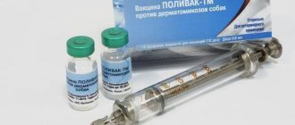
- Vaccinate and deworm on time.
- Avoid contact with sick animals.
- Maintain a nutritious and balanced diet. The dog should receive all the vitamins and microelements necessary for its age.
- Moderate physical activity is beneficial for dogs.
If the dog receives good care and proper treatment, the prognosis of the disease is often favorable. Modern experience in the treatment of pneumonia is quite rich and allows us to cope with even a severe form of the disease.