Uterine prolapse after childbirth
More often the exit appears during or after childbirth. The causes of complications are:
- active labor due to hormonal overloads of the body;
- heavy bleeding during childbirth;
- multiple births - due to the non-standard number of puppies, the muscle layer of the vagina is stretched, the tone of the reproductive organ decreases;
- hydrops fetalis – an increase in the size of the puppy, leading to prolapse or inversion of the uterus;
- improper feeding and care of a pregnant bitch.
Symptoms
The best way to avoid uterine prolapse in a dog is to know the signs of this condition and understand what to do in this situation. Uterine prolapse can occur during childbirth, the difficulty lies in understanding the symptoms in time.
It is especially important after childbirth to pay attention to the following signs:
- The dog may have abdominal pain.
- Unusual tension after childbirth.
- Heavy bleeding.
- The animal begins to lick the vaginal area frequently.
- The tissues in the vaginal area change.
Uterine prolapse usually causes mild pain in dogs, so symptoms are easier to identify after birth.
Causes and symptoms of hair loss
What leads to illness:
- exhaustion or obesity of the bitch;
- lack of walks (at least half an hour 2 times a day);
- poor, unbalanced diet;
- lack of minerals and nutrients;
- advanced age of the female.
It is possible to determine that an animal suffers from prolapse when there are no visible manifestations of the disease yet, based on the following factors:
- the bitch behaves restlessly, strains and constantly licks itself;
- cracks and scabs appear on the organ;
- the horn of the uterus protrudes from the vagina;
- the female experiences pain, restlessness and anxiety, and behaves aggressively.
Main causes of the disease
The causes of uterine prolapse in dogs are directly related to childbirth. Difficult labor is the most common cause of uterine prolapse.
Other causes of uterine prolapse in dogs:
- severe stress during childbirth,
- improper obstetric care,
- heavy bleeding during childbirth.
Treatment at a veterinary clinic
If complete or partial prolapse of the vagina is detected, the veterinarian begins to return it to its anatomically correct position and prevent infections. The bitch is hospitalized regardless of the severity of the condition.
Treatment is performed surgically if damage or contamination of tissues or disruption of the functioning of internal organs is detected. In advanced cases, amputation of the reproductive system is required to avoid death.
The vagina can return to its place if the operation is successful and the tissue walls are in satisfactory condition. If the dog has no breeding value, it is better to get rid of the reproductive organs.
After surgery, follow your doctor's instructions, disinfect and continue antibiotic therapy.
Treatment of uterine prolapse in dogs
Before receiving qualified assistance, the animal owner must be able to provide first aid. First of all, you need to place the dog on a clean bedding. Using sterile wipes, pre-moistened with a cold disinfectant solution, wrap the prolapsed organ. A solution of chlorhexidine, potassium permanganate, furatsilin, Miramistin is used as an antiseptic.
After the animal is diagnosed, the veterinarian, based on the severity of the obstetric and gynecological pathology, will choose the best way to solve the problem.
If the organ has not completely prolapsed and the outer part does not show signs of inflammation, then in some cases they can manage by repositioning the uterus. The tissues are treated with cool antiseptic solutions, and if necessary, a novocaine blockade with an antibiotic is carried out. After irrigation with disinfectant solutions, the uterus is sprinkled with streptocide powder.
Reduction of the uterus followed by sterilization
Manipulation to return the organ to its anatomical position is carried out using anesthesia, for example, under a sacral blockade. To return the uterus to the abdominal cavity, special nylon sticks are used. To prevent the development of inflammation, a sick dog is prescribed broad-spectrum antibacterial drugs, usually cephalosporins. After reduction, the veterinarian applies special loop-shaped sutures to the vulva.
Sutures for holding the vagina: a) loop-shaped; b) roller
If serious tissue damage has occurred and there is a risk of organ necrosis, a veterinary specialist will perform amputation of the uterus. For animals that are not of breed value, ovariohysterectomy is recommended. Sterilization of the dog is also necessary if uterine prolapse is regular.
For information on surgical reduction of the uterus in dogs, watch this video:
CAO and prolapse disease
Uterine rupture is not a breed disease of Central Asian Shepherds (unlike bulldogs and bull terriers). But if we take into account only cases of obstetric pathologies, vaginal protrusion is a common problem. It is often found among Asians of factory or semi-culture breeding, where the blood of indoor kennel dogs is present.
CAOs that have survived vaginal repositioning are not recommended for breeding - there is a high risk of inheriting the disease by offspring.
Care and prevention
Caring for an animal after repositioning the reproductive organ comes down to following the veterinarian’s recommendations. If surgical removal was performed, the postoperative period is reduced to treating the sutures with antiseptic drugs and systemic use of antibacterial agents.
Prevention of uterine prolapse in dogs is possible if the owner follows the following rules and recommendations of veterinary specialists:
- Balanced feeding of a pregnant female eliminates many gynecological problems in the postpartum period, including uterine prolapse.
- Regular dosed physical activity during pregnancy allows you to maintain the smooth muscles of the reproductive organ in good shape.
- Providing qualified assistance to an animal during childbirth.
Partial or complete uterine prolapse is most often diagnosed in dogs immediately after birth. In rare cases, pathology can be observed during estrus. A sick animal needs qualified help. In some cases, a veterinarian performs an ovariohysterectomy to save the dog’s life. In uncomplicated variants of the disease, reduction of the prolapsed organ is used with further antibacterial therapy.
New growth or tumor of the mammary gland in a dog: causes, symptoms, treatment and prognosis. . Causes of neoplasms in dogs. Older animals over 6 years of age are primarily at risk.
Indications for termination of pregnancy. The structure of the uterus in domestic animals is such that removing numerous fetuses from the womb of a dog is impossible without severe injury to the organ.
If the bladder prolapses, anuria is observed. When palpating the protrusion, the dog may experience involuntary urination. If a pregnant uterus is caught in the hernial ring.
The animal experiences loss of function of organs located below the injury to the spinal column. During the early phase of collapse, the dog exhibits the following signs: tachycardia, rapid and uneven breathing
Uterine inversion and prolapse
Uterine inversion and prolapse is a displacement of the uterus in the form of eversion of the horn wall (intussusception) or complete inversion with its prolapse outward.
Etiology. This disease is a complication of childbirth and occurs mainly in cows and goats, less often in mares and other animals. It happens during difficult labor, childbirth with a large fetus, with overdistension of the uterus (hydropsis of the membranes, multiple pregnancy), sagging muscles of the uterus. In practice, uterine prolapse is often caused by rapid fetal extraction, especially during dry labor, when negative pressure is created in the uterus, and close contact between the fetus and the uterine mucosa, when the fetus is pulled out, promotes inversion of the uterus after the fetus is removed.
Uterine inversion can occur at the time of birth, when the fetus has an umbilical cord that is too short and strong, or spontaneously, due to increased intra-abdominal pressure (colic, tympany, when feeding animals with bulky feed). There are isolated cases of spontaneous uterine prolapse immediately or after 1-2 hours and after an easy birth. In private household plots and peasant farms of citizens, when animal owners tie various weights to the afterbirth, uterine prolapse also occurs.
In rabbits, both uteruses or just one fall out. In carnivores, there is predominantly complete loss of one horn during invagination of the second.
Clinical signs. Uterine intussusception is not characterized by any strictly specific signs. Usually the animal is worried, we observe attempts, the animal behaves as if it has colic.
Rectally, a veterinary specialist is sometimes able to palpate the fold formed by the bent walls of the uterus.
With complete prolapse of the uterus, a round or pear-shaped mass protrudes from the external genitalia, which in some cases descends to the hock joint. Cows, sheep and goats have succulent, sometimes bleeding, caruncles hanging in clusters. In pigs, the prolapsed uterus looks like intestinal loops. In a mare, the surface of the prolapsed uterus is smooth or slightly velvety. In carnivores, the fallen horn has the shape of a rounded body with a depressed apex. In case of complete prolapse, a round tube bifurcating at the ends with characteristic indentations of the peripheral ends of the horns protrudes from the genital slit.
Sometimes prolapse of the uterus, rectum and bladder occurs. The bladder in animals can prolapse through a vaginal wound or be everted through the urethra.
With invagination, when it is not accompanied by an inflammatory reaction, the invaginated area can spontaneously straighten out. The areas of the serous membrane touching in the fold usually become soldered to each other due to adhesive inflammation; In the resulting cavities, exudate accumulates, which sometimes resolves. As a result of a chronic course, the animal develops chronic endometritis, which subsequently leads to infertility. In place of the welded folds, thickenings form, which disrupt the normal course of pregnancy, if the animal is fertilized. In the area of intussusception, some animals develop purulent or putrefactive inflammation, which ends in a generalized form of purulent peritonitis or general sepsis.
When the uterus prolapses in the first hours, it has a bright pink or red color. As stagnation develops, the fallen surface becomes blue and even dark gray. The mucous membrane swells and becomes gelatinous; It is easily injured, bleeds, and cracks when dry. After some time, signs of inflammation appear in the uterus, necrosis of the mucous membrane occurs, accompanied by fibrinous deposits, dirty brown scabs, the placentas disintegrate, with the separation of soft, crumbly masses. If the patient is not provided with the necessary veterinary care in a timely manner, gangrene and sepsis develop.
We recommend reading: What to do if your cat has an inflamed eye
Treatment. If the pathological process during uterine intussusception has not started (no more than 2 days have passed), you should try to straighten the uterus by hand or by pouring large quantities of weakly disinfecting solutions into its cavity, with the croup (back of the body) raised. The animal is given general or sacral epidural anesthesia.
If the uterus has prolapsed completely, then before its reduction we perform sacral anesthesia or give the animal anesthesia, and begin cleaning and disinfecting the mucous membrane of the prolapsed uterus. If there is an afterbirth on the uterus, then we separate it. The entire surface of the prolapsed uterus is thoroughly washed with a cold astringent solution (alum, tannin, 0.1% solution of potassium permanganate), if necrotic lesions appear on the uterus as a result of late veterinary care, then we wash it with warm solutions. We lubricate the dead areas with tincture of iodine or cauterize them with lapis. The volume of the uterus enlarged as a result of edema in fresh cases can be slightly reduced by tightly bandaging with a towel or a wide bandage. Practitioners, in order to reduce the volume of a prolapsed uterus, use the injection of an oxytocin solution into the body of the uterus. If there are wounds on the prolapsed uterus, they must be revived and closed with a catgut suture.
Having carried out such preliminary preparation, it is necessary to begin repositioning the uterus. Regardless of the species, we give the animal a position with a highly raised croup. Reduction can be done both from the top of the uterus and its base (vaginal part). In the first case, a towel is wrapped around the fist and the top of the horn is pushed forward with a careful movement. At this time, the assistant helps to reduce the prolapsed uterus by applying pressure on the area of the prolapsed uterus with their hands. When pushed, the body of the uterus or, conversely, its apex returns first into the pelvic cavity. The sequence of reduction is not significant. Depending on the circumstances, various combinations are used when adjusting. In private household plots and peasant farms, clean sheets are used to fuse the prolapsed uterus. If the animals are small, then after raising their rump, a weakly disinfecting solution is poured under pressure into the remaining unturned part of the horn and at the same time the parts of the prolapsed uterus that are located on the outside are straightened with your fingers.
To prevent prolapse from happening again, the reduced uterus must be strengthened. In large animals, in order for the uterus to be kept in a normal position after reduction, we increase the tone of the uterus by stroking the mucous membrane with a hand inserted through the vagina and stimulating the involution of the muscular layer by injecting cold solutions. Sometimes you have to hold the uterus with your hand for 30-60 minutes.
To fix a prolapsed uterus, some veterinarians use a soccer ball camera, which is inserted into the vagina and then inflated with air. Sometimes they resort to suturing the vulva, as with vaginal prolapse.
In dogs and cats, the uterus is adjusted after a laparotomy, and strengthened in place by suturing it to the abdominal wall with several stitches of a seromuscular suture.
After uterine prolapse, animals develop postpartum endometritis, which will need to be treated intensively and comprehensively.
In cases where the prolapsed uterus is severely damaged, contaminated or subject to necrosis, then it is useless to reset it, and sometimes even dangerous for the life of the animal. In these cases, surgery to amputate the uterus is performed.
In rabbits, both uteruses or just one fall out. In carnivores, there is predominantly complete loss of one horn during invagination of the second.
Zinaida Mikhailovna
Prolapse or prolapse of the uterus in a dog - what to do?
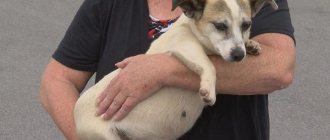
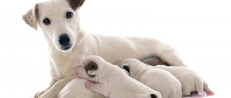
Uterine prolapse and vaginal prolapse (protrusion of the vaginal wall outward from the genital slit) are fairly common occurrences in dogs. Most often found in representatives of small breeds, as well as in multiparous bitches. They are observed much less frequently in young females. As a rule, protrusion of part of the uterus through the cervix occurs immediately after birth and the birth of the last puppy in the litter, but spontaneous miscarriage can also contribute to this.
What does a two-horned one look like? First
uterus? Let's figure out what a normal human uterus looks like and how it differs from some uteruses of representatives of the animal world.
The human uterus has the shape of an inverted pear. The part that is located on top of the uterus is called the fundus, and there is a lower exit from the uterus: the cervix and so. The vagina is designed by nature that there should be one child in the human uterus, its shape is precisely intended for bearing one fetus. Of course, multiple pregnancies do happen, and pregnancies with twins do not, however, such pregnancies are always rare, most complicated for a woman and her babies, since the uterus has to stretch more strongly to accommodate more, or even two, children.
The uterus of many animals is completely different. For example, the uterus of a cat or a dog, which is destined by nature to bear several babies at once, is just a two-horned one. pear-shaped, like the human uterus, the uterus of animals is divided in two, forming “two” horns on the right and left sides. During pregnancy, a cat's uterus can be felt from the sides, and the kittens are located in it, like peas in a pod, one after another. During pregnancy, the human uterus protrudes forward and is located in the middle of the abdomen. The same arrangement of the uterus can be found in anthropoids, which, monkeys, also usually bear one So.
We recommend reading: Causes of Pyometra in Dogs
a dog's uterus looks like a baby
It’s not difficult to guess that if there are anomalies in the development of the uterus, we will get a human uterus, similar in shape to Takaya’s uterus. In animals, the pathology of the development of the uterus, like a bicornuate uterus, occurs as a result of disturbances in the fusion of the Müllerian ducts during the period of intrauterine formation of the fetus. double A uterus is formed that has one exit, the cervix through the uterus and vagina, but is fused with two cavities below.
Preventive measures to prevent uterine prolapse
If the female is not intended for breeding, the best method of prevention is to sterilize her.
Prevention measures that can prevent uterine prolapse include:
pregnant females should lie on a flat floor without a slope;
the litter should always be dry;
you need to walk with the female every day and not miss walks;
nutrition should be balanced and complete;
regular examination and monitoring of the pet’s health, consultation with a specialist is required;
with partial eversion, timely first aid is necessary.
It is advisable that a veterinarian attend the birth of the dog. In case of multiple pregnancy and some other factors, a cesarean section is indicated.
If the owner does not have the opportunity to resort to qualified help from a specialist, he can give birth on his own at home, but in this case one should not be too persistent when the fetus is delivered.