Oncological processes represented by newly formed tissue are becoming increasingly common among animals. Breast cancer in dogs can occur for a variety of reasons. When pathology occurs, the owners note the development of seals in the nipple area, resembling balls. If the pathological process is stopped in a timely manner, you can do without euthanizing your pet.
General information about breast cancer
Mammary gland tumors in dogs can occur in both females and males. According to statistics, 2-3 male dogs are at risk of developing this type of cancer. Usually the pathology occurs in uncastrated animals over five years old. The neoplasm can be either malignant or benign. There are a large number of photos of this terrible disease on every website about pets.
A benign pathological process is caused by the formation of atypical cells in tissue structures. The growth and development of the tumor is quite slow. Its progression is influenced by endogenous steroids and progestogenic sex hormones of the dog. However, removing reproductive organs from animals under two years of age significantly reduces the risk of developing tumors in the future. Without surgery, the number of sexual cycles and the release of estrogen and progesterone increases. Males have low levels of steroids in the blood. Therefore, compared to bitches, they are very rarely affected by breast cancer.
Malignant formation is an uncontrolled cell division, which leads to the formation of secondary foci of tumor growth. There are many versions of cancer, such as:
- poor nutrition and obesity;
- radiation;
- genetic predisposition;
- viruses;
- severely weakened immunity.
If breast cancer is detected in a dog, you cannot independently determine its breed and self-medicate. You need to contact a veterinarian as soon as possible about this problem. Be prepared for the possibility that your pet will require surgery.
Changes in a pet's body leading to a tumor (malignant tumor) were previously diagnosed after the age of 8 years. Recently, it has been proven that malignant neoplasms are diagnosed in pets at a young age.
Therapy for papillomatosis
There are several ways to treat this disease.
Surgical
If warts cause pain and discomfort to the animal, they can be removed surgically. Currently, modern medicine uses minimally invasive (gentle) methods that allow operations to be performed without the use of general anesthesia. These are local anesthesia, laser removal of warts, cryosurgery (exposure to low temperatures, causing cell death and rejection of papilloma), electrocoagulation (exposure to weak currents).
If the growths are multiple, located in hard-to-reach parts of the animal’s body (throat, genitals, ears), and are very bothersome (pain, bleed, itch), then general anesthesia and standard surgery cannot be avoided.
But sometimes surgical removal of tumors in a dog is not always necessary. Many veterinarians are inclined to believe that radical measures contribute more to the spread of the disease.
Drug treatment
Drug treatment of papillomatosis is mainly based on the administration (intravenously, locally or at the base of the papule) of a solution of novocaine, the use of immunomodulatory drugs or the use of antiviral ointments.
When treating with ointments or pastes, it is necessary to put a protective collar on your pet so that he cannot lick off the medicine.
Under no circumstances should you engage in drug treatment on your own. Only a veterinarian should make a diagnosis and, if necessary, prescribe treatment.
Traditional methods
Some “craftsmen”, having discovered papilloma in a dog, try to get rid of them on their own. What “benevolent” owners don’t do: they tie up papillomas with thread, rub celandine or dandelion juice into the affected areas, burn the growths with vinegar, and so on.
Vaccination
To date, vaccination is the most effective means of combating the papillomatosis virus. To make the vaccine, papillomatous tissue is taken from an animal infected with the virus and injected subcutaneously at intervals of 7-10 days. The effect occurs after 3-4 weeks.
There is a wonderful proverb: If you don’t treat a runny nose, it will go away only after two weeks, and if you treat it, it will go away after 14 days. In the case of papillomas, the situation is exactly the same. If your dog has a good immune system, the warts should resolve spontaneously within 2-3 months.
The most likely danger is that any other neoplasm can be mistaken for papilloma.
If you are not sure that the new growths that have arisen are precisely papillomas and they have not disappeared after 2-3 months and cause discomfort to the four-legged animal (they hurt, bleed), the animal should be shown to a veterinarian.
There is a wonderful proverb: If you don’t treat a runny nose, it will go away only after two weeks, and if you treat it, it will go away after 14 days. In the case of papillomas, the situation is exactly the same. If your dog has a good immune system, the warts should resolve spontaneously within 2-3 months.
Where do tumors appear?
The inflammatory process from the newly formed tissue manifests itself in the form of a seal on the dog’s mammary glands. It is a dense fibrous node or ball with a bumpy surface on the mammary gland. The neoplasm can be of various sizes: from a single compaction to a scattering of small cones. In this case, it can be motionless or wandering. In any case, in comparison with healthy tissues, the affected area in the abdominal cavity will be compacted and have a different shape.
How many mammary glands in a dog can cancer appear in? The number of external parts of the mammary gland in dogs ranges from 8 to 12, 4–6 pieces each, located on parallel lateral lines. Five pairs is the usual number for large breeds, four for small ones. They are located in two ridges from the axillary recesses to the groin area. A local cell population may develop at or near any nipple. However, most often, dysregulation of cell growth and differentiation is observed in the posterior paired organs.
At-risk groups
According to statistics, the risk of malignant or benign neoplasia of the mammary glands of dogs is significantly reduced when females are castrated before two and a half years of age. Delayed removal of reproductive organs reduces the occurrence of benign neoplasm. However, it does not prevent tissue damage from malignant neoplasms.
In dogs that have undergone surgery to remove the testicles and ovaries before the age of one and a half years, a malignant tumor of the mammary gland occurs very rarely. In animals that are not emasculated between 5 and 12 years of age, the risk of cancer is increased several times.
In addition, some breeds at risk for canine mammary gland cancer include:
- Rottweiler;
- Great Dane;
- Irish Wolfhound;
- small poodle;
- greyhound;
- boxer;
- retriever;
- pug;
- bull terrier;
- basset;
- dachshund.
What to do if papilloma is detected
The main thing to do when you discover papilloma in a dog is not to panic and observe calmly. Since we are talking about a benign formation, which, if the disease emerges from its latent form, has a completely characteristic clinical picture, which makes it possible to almost accurately differentiate plaques and nodules that appear on the skin or mucous membranes. In most cases, emergency medical care is completely unnecessary for your pet.
Papillomaviridae has been said to be present in many animals. It has been noted that if a dog’s papillomas or warts resolve (disappear) on their own, without the intervention of a veterinarian, the pet will most likely not get sick again. True, having defeated the virus, a dog can retain the pathogenic genome in some of its cells and, accordingly, pose a potential danger to relatives susceptible to the disease, but objectively nothing can be done about this problem.
You should only contact a specialist about papillomas if:
- the number of neoplasms is constantly increasing;
- warts or plaques cause real suffering to the animal (lameness, problems with eating, etc.);
- papillomas are located in places where there is a high risk of damage (in addition to the addition of a bacterial infection to the disease, violating the integrity of the neoplasm increases the risk of its degeneration into squamous cell carcinoma);
- after a long, stable course, the clinical picture of the disease showed noticeable changes towards activation (may indicate malignancy of the tumor).
Causes of neoplasms
Tumors of the paired mammary organs in dogs are a common disease diagnosed in pets that have not been sterilized. There can be many reasons for the occurrence of neoplasms. However, the main factors that trigger the mutation of cells of paired mammary organs are:
- a condition of an animal in which there is too much or too little of the steroid estrogen in its bloodstream;
- aging processes, during which the cardiovascular, digestive, excretory, nervous and reproductive systems become vulnerable. Areas of paired organs may swell in bitches older than 8 years;
- pseudo-puppyness. Due to the psychophysical state, which is triggered by a hormonal imbalance, the dog often develops tumors of the mammary gland;
- absence of mating upon the onset of puberty of the animal;
- mastitis and natural feeding of offspring. In some cases, congestive mastitis can develop into acute mastitis, and after that into cancer. Pathology occurs due to inflammation of one or more nipples. If a dog does not feed its puppies, its mammary glands become swollen and red, a nuisance that can lead to the formation of tissue nodules and neoplasia;
- pathological conditions that occur as a result of improper activity of the endocrine glands or endocrine glands;
- carcinogens contained in the environment and entering the dog’s body with low-quality food.
The structure of breast tumors looks like modified tissue cells. Mutated elementary units have individual DNA, which they pass on to their daughter cells. Therefore, the cells formed during the budding of a cancerous mother rapidly grow and develop.
Phenomena that aggravate the development of the inflammatory process in milk bags. And also provoking the suppression of the body’s defenses are:
- eating disorders that occur if the pet receives low-quality moldy food;
- long-term, untreated parasitic diseases caused by helminths;
- natural predisposition to the development of malignant tumors at the genetic level.
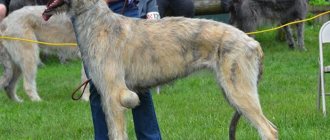
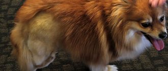
growth on a dog's nipple
Growths on a dog's skin
A growth or growth on a dog's skin is called a tumor. Tumor is a pathological growth of tissue. There are two types of tumors - malignant and benign. Malignant tumors are fast-growing and easily spread to other tissues, giving metastasis. Benign tumors grow slowly and do not spread beyond the type of tissue on which they appeared.
Growth in a dog
A wart-like growth on a dog's skin is often found in domestic animals; in young dogs it appears mostly in the mouth, on the tongue, on the inside of the cheeks, on the soft palate and on the lips. It looks like single formations of different sizes and shapes. These tumors are benign, despite their viral nature.
With age, papillomas appear on the dog's body and, less commonly, on the stomach. They look like dark plaques rising above the skin. These are exophytic keratopapillomas that occur more often in male dogs. The formations initially appear in the form of white papules, gradually turning into hyperkeratotic neoplasms similar to cauliflower. There is information in the literature about the degeneration of keratopapilloma into a malignant tumor. With reverse development, the papilloma becomes soft and dark red.
Keratopapilloma itself does not pose any danger, but it can degenerate into a malignant tumor and its very appearance is a signal of weakening of the body. Therefore, the question of its treatment has been around for a long time.
The search for a treatment for papillomas has been unsuccessful due to a lack of information. At different times, ammonia, celandine, lapis, formaldehyde, iodine and much more were used. Good results were obtained with intravenous injections of novocaine and administration of novocaine under the papilloma. But the result was not stable.
Nowadays a combination of hir is used
Source
The following types of growths and tumors are common in dogs: abscesses, cysts, tumors, hernias, enlargements of certain glands and warts.
An abscess is a pus-filled swelling commonly called an abscess. It can appear anywhere on the body, most often where there was a scratch, bite or other damage to the skin through which the infection entered the body.
An abscess cannot be squeezed out or opened until it is “ripe,” that is, until it is ready to burst on its own. If it is crushed or opened before a pouch has formed around the pus, the infection can enter the bloodstream and cause serious complications. Hot compresses help the abscess quickly form into a purulent “head”.
Sometimes it happens that the abscess looks quite mature, but there is no purulent head. In this case, it must be opened - cut with a sharp lancet or razor, while taking extreme care to cut only the outer surface covering the abscess. If you cut deeper and rupture the pus sac, germs will likely enter the bloodstream and the animal may die.
After removing the pus, the wound should be treated with a good antiseptic, such as iodine or zonite. The wound must be kept open so that it heals first from the inside and then from the outside.
A cyst is a tumor similar to a tumor, but filled with a liquid or semi-liquid substance. There are several types of cysts, but the most common in dogs is the blood cyst, or ear hematoma. Sometimes it is caused by accidental damage to the ear or scratching, but most often it is the result of private contact (for example, while running) with the iron parts of the collar.
Ear hematomas usually appear on the inner surfaces of the ears and are blood-filled cavities between layers of skin tissue. Small hematomas, called blood plaques, usually resolve on their own. Large neo
Source
Benign
The disease, which occurs as a result of a violation of the mechanism of cell division and growth, consists of different types, one of which after a certain period of time can transform into cancer. Other types of benign neoplasms include:
- neoplasia of the glandular epithelium. Mammary adenoma in dogs increases with poor nutrition and obesity;
- intraductal papillomas, which look like papillary growths, sometimes resembling cauliflower in shape.;
- basal cell adenoma, which is formed from glandular tissue cells. However, in some cases, cylindrical, stromal, and epithelial cells are mixed with the tumor. The density and structure of the node depends on this;
- organ-specific fibroadenoma, which is characterized as a dense, elastic, often round formation in a milk bag.
The exact type of benign neoplasm can only be determined in a veterinary clinic using ultrasound diagnostics.
A lump near a dog’s nipple: what is it and how to deal with it?
Normally, dogs do not have formations, lumps or swelling in the nipple area. Their appearance should alert the owner and become a reason to visit a veterinarian. Most often, a lump appears near a dog’s nipple for the following reasons:
- cancerous tumors;
- benign tumors;
- mastitis;
- mastopathy.
Injuries and an overgrown tick head can also trigger the formation of seals. For these reasons, bumps appear not only in the nipple area, but also on any part of the animal’s body.
Malignant
The process of development of pathology of uncontrolled cell division of a progressive nature develops much more intensively. Even after treatment for a mammary tumor, your dog's symptoms will worsen daily. A pet simply cannot live normally with excruciating pain.
This form of pathology occurs in 45%-55% of cases and usually leads to the death of the animal. The development of neoplasm occurs in a disorderly manner and rapidly grows further throughout all organs. Rapid metastasis leads to the appearance of new foci. Infected cells penetrate deeper into the tissue. In this case, it is impossible to do without removing the dog’s mammary tumor through surgery. There are the following types of canine mammary sarcoma:
- An inflammatory carcinoma that develops from epithelial tissue cells of the dog's mammary gland. Hidden and initial stage of the onset of cancer, when signs of the disease are still invisible;
- carcinosarcoma. The structure of this neoplasm of the mammary gland in a dog is filled with sarcoma and adenocarcinoma cells. It is the rarest, but very aggressive cancer disease;
- serous or mucinous cystocarcinoma. This type of dog mammary cancer has 4 stages of development. With the 1st and 2nd degree of development of the pathology and timely surgical intervention, a complete recovery of the pet can be achieved;
With stage 4 breast cancer - cystocarcinoma in a dog, the neoplasm disintegrates. After this there is no way to operate on him. Only supportive therapy is carried out aimed at alleviating the animal’s condition.
How to detect papillomas on an animal’s body and how they can be dangerous
If you have a smooth-haired pet, then detecting papilloma in a dog is quite simple. When carrying out hygiene procedures, you will immediately be able to notice foreign growths on the body of the four-legged animal. But there are cases when sometimes even experienced dog breeders may not notice this disease for years.
It is especially difficult to detect the virus in long-haired dogs. Lesions on the genitals are also not noticed by all owners, except perhaps professional breeders who regularly conduct preventive examinations of their animals. It is also difficult to detect warts in the oral cavity and in the ears.
Only when the disease becomes significantly more complicated do multiple growths appear, causing pain when chewing food. Fearing pain, the dog may refuse to eat, and when it eats food, the bitten growths may bleed. Increased salivation and an unpleasant odor from the mouth appear.
Papilloma on the paws, between the toes, can cause big trouble. In this case, the pet will just as quickly make it clear about the danger: it begins to limp, tuck its paw, and constantly lick and chew its fingers. Fresh wounds that appear can easily become infected or fungus.
Very often, papillomas may not bother your pet for years. And in the case of spontaneous remission, you can guess that the dog once had a wart by the remaining scars. In some animals, warts that appear may spontaneously disappear, while in others they persist for life.
What areas of the body should you pay attention to? As a rule, warts appear around the eyes, on the lips, in the mouth, and near the ears. They often cover the limbs. Please note that hair does not grow on the growths. If the papillomas are of the “cauliflower” type or resemble a honey mushroom colony, then you will immediately identify them.
But they can often look like a simple pinkish pimple. In this case (and in any other case, if you discover any neoplasm of unknown origin), you should not try to remove it yourself. After all, as we mentioned earlier, this could be an ordinary papilla.
We recommend reading: Treatment of Severe Dog Poisoning
Papilloma in dogs is a benign neoplasm. But some varieties can degenerate into malignant ones. This happens quite rarely. But such cases have happened in world veterinary practice.
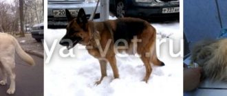
Symptoms of pathology
Neoplasia of the paired organs of the animal’s reproductive system has one negative feature: in the first stages of development, the disease is not recognized. However, if a dog has a swollen mammary gland due to a malignant neoplasm, symptoms will appear very quickly. Nodules and bumps protruding through the skin are felt when touching the abdominal cavity. When pressed, the animal immediately demonstrates that it is in pain.
In addition, areas of the mammary glands swell. The area around the dog's red, swollen nipple is inflamed and purulent discharge appears. And enlarged lymph nodes indicate that metastases are beginning to actively grow. In order to recognize the symptoms in time, you need to examine the bitch weekly during simple and false pregnancy, and feeding puppies. If a benign tumor of the dog's mammary gland has passed all stages and developed into cancer, metastases and other signs of the disease will appear. So, for example, if secondary foci of the pathological process affect the armpits, the animal will develop lameness. And over time, her general condition will worsen.
With a diffuse benign tumor in a dog, all tissues of the external abdominal cavity swell. The fibrous cover becomes inflamed, the mammary glands increase in size. However, a neoplasmic node is not always present.
Malignant neoplasms have an adenocarcinoma, armored and mastitis-like appearance. During the oncological process, leading to the formation of a malignant tumor in epithelial and glandular cells, the skin temperature rises. Neoplasmic formation knows no boundaries, the pet constantly experiences pain, the tumor quickly metastasizes into the skin.
A mastitis-like malignant formation is characterized by a rapid growth rate, a voluminous tumor on the gland, and does not have clear boundaries and contours. When palpated, an accumulation with an admixture of blood and lymph is felt. The temperature of the skin and body rises. With a long process of growth, the neoplasm can open. When making a diagnosis, the doctor excludes an abscess of the paired reproductive organ.
With an armored tumor of the mammary gland, the dog's skin becomes hyperemic and thickens. This pathology is not amenable to therapeutic treatment. To prevent metastases from spreading to neighboring organs, urgent surgery is needed. The pathology is very acute, mainly affecting the area of the last paired organs.
Papillomatosis in dogs: causes and treatment
These growths regularly make beginners faint. They look really unpleasant and make you think of the worst. However, papilloma is just a benign neoplasm from the category of fibroepitheliomas in the form of a small papilla or polyp.
Localization and appearance
Mostly papillomas, which are a soft and loose structure with an abundant blood supply, are observed in dogs in the oral cavity (tongue, cheek gums, palate), as well as on the lips. They can be located separately from each other or crowded. In serious cases, one growth appears first, around which more and more are formed over time.
It is believed that canine papillomatosis is not a terrible disease and does not require close attention. In the overwhelming majority of cases, this is true, and with satisfactory immunity, self-recovery occurs.
But it is still worth understanding that warts make it difficult to chew, they can get between the teeth as a result of which they will be crushed, and in their place an ulcer will form - not far from stomatitis or even worse - to exhaustion due to the inability to eat normally.
The second unpleasant moment in papillomatosis is the possibility of warts degenerating into a malignant tumor, which will inevitably lead to the death of the dog. However, all these consequences are so rare that you don’t even have to think about them.
Where do papillomas come from?
The reasons have not been reliably established. Thus, in the past, many experts argued that neoplasms appear as a result of disruption of nerve conduction to tissues. Others considered them the result of some kind of chronic inflammation, while others even considered warts to be congenital defects.
Today, most veterinarians are inclined to believe that the origin of papillomas is viral, especially since they act
Source
Diagnostics
To make a correct conclusion about the nature of the disease and the pet’s condition, it is important to contact a veterinarian in a timely manner. What to do and how to treat the dog will depend on the type of neoplasm and the stage of development of the inflammatory process. In this case, it is very important to establish the tumor nature of her breast cancer.
The specialist must examine the animal and feel the diseased organ with his fingers. This is necessary in order to accurately determine which areas of the paired reproductive mammary organs are affected. And:
- compare the temperature of healthy and diseased skin areas;
- determine the boundaries and contours of the neoplasm;
- adhesion of the skin to surrounding tissues;
- dilation of lymphatic vessels;
- the presence of necrosis zones.
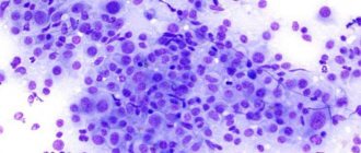
Most often, malignant neoplasm develops on glands that actively secrete milk. To make an accurate diagnosis, the veterinarian will order an x-ray to check for metastases. And also: conduct ultrasound and cytological examination. As an additional examination, the doctor may prescribe a computer or magnetic resonance imaging scan.
The doctor makes the final conclusion after a histological analysis, for which a piece of the tumor itself will be needed. This procedure is performed under general anesthesia.
Treatment
After the results of the studies and confirmation of the symptoms of dog mammary cancer, the veterinarian selects treatment for the animal. Its tactics are selected depending on the stage of the inflammatory process, the growth of neoplasm and the condition of the peripheral organs of the lymphatic system.
Diffuse lesions are considered to have a poor prognosis. Therefore, the main method of treating a dog mammary tumor is excision of mammary neoplasia through surgery. The use of a method of introducing chemicals into the pet’s body to suppress cancer cells is prescribed only in cases where the tumor cannot be removed by surgery. In addition, mammary glands are removed:
- simple;
- regional;
- half with adjacent lymph node;
- unilateral;
- bilateral.
To carry out such operations, the oncologist-surgeon must be proficient in the ablastic technique, using which it is possible to completely disperse tumor cells. Be able to remove tumors without exposing their surface, know the increase in volume and speed of movement of lymph to milk bags. For dogs with terminal malignant neoplasia, the veterinarian prescribes only chemical therapy to prolong the animal's life.
Operation
The most important treatment for lactation neoplasia in dogs is mastectomy. It involves removing the tumor using surgical instruments, a laser beam or freezing. To prevent cancer cells from resuming their “dirty work” after the operation, sometimes the surgeon removes two ridges of nipples at once. Almost any neoplasia requires surgical removal. The only exception is severe inflammation and changes in the epithelial layer of neoplasm cells.
If the neoplasia has opened and is bleeding, immediate surgery will be required to remove it. Suppuration and bleeding of a cancer focus is very dangerous, since tumor cells break out and grow rapidly. Antibiotics are practically powerless in such lesions and are unable to suppress inflammation. This will lead to rapid proliferation of bacilli and intoxication.
In order for the operation to remove the tumor to have a successful outcome, you need to take into account the pet’s age and state of health. Take into account all possible contraindications.
Chemotherapy
The effectiveness of therapy by introducing chemicals into the blood depends on the type of therapy, the goals of the owner and the veterinarian. The technique is most effective when a combination of various means is used simultaneously. And also if surgical intervention is necessary. The effect of such therapy persists in those areas where tumor cells are destroyed. In cases where the animal is diagnosed with the last stage of pathology, chemotherapy will simply temporarily alleviate the dog’s condition. It will also ease the course of the pathology.
The technique involves a surgical operation to remove parts or an entire organ and the impact of ionizing radiation on the site of tissue damage in order to suppress the activity of pathogenic cells. Resection is carried out to neutralize most of the tumor, which cannot be stopped completely. Palliative therapy removes cancer cells from remaining lesions. When removing large tumors, the radiation technique is not effective on its own. Therefore, when treating cancer, several methods are combined, each with its own strengths and weaknesses.
Can it be cured without surgery?
Many dog owners are interested in whether animals with mammary tumors can live without visiting a veterinarian. And how long can they “last” with this disease. The lifespan of a pet depends on the stage of development of oncology. And also on whether the neoplasm is benign or malignant.
If it is not possible to contact a veterinarian, in order to prolong the dog’s life, it needs to be switched to a different diet. Besides:
- Animals with cancer need folic acid. Therefore, it is recommended that they be given 50 g of chicken or beef liver daily;
- If possible, feed your pet only organic meat;
- «black cells hate raspberries. They do not like fruits, vegetables, leaves and herbs. Therefore, if the pet’s diet did not include this assorted plant food, then it is necessary to accustom it to it. You can throw all the greens along with the liver or meat into a blender and puree. The dog will eat the mixed mass with pleasure;
- in addition to ordinary vegetables and herbs, the dog can and should be given medicinal plants such as: burdock, dandelion, yarrow, stinging nettle, St. John's wort, knotweed;
- A variety of nuts are good at fighting cancer cells.
Medicinal herbs and nuts contain not only antioxidants, enzymes and vitamins in sufficient quantities. But minor substances with anticarcinogenic and antitumor properties such as: polyphenols, phytoestrogens, sulfides, flavonoids and carotenoids.
If your pet does not tolerate fruit well due to irritation of the gastric mucosa, you can give some freshly squeezed fruits and vegetables. If the tumor is small, the affected area is insignificant. The initial stage without the formation of secondary lesions, the dog has every chance of beating the disease without surgery or intensive care.
ZooForum: Growths on the tongue. — ZooForum
(others can give advice in a PM, indicating that the advice is not given by a veterinarian. But the forum is not responsible for THESE advice.)
Growths on the tongue. I don’t know what I encountered.
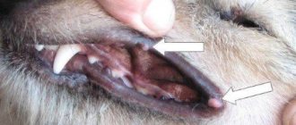
Recently I noticed two white growths on my dog’s tongue. Outwardly they look like two pieces of semolina porridge with a diameter of 5-7 mm stuck to the tongue. They are pure white in color without any additional colors, and there are no changes on the tongue around them. I just can’t imagine what it could be, their appearance worries me.
We recommend reading: Cat Yells After Moving
Take a photo - the description looks like papillomatosis - a mild viral disease that usually goes away on its own without treatment. But it may pass in a couple of months. Treatment is required only if there are a lot of papillomas, they are large and bother the dog.
I have another dog (a 4 month old puppy) and they eat from the same bowl. And if this is the disease that you suspect, and it is viral, then maybe it is worth temporarily limiting their communication?
This makes sense, although complete separation and prevention of contamination is practically impossible. But not all dogs get sick with this virus; it depends on many factors.
Can't take a photo. the growths are located under the tongue (I noticed them quite by accident when the dog pulled out its tongue due to the heat). I open her mouth, but she hides her tongue. I only noticed when I licked my hand that one new growth had appeared (now there are 3 of them). I’ll try to take a photo this weekend, when during the day my tongue sticks out in the sun, to accurately determine the disease.
You said that the appearance of papillomatosis is influenced by various factors. If you can clarify which ones, in order to avoid them later. Could this have occurred due to a tick bite (I removed it 3 times)? I have a “street” dog, so to speak, lives in the yard, constantly gnaws on twigs (plucks bushes), drags around with bones (buries
Source
In most cases, warts in dogs are benign and do not require removal. Removing warts can be stressful for your dog and can also cause new ones to appear in the near future. If you do want to remove your pet's warts, there are several traditional and alternative treatments you can try before going to the vet and paying for expensive procedures.
Make sure that the growth is really a wart.
Warts or sebaceous cysts in dogs are actually benign growths that appear in aging dogs, similar to the appearance of moles in humans. However, you may notice other growths on your dog's body. These may include papillomas, mast cell tumors, fibrous histiocytoma, hair follicle tumors, collagen nevus and fibroids.
If you are unable to determine what kind of growth has appeared on your pet's body, or if you doubt that it is a common wart, consult your veterinarian before proceeding with treatment. The doctor will conduct a microscopic assessment of the cells taken from the tumor and make an accurate diagnosis.
Examine the wart.
Warts are caused by the human papilloma virus and usually appear in puppies or adult dogs with weakened immune systems. These warts have the appearance of an expanding cauliflower. Warts are more common near the lips, nose, or gums.
These warts usually go away after a few months as the immune system strengthens. However, please note that they can be contagious and cause breathing and swallowing difficulties for your pet.
Common benign warts are usually flesh-colored and small in size. They resemble tiny mushrooms.
If you notice growth or inflammation of the benign
Source
If the tumor has opened
If a pathological neoplasm is revealed, then most likely the animal is already in the third or fourth stage of oncology. With them, the spread of “black” cells occurs outside the affected mammary gland. At these stages, simple surgery may not help. Therefore, chemotherapy is also necessary.
If such a problem is detected, the dog must be carefully examined. If there is fur on the affected area, you should try to shave it off. After this, it will be easier to assess the nature of the exposed neoplasm. If the ulcer begins to bleed, then the blood clots, and then flows again with renewed vigor, you should immediately contact the clinic. If a simple ichor comes out of the opened tumor, there is still time to show the dog to the veterinarian. At the same time, whether to treat your dog with such symptoms or not to treat it is an individual choice of the owner.
Gum tumors (epulids) in dogs
Epulids are tumors or tumor-like formations on the gums of an animal. They appear in the form of small formations growing from the gums (as if on stalks). As they grow, they often shift their teeth. Most epulids are attached to bone and do not have a capsule. They have a smooth or slightly rough surface. These tumors do not spread, but can deform the dog's face.
Epulide is the fourth most common tumor in dogs (it is rare in cats). The Boxer is more susceptible to fibromatous tumors than other breeds.
Symptoms and types
There are three types of epulids: fibromatous, ossifying and acanthomatous. Acanthomatous epulids often penetrate the bone and are usually located in the anterior part of the mandible. Sometimes symptoms do not appear outwardly, so it is important to examine your dog's mouth if you suspect he has this condition. Symptoms of epulide include:
Causes
Diagnostics
After you provide your veterinarian with a detailed medical history, he or she will examine your pet's mouth. If the presence of epulide is confirmed, the doctor will perform an X-ray test to determine the type of tumor and assess the health of the teeth. Part of the tumor needs to be separated and sent for laboratory testing. This procedure is best performed under anesthesia.
Treatment
The veterinarian will remove the tumor surgically using anesthesia. Severely damaged teeth will also have to be removed, and the dental cells will need to be cleaned with special tools.
If the tumor is acanthomatous (the most aggressive and sometimes contains precancerous cells), part of the upper or lower jaw may need to be removed. This is followed by radiation therapy to prevent the recurrence of epulide. Sometimes three
The following types of growths and tumors are common in dogs: abscesses, cysts, tumors, hernias, enlargements of certain glands and warts.
Prevention
There is no set of measures aimed at completely eliminating cancer. However, you can monitor your pet’s health and lifestyle. It is necessary to provide your pet with high-quality and balanced nutrition. Make sure your dog goes for walks more often and show it to the vet in a timely manner.
In addition, as a preventive measure, you need to protect your pet from the burning effects of ultraviolet rays. It has been proven that malignant neoplasms most often arise from systematic irradiation and sunburn. As an effective prevention, it is recommended to carry out timely removal of the dog’s reproductive genitals. Studies have shown that sterilized animals are 60% less affected by benign and malignant neoplasms.
The prognosis and outcome of cancer depends on the stage of the neoplasmic process. With timely contact with a veterinary oncologist with the first and second stages of cancer, dogs that have undergone therapy live happily ever after. And most often there is no need for repeated treatment.