Hypoalbuminemia as a cause of ELD
A decrease in albumin levels (hypoalbuminemia) is determined by symptoms such as chronic diarrhea, vomiting, weight loss, abdominal enlargement, and swelling of the extremities. The animal may also suffer from shortness of breath. Once hypoalbunemia is detected in a dog, it is necessary to immediately determine how much protein synthesis is reduced (liver failure) or the extent of protein loss (renal failure).
Loss of proteins can occur through the kidneys, intestinal mucosa, with severe purulent peritonitis, purulent pleurisy, or through the skin as a result of severe mechanical damage (for example, burns).
Standard laboratory tests - urinalysis, complete blood count, bile acid analysis, biochemical blood test - can exclude liver failure or protein-losing nephropathy from the list of causes.
Protein-losing enteropathy or protein enteropathy in dogs
Content
- Symptoms of enteropathy
- Causes
- Diagnostics
- Treatment
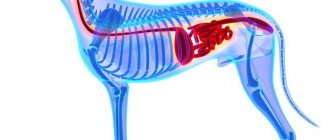
We all know that a lot depends on the health of the intestines and that the body receives all the necessary nutrients by breaking down and absorbing food through the intestinal wall.
If intestinal pathology develops, a disruption in the processes of assimilation of food components occurs, which in turn can lead to a cascade of subsequent undesirable reactions.
Protein-losing enteropathy in dogs is one of the conditions that affects the animal's ability to function fully.
Enteropathy is any abnormal (disturbed) condition of the intestines.
There are a number of diseases that can damage the intestines so much that they cause protein to be lost through the lymphatic vessels.
Normally, nutrients, after being broken down by digestive juices, enzymes and bacterial microflora, are absorbed through the villi of the small intestine and ultimately enter the bloodstream, where they are distributed throughout the animal’s body and converted into various biological substances and types of energy. Metabolic products not required by the body are excreted transiently with feces.
When the process of absorption of nutrients into the blood vascular bed of the intestine occurs, a small amount of protein can leak from the blood vessels back into the intestinal lumen. Typically, these proteins are digested in the intestines and absorbed back into the bloodstream for subsequent use. In the case when there is inflammation and “damage” to the intestines, a large “leakage” of protein into the intestinal lumen through the vessels can occur, which the body can replace/replenish.
This pathology can generally develop in any breed of dog, but some are more predisposed. For example, the Wheaten Terrier, Basenji, Yorkshire Terrier and Norwegian Lundehund are more likely to suffer from this pathology compared to other breeds. Symptoms. Clinical picture
- Episodic diarrhea
- Chronic diarrhea
- Weight loss
- Apathy associated with lack of energy
- Shortness of breath = Difficulty breathing
- Increased abdominal volume
- Edema of the extremities
Causes that can cause protein-losing enteropathy:
- Tumor diseases of the intestine
- Infectious bowel pathology
- Bacterial microflora (for example salmonella)
- Fungal infections of the intestines
- Intestinal parasites (hookworms and whipworms)
- Inflammatory bowel disease described as IBD (inflammatory bowel disease = intestinal bowel disease)
- Food hypersensitivity = allergy to food components
- Ulcerative lesions of the stomach or intestines
- Chronic heart failure
- Lymphangiectasia. Dilatation of intestinal lymphatic vessels, disruption of lymph movement.
Diagnostics
When communicating with your veterinarian, it is very important to talk in detail about the state of your pet’s health and the order in which the symptoms of the disease appear sequentially.
A careful physical (external) examination and standard laboratory tests are required, including a general blood count and biochemical blood tests with electrolytes, as well as a general urine test.
Based on the tests, the level of protein in the blood serum and the level of calcium are assessed. In some cases, it may be necessary to study the concentration of B vitamins in the serum, which can be reduced if the intestinal absorption of these vitamins is impaired.
Stool examinations may also be required to exclude parasitic or infectious intestinal pathologies.
X-rays and ultrasound examinations of the chest and abdominal cavity of an animal allow you to visually assess the internal organs for the presence of internal damage or a tumor process, and will also allow you to assess the condition of the heart and its performance.
If necessary, an endoscopic examination of the stomach and intestines – gastroduodenoscopy/colonoscopy – may be offered.
This study allows you to assess the condition of the gastric and intestinal mucosa for ulcerative lesions, tumor process and assess the condition of the mucous membrane and lymphatic vessels. If abnormalities are detected, a tissue biopsy is taken for histological examination and morphological diagnosis, which can help make a final diagnosis. Treatment
Treatment will depend on the underlying disease that is causing the protein loss. If protein levels are very low, a transfusion may be needed to replace some of the blood protein. Having identified the main diagnosis, it is possible to control the underlying disease and avoid complications.
Condition monitoring. Lifestyle
In most cases, there is no specific cure for intestinal protein loss.
A joint treatment plan is required to help you manage your symptoms. This includes exercise, special dietary therapy to ensure that as many nutrients are absorbed as possible, and medication to treat the underlying condition.
After stabilization, subsequent visits will require monitoring of blood tests to assess the dynamics and determination of serum protein levels and the state of the gastrointestinal tract, as well as assessment for the absence of accumulation of free fluid in the abdominal cavity, which may occur due to protein loss.
Assess your pet's condition. You may need to change the schedule and intensity of your walks depending on your physical needs. Maintain a rest routine.
Health to you and your pets.
The article was prepared by doctors of the therapeutic department "MEDVET" © 2013 SEC "MEDVET"
Diagnostics
If an ultrasound reveals thickening of the intestinal walls, it is necessary to perform a puncture biopsy of the damaged organs or lymph nodes. This method allows us to exclude a neoplasm and give the dog a final diagnosis of EPD. If an abdominal ultrasound shows little or no changes, an endoscopic examination may be indicated. An internal examination of the stomach or intestines reveals ulcers, tumors or other abnormalities in the structure of the walls. In addition, tissue samples (biopsy) can be obtained during gastroscopy.
Recovery prognosis
In most cases, EPB, which develops against the background of intestinal inflammation, is successfully treated. But even with intensive treatment, the condition of animals does not always improve. If the dog does not improve 2 weeks after starting therapy, the prognosis is usually poor. If coagulation is impaired or thrombosis occurs, the dog’s chances of recovery are significantly reduced.
Protein-losing enteropathy in dogs is a dangerous condition that significantly reduces the standard of living. This syndrome occurs more often against the background of various infections and inflammatory processes of the digestive organs. To avoid complications and preserve the health of the animal as much as possible, it is necessary to regularly monitor the state of the gastrointestinal tract and provide proper care for the pet.
Share this article with your friends:
Treatment
Treatment for the dog will directly depend on the cause of the disease. If protein levels are dangerously low, blood or plasma transfusions may be required to correct the deficiency. Albumin preparations can also be used in animals.
Due to low protein levels, the animal must be given a special high-carbohydrate diet with low fat and fiber content. For better digestibility, it is recommended to eat in small portions, but more often.
In the case of chronic manifestations of enteropathy, treatment with corkisteroids (prednisolone) or other immunosuppressive drugs (cyclosporine) is recommended.
Possible complications and consequences of chronic intestinal disease in dogs
Hypocobalaminemia occurs in almost all dogs with enteropathy
Therefore, it must be reflected in the patient’s chart.
Hypocobalaminemia is a prognostic factor for the disease. In some cases, hypocobalaminemia can be extremely severe and contribute to further deterioration of the intestines, since cobalamin is very important for the rapid division of cells such as enterocytes. Therefore, for dogs with intestinal disease, cobalamin support is recommended as long as cobalamin levels in the blood are below normal levels. You can give one injection of cobalamin while waiting for test results (250 - 1500 mcg depending on the dog's weight).
Hypocalcemia is sometimes observed in dogs with canine enteropathy. Decreased ionized calcium can cause seizures, especially in Yorkshire terriers, hence the need for intravenous calcium injection. The simultaneous development of hypomagnesemia is possible due to impaired absorption of magnesium in the intestine and, probably, due to its increased excretion from the intestinal cavity. Dogs with intestinal disease also have decreased vitamin D concentrations, which is likely contributed to by hypocalcemia. Pleural effusions sometimes complicate cases of enteropathy in dogs, so they should always be documented before anesthesia is administered for procedures such as endoscopy and surgery to take biopsies of the intestinal mucosa.
Dogs with intestinal disease may exhibit a hypercoagulable state, which is associated with decreased plasma antithrombin III concentrations as well as increased thrombin-antithrombin complexes and possibly other complex mechanisms.
Thromboembolic complications are reported in 10% of dogs with canine enteropathy. Sudden death associated with pulmonary thromboembolism is a possible fatal complication of chronic intestinal disease in dogs.
Further measures
After the dog recovers, it will be necessary to carry out monthly observations at the veterinary clinic. These examinations make sure that the level of protein in the animal’s body is stable, there is no fluid in the abdominal cavity, and the dog does not have breathing problems. Specialists will also be able to select a special diet. When changing the diet, it is necessary to conduct regular examinations with a veterinarian to avoid relapse.
Practice shows that with timely detection and initiation of treatment for EPD, the prognosis will be favorable.
Symptoms and diagnosis
And now - symptoms and diagnosis. First you need to understand the symptoms of the disease:
- Sick dogs almost always suffer from chronic diarrhea.
- Of course, in all cases there is pronounced exhaustion.
- Retching and vomiting itself. Again, this sign is controversial. About half of the sick dogs have it, while others do not vomit at all. In addition, in many cases it begins to manifest itself only in the terminal stage of the pathology.
- Appetite first worsens and then disappears altogether.
- A characteristic symptom is swelling in the chest, abdomen, and limbs (there is no protein in the blood, its liquid fraction begins to seep through the walls of blood vessels).
- Dropsy of the abdominal cavity (ascites). Cases of dropsy of the chest cavity (hydrothorax) have also been described, but this happens much less frequently.
Note that swelling and various types of dropsy are a direct consequence of a strong drop in oncotic pressure. If a pet suffers from constant diarrhea and vomiting, then the animal (provided that the owners did not contact the veterinarian in time) will certainly develop anemia, accompanied by severe pallor of all visible mucous membranes.
Progressive nephropathy in dogs.
Progressive nephropathy is a constantly worsening disorder of the structure of renal tissue. The disease can be hereditary (due to defective parental genes) or congenital (due to some teratogenic factor that “broke” the normal formation of the organ). As a rule, the first signs of chronic renal failure with this pathology appear in dogs between the ages of 4 months and two years. In addition, it can lead to secondary hyperparathyroidism.