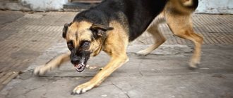
Many owners can argue with this statement, but animal psychologists and veterinarians are confident that animals cannot cry due to excess feelings. If your dog has watery eyes or one eye, discharge or redness appears, this is a reason to pay close attention to the pet’s condition and consult a doctor.
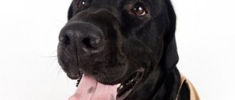
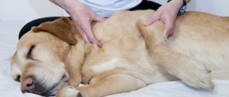
A dog being examined by a veterinarian with a complaint of excessive lacrimation
Excessive lacrimation is one of the most common complaints when visiting a veterinary ophthalmologist. In medicine, the term epiphora is used. It can be one-sided or two-sided, abundant or insignificant, present throughout the dog’s life, for a long period or short-term.
There are many reasons for lacrimation, but they are all divided into two types:
- increased tear production;
- problem of tear drainage.
In addition to clear tears, discharge may appear in the eyes:
- Mucous (transparent or white);
- Purulent (rather thick greenish or yellowish in color).
About eyes and tears
Tears perform important functions: they prevent the conjunctiva and cornea from drying out, freezing the eye, wash away foreign objects and substances (viruses, bacteria, dust, allergens, own epithelial cells, wool), reduce friction, deliver oxygen and nutrients to the cornea.
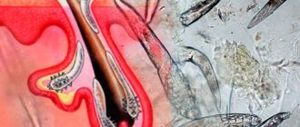
70% of tears are produced by the main lacrimal gland, located behind the upper eyelid, 30% by the additional one, which is located in the thickness of the third eyelid. Through the lacrimal ducts, fluids enter the conjunctiva, mix and spread over the cornea when blinking. Then they flow down and collect near the inner corner of the eye. The lacrimal openings are located here. Through these holes, the tear goes into the nasolacrimal canal, through which it enters the nasal passages and the oral cavity.
Accumulating in excess, tears begin to flow down the back of the nose, forming a tear track, and also moisturize the skin and fur around the eye, creating a favorable environment for the development of viruses, bacteria and fungi.
In light-colored dogs, the tracks take on a red-brown tint due to the porphyrin pigment contained in the tears. In open air, the substance oxidizes, changes color and stains the hair.
Lacrimal drainage system in dogs
When is excessive tearing normal?
If your dog tears a little when yawning, after sleep, in the cold, in the wind, when a hair gets caught, but after the irritating factor disappears everything goes away, then there is nothing to worry about. The eyes almost always water in brachycephalic dogs (Pekingese, pugs, boxers, etc.), in dogs with an abundance of folds on the muzzle (Neapolitan mastiffs, Shar-Peis, etc.), in those with long hair that gets into the eyes (poodles, shivers). tzu, Yorkshire terriers, etc.). These breeds require special care for their eyes. Their tear ducts need to be kept clean and dry. Wipe daily or several times a day. If necessary, use bleaching agents for wool.
Useful video
When carrying out the procedure for washing the lacrimal duct, extreme caution is necessary, since during the procedure, injuries to the nasolacrimal duct are possible. Based on the manipulation, it becomes possible to determine the area of blockage and its degree.
Author's rating
Author of the article
Alexandrova O.M.
Articles written
2029
about the author
Was the article helpful?
Rate the material on a five-point scale!
( 4 ratings, average: 4.25 out of 5)
If you have any questions or want to share your opinion or experience, write a comment below.
Lacrimation due to impaired tear outflow: causes, treatment
This group of pathologies includes cases when the outflow of tears is disrupted when they are produced in a normal volume.
Lacrimal punctal atresia
Hereditary pathology. It is characterized by fusion/narrowing of one or two lacrimal openings, as a result of which full tear drainage becomes impossible. Can be one-sided or two-sided. Infection of the superior lacrimal openings is more common in English bulldogs. Overgrowth of the lower parts is common in American Cocker Spaniels and Hairless Chinese Cresteds. With atresia, lacrimation may increase or decrease, but does not stop.
Narrowing of the tear ducts or nasolacrimal duct
It develops against the background of chronic ophthalmological diseases, clogging them with foreign objects, dust or sand (often found in hunting dogs during active work), allergies, infectious diseases (swelling of the nasal mucosa develops and mechanical compression of the nasolacrimal duct occurs), the presence of neoplasms.
Treatment
Overgrowth of lacrimal openings or canals in the tear drainage system can only be eliminated surgically, using the method of bougienage and rinsing. The operation is performed under general anesthesia. If there are inflammatory processes in the immediate vicinity of the lacrimal system, antimicrobial and anti-inflammatory eye ointments and drops are prescribed before surgery. The course of treatment is continued after surgery to prevent infection.
Diagnosis and treatment
If a small amount of exudate is released during the disease process, conservative treatment remains a priority. It is aimed at restoring the impaired patency of the paths and prompt removal of the contents. For this purpose, rinsing is done through the nasal openings using astringents and disinfectants. At the same time, the bag should be washed through the lacrimal openings. To effectively perform the procedure, solutions of silver nitrate, furatsilin, protargol, boric acid, zinc sulfate, penicillin along with novocaine are used.
If conservative tactics do not help, then they resort to excision of the lacrimal sac, after which the wound heals. There is a danger of obstruction of the outflow tract over time due to the appearance of a postoperative scar, because the wound after excision heals by secondary intention. This operation is not radical, although after a short time the production of tears is reduced. To completely stop lacrimation, removal of the lacrimal gland is required.
Surgery on the lacrimal sac is performed under local anesthesia. The veterinarian makes a skin incision down to the internal ligament directly along the convex component inward from the horseshoe angle of the eye slit. The wall of the sac is then grabbed with tweezers and carefully bluntly removed from nearby tissues. The manipulation is easier if infiltration anesthesia is applied around the circumference of the bag in advance using 0.5% novocaine. The remaining junction points and lacrimal canaliculi are carefully cut off with scissors. All that remains is to apply stitches.
The lacrimal gland is removed while the animal is securely fixed under local anesthesia. Extirpation of the lacrimal gland occurs sequentially by cutting the skin up to 6 cm long in the area of the outer half of the upper-inferior edge of the orbit, fascia, penetration between the aponeurosis of the levator eyelid from above and the edge of the orbit. The wound expands, the edge of the gland is grabbed with wide tweezers and prepared by lightly pulling away from the surrounding orbital tissue. The resulting wound cavity is powdered with Zhitnyuk powder, filled with gauze and the edges of the wound are connected with provisional sutures, several of which are removed on the 2nd day to remove the gauze. Subsequently, treatment occurs using the open method.
Lacrimation associated with excessive tear production: causes, treatment
Much more often, excessive tearing is caused by increased tear production.
Breed characteristics
Often found in dogs with round, wide-open eyes and/or lots of fur around the eyes. Constant irritation of the cornea by dust particles and hairs provokes increased production of tears, which should quickly wash away foreign objects. It is very important for owners to keep these eyes clean and regularly remove excess moisture from the corners and tear ducts with a gauze swab dipped in lotion. And also prevent hair from getting on the cornea.
Entropion and eversion of eyelids
In the first case, the eyelashes of the upper or lower eyelid are adjacent to the cornea and constantly irritate it. Hereditary predisposition is noted in many breeds: Shar Pei, Chow Chow, Alabai, Caucasian Shepherd, Akita, Cane Corso, AST, Bulldog, Pekingese, Spitz, Pug, York, Bull Terrier, Dalmatian, Rottweiler, Husky, Great Dane, Poodle and others. Eversion is the formation of excess space between the lower eyelid and the eye. The eyelid droops downwards or to the side. More common in basset hounds, bulldogs, bull terriers, cocker spaniels, Labrador retrievers, and Shih Tzus. It is very rare that these two pathologies are observed simultaneously in giant breed dogs.
Cunning breeders often sell puppies with such defects, convincing buyers that they are cosmetic. But without surgical intervention, any of them will lead to decreased or complete loss of vision.
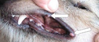
Distichiasis
Pathological eyelashes sometimes grow on a dog’s eyelids, which “lie” on the cornea. The disease is called districhiasis. It is hereditary, the average age of manifestation is 3 years. Spaniels, Bulldogs, Shelties, Shih Tzus, Dachshunds, Poodles, Pekingese and others are predisposed.
There are three treatment methods. Epilation brings only short-term results. Surgical excision of “irregular” hair follicles is dangerous due to the formation of scars, which will further injure the cornea. Cryo-epilation is considered the most gentle option, but is carried out in few clinics.
Districhiasis in a dog. An eyelash growing downward irritates the cornea, causing constant tearing.
Injuries, irritation
Excessive lacrimation that appears suddenly can be the result of injury, dust, a foreign object, or your own fur getting into the eyes. This can often be observed after walking, running through the bushes, on the beach or in tall grass. Watery eyes usually occur in one eye.
Allergy
Watery eyes from one or both eyes can be caused by allergies to food, air freshener, cosmetics, a new bed, cigarette smoke and much, much more. The eyes can be the site of development of an allergic reaction in many systemic immunological disorders: rhinitis, asthma, atopic dermatitis. And also in case of systemic interaction with the toxin when affected by worms or external parasites. The veterinarian will prescribe anti-allergy medications that block the action of histamine, in addition to eye drops.
Infections
If your dog has gassy tears, this may be a symptom of a local or general infection caused by a virus, bacteria, or fungi. For example, epiphora occurs against the background of conjunctivitis and other ophthalmological diseases, acute viral infection (aviary cough, plague, enteritis), food poisoning, pneumonia, etc. Often, lacrimation accompanies otitis media and various diseases of the oral cavity.
Diet
Watery eyes may indicate a lack of nutrients, in particular vitamin A, B1, B6, and lutein. This happens when a dog eats economy-class food or table scraps for a long time. Improving the menu and introducing vitamin complexes can significantly improve the condition of the pet and his eyes.
What to do
First of all, you need to thoroughly wash your hands and carefully examine the dog's eyes. If the cause of lacrimation is a foreign object (fur, dust, lint), it must be carefully washed off with hygienic eye lotion (diamond eyes, Apicenna Dewdrop, Veda and Calendula/Chamomile) or clean boiled water, heated to a temperature of 40°C. If there is inflammation (redness of the conjunctiva), apply anti-inflammatory antibacterial drops. This can be regular albucid (sulfacyl) or chloramphenicol eye drops.
If no visible cause of lacrimation is found, the pet’s eyes should be cleaned and the area wiped dry. Treat with hygienic lotion or apply anti-inflammatory drops to prevent infection.
You also need to analyze the parameters of keeping and feeding dogs. If an animal lives outside in damp conditions and tries to unbalance its immune system, it weakens and various diseases develop, one of the symptoms of which may be lacrimation. The eyes of pets living in an apartment may encounter various irritants, which are mentioned above. And as long as they affect the cornea, treatment of epiphora will be ineffective.
If you suspect a corneal injury, you should immediately contact the clinic. If possible, apply a bandage and do not allow the dog to rub its face with its paws.
Treatment
Therapy is aimed at eliminating the cause of epiphora. In the initial stages of viral or bacterial conjunctivitis, when foreign bodies, liquid or gaseous irritants enter, veterinarians often recommend the drug diamond eyes, ophthalmosan or similar (bactericidal and anti-inflammatory drops with chlorhexidine and succinic acid, used for the prevention and treatment of acute and chronic ophthalmological diseases, hygienic eye treatment for age-related degenerative and dystrophic diseases of the retina and cornea).
In advanced cases, in the presence of a bacterial infection, stronger antibiotic-based agents are used: iris, dekta-2, ciprolet, leopard, chloramphenicol drops, sofradex, gramicidin, tobrex, erythromycin or tetracycline eye ointment.
If lacrimation is a symptom of a disease in other organs or systems, the treatment regimen will include eye drops or ointments that will help prevent the development of serious ophthalmological diseases.
Treatment of eyes in cats - cataracts, conjunctivitis, nasolacrimal duct obstruction
Inflammation of the tear duct in cats is a fairly common phenomenon that can be caused by a number of different pathologies. Some of them are dangerous and can cause serious deterioration in visual function or even cause complete loss of vision. That is why, when there is inflammation of the visual organs, it is important not to self-treat and visit a veterinarian.
How to diagnose nasolacrimal duct obstruction?
To determine the patency of the nasolacrimal duct in animals, a fluorescein test is performed. Before the test, the eye is thoroughly washed with an antibiotic to evacuate purulent and mucous secretions, which may adversely affect the test result.
One or two drops of fluorescein solution are dropped into the eye or a special fluorescein strip is placed into the conjunctival sac. Then the animal’s head drops slightly downward (otherwise fluorescein will not go into the nose simply according to the laws of physics).
After 1-2 minutes, a green color will normally appear from the nasal cavity in dogs, and from the nasal and oral cavities in cats. If the canal is partially obstructed, fluorescein will appear in 5-10 minutes and in small quantities. Complete obstruction is characterized by the absence of green coloration of the nasal cavity.
It should be noted that the fluorescein test is correct only in 70% of animals. In 30% of cases, with complete patency of the nasolacrimal duct, fluorescein may not appear in the nasal cavity.
Keratitis and inflammation
The pathology is characterized by inflammatory processes of the cornea, during which it becomes cloudy.
The disease can be triggered by injuries, conjunctivitis, burns, allergies, as well as autoimmune and infectious diseases. The pathology can affect either one or two eyes at once.
Swelling of the mucous membrane of the eye often occurs, tear tracks are observed, and sometimes there are impurities of pus in the discharge.
Development of iritis
It is an inflammation of the iris of the eyeball. The pathology is caused by bacterial and viral diseases, fungi, and traumatic eye injuries. The following symptoms are characteristic of iritis:
- photophobia;
- constriction of the pupil;
- pet's irritation.
Panophthalmitis
This dangerous disease is often provoked by Pseudomonas aeruginosa.
Total purulent inflammation and melting of all structures and membranes of the eyeball is caused by infection with pneumococcus, staphylococcus, and Pseudomonas aeruginosa.
Pets experience severe pain, which causes them to scream and roll on the floor. The cat becomes restless, aggressive, and reacts painfully to light. The lacrimal gland is inflamed, the eyelids are swollen and hot.
The formation of pus is observed in the anterior chamber of the visual organ, and a rupture of the sclera can often be seen.
Dacryocystitis
This is an inflammatory process in the lacrimal sac, which is mainly diagnosed in a chronic form. In addition to the fact that inflammation and obstruction of the nasolacrimal duct is observed, there are the following symptoms:
- swelling of the conjunctiva;
- redness of the mucous membrane;
- discharge of different consistency and shade.
Blepharitis in cats
A characteristic feature of the disease is the appearance of swelling of the eyelids.
The disease is characterized by inflammation of the edges of the eyelids. Animals experience severe itching of the visual organs, as a result of which they can scratch their eyes and cause injury to both the eyelid and the cornea. In cats, a tear track is noticeable, and the discharge may be purulent in nature. There are also the following symptoms:
- swelling of the eyelids;
- corneal clouding;
- change in iris shade;
- aggressiveness;
- fear of light.
Third eyelid
The visual anatomy of cats is different from that of humans. They have a nictitating membrane that protects against a variety of injuries. The film is a thin layer of light skin that is located near the inner corner of the organ of vision.
If there is prolapse of the nictitating membrane, there is an increased secretion of tears, and sometimes pus may be noticeable. This pathology is caused by conjunctivitis, disruption of the gastrointestinal tract, bacterial damage, and allergic reactions.
Cataract
With this pathology, the lens visually becomes cloudy.
During the development of this disease, clouding of the lens occurs. Older cats are more susceptible to the pathology, however, the disease can also be diagnosed in kittens due to infection. The main symptom is deterioration in visual function. The pet begins to have difficulty navigating in a room that is familiar to it, and bumps into people and objects.
Entropion and injuries
Turning inward or outward occurs. The pathological condition is caused by burns, inflammatory processes, as well as traumatic injuries.
In addition to inflammation of the tear duct, the cat suffers from pain, which occurs due to constant rubbing of internal tissues by hairs. As for eye injuries, they can be both superficial and deep.
Cats that roam outside often get into fights with other animals and get seriously injured. In this case, there is an increased secretion of tears, redness of the mucous membrane of the organs of vision and inflammatory processes.
Epiphora and lacrimation
This symptom can accompany many eye pathologies in an animal. As the disease progresses, cats suffer from constant and uncontrollable production of tears. Tracks are visible on the cheeks, the coat becomes brown.
This condition can be provoked by the presence of foreign objects in the eye, a corneal ulcer, or conjunctivitis. In addition, epiphora in cats can also be caused by improper development of the lacrimal lines.
Often there is a tight fit of the lower eyelid to the eye, and a small size of the lacrimal lake.
How to treat nasolacrimal duct stenosis?
Stenosis of the nasolacrimal duct causes chronic conjunctivitis and ulcerative blepharitis, requiring periodic antimicrobial and anti-inflammatory therapy.
To reduce tear production and dry out affected areas of the skin, ophthalmic antiseptics are primarily used due to their astringent and tanning effects, local antiallergic and anti-inflammatory drugs.
If the stenosis is associated with fusion or blockage of the nasolacrimal canal, then surgical bougienage of the canal with subsequent hygienic treatment of the eyes is possible.
Diagnosis of eye diseases in cats
Diarrhea in a cat: how to treat it at home
If this misfortune has befallen your pet, you should immediately consult a veterinarian. The organ of vision is examined and its general condition is assessed according to three parameters:
- appearance of the eyeball and surrounding tissues;
- general condition: size, shape, presence of injuries;
- state of vision.
After a superficial diagnosis, a study is carried out using special devices. The fundus of the eye is examined, the sensitivity of the microflora to antibiotics is determined, and the pressure inside is measured. A specialist may prescribe a biochemical blood and urine test.
Fundus examination by a veterinarian
Source: https://prokamen.com/zabolevaniya/dakriocistit-u-koshek-lechenie.html
How to properly apply eye drops to a dog
It’s great if you have a helper to hold the dog. Otherwise, you need to sit her in front of you. With one hand, fix your head in a raised position, holding the lower jaw. Pressing the bottle with your thumb and forefinger, drop the number of drops indicated in the instructions onto the visible part of the sclera. In order for the drug to spread better after instillation, you need to close your eye for a few seconds.
Do not use cotton wool to wipe your eyes. Villi from it may remain on the cornea.