Prosthetic eye replacement for dogs and cats. Enucleation
Evisceration of the eye of dogs and cats with installation of an intraocular prosthesis.
In the network of our veterinary clinics "Vasilek" they carry out evisceration of the eyeball
with the installation
of an intraocular prosthesis.
For our veterinary ophthalmologists, this method has become an attractive alternative to enucleation.
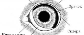
What is evisceration and enucleation of the eyeball in dogs and cats?
Enucleation
eyeball (from the Latin word enucleo - I take out the nucleus, clear the shell) - one of the ways to remove organs. Enucleation is the removal of the eye without removing the surrounding organs - the eye muscles.
Evisceration of the eye
(evisceratio oculi;) surgical operation: removal of the contents of the eyeball while preserving the fibrous membrane - the cornea and sclera.
In recent years, evisceration of the eyeball in dogs and cats has been carried out with the placement of an intraocular silicone prosthesis.
When is evisceration performed and when is enucleation of the eye performed?
Despite the many modern methods of treating eye diseases in dogs and cats, veterinary ophthalmologists from time to time encounter situations where it is not possible to restore the functions of the affected eye, and the only possible method of treatment is evisceration or enucleation of the eye.
Enucleation of the eyeball is an extensive surgical procedure in which the entire eye is removed and the skin is sutured closed. This method requires deep general anesthesia, since the operation is very painful and is often accompanied by blood loss. As a result of the operation, the animal appears externally as if it had a “squinted eye.”
In some cases, it is impossible to perform enucleation of the eye with intraocular prosthetics, but only enucleation can be performed:
- Neoplasms (tumors) of the eye and surrounding structures.
- Extensive corneal damage and low tear production. The affected cornea may be unable to contact the intraocular prosthesis. Also, the affected cornea may not have enough nutrition, since during evisceration there is no aqueous humor that nourishes the cornea from the inside.
- Panophthalmitis (purulent inflammation of all structures of the eye).
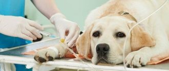
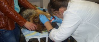
Preparation for the operation
Before surgery, a series of examinations of the dog’s condition are required. The list includes a clinical blood test to determine the rate of blood clotting. Additional examinations of the dog are also carried out if its condition and characteristics of the course of the disease require it.
The area in the eye area where the operation is planned is completely shaved and treated with an antiseptic composition. Retrobulbar administration of an anesthetic drug is carried out. The choice of drugs and doses for pain relief is determined individually depending on the weight, age and other parameters of the dog’s condition.
Since during eye surgery the anesthesiologist has limited access to the dog's head, assessing a number of reflexes seems impossible. Therefore, anesthesiological monitoring necessarily includes measures such as capnography, pulsometry, and ECG.
Ocular evisceration is an attractive alternative to enucleation.
The procedure, like enucleation, is performed on completely diseased eyes. This operation eliminates the need to use local and systemic medications and provides a good cosmetic effect.
Indications for surgery (evisceration):
- The end stage of primary glaucoma, in which the response to drug therapy disappears, the eyeball becomes enlarged and painful, and the visual functions of the eye are lost.
- Severe eye injuries without the development of purulent inflammation.
- Traumatic and endogenous uveitis with vision loss when drug treatment is ineffective.
The surgical technique is evisceration of the eye with placement of an intraocular prosthesis in dogs and cats.
The essence of the method is to remove the contents of the eye through a scleral incision, leaving only the fibrous membrane. A sterile silicone bead is inserted into this corneoscleral membrane and the scleral wound is sutured. The usual result is a painless, mobile and cosmetically good-looking eye that does not require drug therapy. There are no visible seams; the seams are located under the upper eyelid.
What is a prosthetic eye for dogs and cats?
The use of ophthalmic prostheses in animals has been of interest to veterinary scientists since the 19th century.
Previously, veterinarians tried to place prosthetic eyes in the conjunctival space or in the orbit. As a result, we realized that these prostheses require daily care; orbital and conjunctival tissues undergo contracture over time, which leads to the prosthesis being pushed out.
In recent decades, scientists have decided that it is best to use silicone prostheses and place them inside the fibrous membrane of the eye.
These prostheses do not cause pain or discomfort, look good, are non-toxic, do not have antigenic activity, are easy to install, are not expensive and fill the intraocular volume.
Our network of veterinary clinics “Vasilyok” uses prosthetics from the German company Acrivet. They come in different diameters (from 12 to 26 mm) for dogs and cats.
During the preoperative examination, our veterinarian ophthalmologist takes measurements and determines the required diameter of the intraocular prosthesis for a given animal.
How long does the operation - evisceration of the eye with placement of an intraocular prosthesis - take and is postoperative care difficult?
This surgery is performed under general anesthesia, the dog does not experience pain. The duration of the operation is no more than an hour.
After this operation, the animal needs to wear a protective collar for 2-3 weeks, the owner needs to treat the sutures on the skin and instill drops into the eye. Two stitches in the skin are the only visible stitches and are removed after 2 weeks.
At our veterinary clinic “Vasilyok”, ophthalmologists try to save the eyes “to the last.” If it is impossible to save the eye, then our veterinary ophthalmologists, if possible, prefer to perform evisceration of the eye with the installation of an intraocular prosthesis rather than enucleation.
Make an appointment with a veterinarian ophthalmologist
Discounts on veterinary ophthalmologist services
Don't miss the opportunity to take advantage of our seasonal discounts on veterinary services, as well as receive a veterinary discount card PLUS VIP status as a regular customer: Discounts on veterinary services.
Veterinarians will answer your questions: 8 (499) 110-01015
Removal of the eyeball in veterinary practice
Author:
Luzhetsky S. A., veterinary ophthalmologist at the Clinic of Neurology, Traumatology and Intensive Care of Dr. Sotnikov.
Unfortunately, removal of the eyeball
– the most common orbital surgery in animals. This situation arises for many reasons. The main reason is that even the most attentive owner cannot notice in time a developing disease inside the eye of his animal and turns to a specialist, as a rule, if the pet has obvious signs of the disease. By this point, the eyeball may already have irreversible changes. Removal of the eyeball is indicated if its functions are irreversibly lost, if it is painful or poses a threat to the life of the animal. Surgery to remove the eyeball should not be performed in place of diagnostic procedures to make a correct diagnosis. It can only be recommended after a preliminary or final diagnosis has been made.
Examples:
Suspicion of an intraocular neoplasm in an animal. Additional studies include ophthalmoscopy, examination of the anterior segment of the eyeball using a slit lamp, and ultrasound of the eyeball. In the case where the detected neoplasm is inoperable, a preliminary diagnosis is made: inoperable intraocular neoplasm. Removal of the eyeball is recommended. The final diagnosis is made based on histological examination of the material. (Fig. 1, 2).
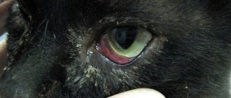
All of the above pathologies lead to the fact that the eyeball becomes a source of suffering for the animal, and the process developing in it can threaten not only the health, but also the life of the animal. We are talking about conditions when the intraocular structures are severely damaged, and visual functions cannot be restored. Once the veterinarian has determined that the eyeball has lost visual function and requires removal, a method for removing the eyeball must be selected.
Purulent discharge after surgery to remove the eyeball in a dog
Hello. Please specify what method was used to remove the eye (evisceration, enucleation, exenteration).
It is worth clarifying that there should not be purulent discharge in any case, but with some types of eye removal, mucous discharge from the palpebral fissure is acceptable.
Only registered users have the ability to start new topics. Register and log in to the site by entering your username and password on the right side of the window, and you can start a new topic.
Before visiting the forum, read the topic: “How to properly consult a veterinarian,” as well as the list of answers to frequently asked questions, this will help you save your time and get an answer to your question faster. Pay special attention to the document: Symptoms of animal diseases. Perhaps in your situation you cannot expect an answer on the forum, but you need to urgently call a doctor or take the animal to a veterinary clinic!
Before joining the forum, read the following sections, this will help save your time and quickly get an answer to your question:
Attention! Pay special attention to the document “Symptoms of Animal Diseases”. Perhaps in your situation you cannot expect an answer on the forum, but you need to urgently call a doctor or take the animal to a veterinary clinic!
Hello. Please specify what method was used to remove the eye (evisceration, enucleation, exenteration).
Enucleation (removal) of the eyeball
Enucleation of the eye is an operation to remove the eyeball. Unfortunately, it is not always possible to save an animal’s eyeball, especially in cases of late contact with an ophthalmologist or in cases of severe eye injury, tumors and complicated cases of eyeball prolapse. The eyeball is removed only when vision is irretrievably lost and when the eye causes suffering to the animal, as well as in cases of risk of involvement of organs and tissues adjacent to the orbit in the pathological process.
The operation is performed under general anesthesia in an operating room and with the necessary equipment. It consists of removing the eyeball, periocular muscles and lacrimal glands and the edges of the eyelids. After that, stitches are placed on the eyelids. Later, the operation site is hidden by fur, and the defect does not seem repulsively ugly.
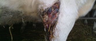
Eyeball loss in a foundling dog
The same dog after enucleation. Drainage for outflow is visible in the corner of the eye
Prices, rub.
| Enucleation of the eyeball of a cat, small breeds of dogs | 8000 |
| Enucleation of the eyeball large breed dogs | 8000 |
| Enucleation of the eyeball medium breeds of dogs | 8000 |
The cat coughs as if he is getting rid of fur (fluffy). Appetite, stool, I don’t know - there are two of them. The hotter it gets, the more frequent the attacks. I don't remember this in winter. Elena
We recommend reading: Kamyshov's Cat
Question: the cat started coughing, what could be causing this?
Hello. Please tell me which specialist I can contact with the following problem. In January, our dog (2.5 years old at that time), a German shepherd, lost his hind legs. An x-ray was taken to check for dysplasia. The first doctor we contacted told us to do a massage and prescribed injections (vitamins and painkillers). In the end, it didn’t get better, but only the paw became swollen. Then a veterinarian friend of mine recommended that we see a neurologist, and criticized this treatment. Unfortunately, we lost about 2 weeks. Next, the neurologist gave us the drug “Propofol Lipuro” and within a week our boy was back on his feet, but lameness and fecal and urinary incontinence remained. Weeks later the injection was repeated, but there was no progress. Then we underwent a CT scan and it was decided to perform an operation with the installation of bone cement and a screw. But the operation did not produce any results. They repeated the CT scan, the doctor did not see what the problem was and recommended that we contact you, because... You have more experience in these matters. CT scans on disks and doctor's notes are in our hands. But we are from another city, and our baby also does not tolerate the road well. Therefore, at least an initial online consultation is very necessary. Anna
Question: My dog’s hind legs are paralyzed, is it possible to get a consultation online?
The operation is performed under general anesthesia in an operating room and with the necessary equipment. It consists of removing the eyeball, periocular muscles and lacrimal glands and the edges of the eyelids. After that, stitches are placed on the eyelids. Later, the operation site is hidden by fur, and the defect does not seem repulsively ugly.
There are several methods for removing the eyeball
Evisceration
– removal of the contents of the eyeball (vitreous body, choroid, retina, lens).
This operation is low-traumatic and does not require special equipment. Its disadvantage is the unsatisfactory cosmetic result in the long postoperative period, the possibility of developing complications (entropion of the eyelids, chronic conjunctivitis). A satisfactory cosmetic result can be achieved using the evisceration method followed by placement of an intraocular prosthesis (IOP). However, the disadvantage of this method is the narrow range of indications for placement of an intraocular prosthesis and the likelihood of developing additional complications. Enucleation
is the removal of the eyeball and part of its supporting apparatus.
Exenteration
- removal of the eyeball and all auxiliary apparatus. Radical surgery used for panophthalmitis with damage to orbital tissue, intraocular neoplasms with invasion of the sclera. The choice of technique depends on each specific case and the condition of the animal. The main task when choosing a technique is to ensure the stable condition of the operated eyeball throughout the rest of the animal’s life. Upon careful consideration of all the options for removing the eyeball, it becomes clear that the most practical of them are two options: placement of an intraocular prosthesis and enucleation of the eyeball with extensive eyelid coverage . Placing an intraocular prosthesis has its pros and cons. The advantages of this operation are obvious - good cosmetic results and relatively low trauma. The disadvantages include the need for careful selection of candidates for placement of an intraocular prosthesis. An intraocular prosthesis should not be placed if an intraocular neoplasm is suspected, panophthalmitis, panuveitis and uveitis against the background of infectious diseases. Enucleation of the eyeball with extensive involvement of the eyelids combines all the requirements that can be presented for surgery to remove the eyeball, and can be transpalpebral and transconjunctival. Transpalpebral enucleation of the eyeball is a “cleaner” operation compared to the transconjunctival technique. The advantage of this method is the removal of the orbicularis eyelid muscle and a significant reduction in skin prominence deep into the orbit in the postoperative period. The downside is significant bleeding caused by damaged eyelid vessels, so in some cases the vessels need ligation. During this operation, the entire eyeball and the entire auxiliary apparatus, including the eyelids, are removed. This ensures a stable result and an extremely low percentage of postoperative complications.
We recommend reading: Drugs for False Pregnancy in Dogs
Preoperative preparation includes:
- Clinical blood test.
- Determination of blood clotting time.
- Additional examination of the animal depending on its condition and the reason for removal of the eyeball.
Preparing for surgery
The surgical field must be completely shaved and antiseptically treated. The conjunctival sac is treated with a 5% Povidone solution. Retrobulbar administration of anesthetic is carried out. Anesthesiological support is provided by an anesthesiologist. Induction: Propofol 4.0 mg/kg with premedication. The use of low doses of opioid drugs (Fentanyl) in induction reduces the risk of coughing.
Anesthesia:
- Propofol 6-12 mg/kg/hour, or Isoflurane.
- Fentanyl 2.5-5 mcg/kg/hour.
- Propofol 6-12 mg/kg/hour, or Isoflurane.
- Fentanyl 1.5-2.5 mcg/kg/hour.
- Ketamine 3-5 mg/kg/hour. Infusion therapy is used in a volume sufficient to relieve hypovolemia and correct electrolyte disturbances using crystalloid solutions (NaCl 0.9%, 5%) and colloid solutions (HES 6% - ny).
Anesthetic monitoring
During ophthalmic surgery, the anesthesiologist has limited access to the patient's head. Accordingly, it becomes impossible to assess some reflexes (corneal), the rate of capillary filling, and the color of the oral mucosa.
In accordance with this, monitoring should include: 1. Pulse oximetry. 2. Capnography. 3. ECG (for diagnosing arrhythmias caused by the oculocardial reflex).