What is phlegmon
Phlegmon (from the Greek “phlegmone” - heat, inflammation) is a purulent inflammation of loose tissue, covering a wide area and having no clear boundaries.
Cellulitis in dogs is a common disease. Sometimes a fight may occur between dogs while walking, or the dog may accidentally injure itself in some other way. A huge amount of pyogenic microflora can get into the wounds, for example, Staphylococcus aureus or Proteus and others. Much attention should be paid to small wounds; they should not be left unchecked; a seemingly small bite often leads to serious consequences. You will ask why? In the area of a small bite, a rather large subfascial cavity is formed, where conditions favorable for putrefactive microflora develop. After a fight between dogs or other injuries, it is better to immediately contact a veterinary clinic to receive first aid. If this is not done or first aid is provided to the dog in the wrong way, then phlegmon often forms at the site of the bite.
Treatment of bronchial asthma in dogs
There is no specialized treatment course to rid an animal of asthma. To normalize the pet’s condition, medications intended for humans are often used.
The veterinarian selects the most effective treatment only after identifying the root cause of the disease - in most cases, its elimination leads to the relief of the disease in question.
The doctor may prescribe medications of various categories depending on the severity of the pet’s condition:
- Antihistamines - help expand the airways and significantly reduce susceptibility to most allergens. Suprastin and Diphenhydramine are considered the most effective.
- Antibiotics – prescribed in case of secondary respiratory tract infection.
- Bronchodilators - lead to relaxation of the bronchial wall. These include Ephedrine and Solutan.
- Inhaled steroids – prevent further development of inflammatory processes. Prednisolone is most often used.
- Oxygen masks – used in case of a severe attack of suffocation.
In case of asthma attacks, an oxygen mask is used to facilitate breathing.
The dosage of drugs is also selected by the veterinarian, taking into account the severity of the condition and the age of the animal. To quickly normalize the dog’s condition, along with effective treatment, the doctor selects a suitable vitamin complex. It helps strengthen the immune system, as well as filling the pet's body with essential nutrients and minerals.
Clinical signs of cellulitis in dogs
Every day the dog begins to feel increasing discomfort in the area of the formed phlegmon. In the shortest possible time, characteristic clinical signs of phlegmon appear, such as refusal to feed, increased temperature of the tissues in the wound area and the whole body. Increased heart rate and breathing. There is a deterioration in general condition. In the area of the wound, skin tension is noted, its borders are smoothed. Subsequently, the swelling becomes limited, and foci of necrosis appear. At the same time, the amount of infiltrate increases - cellular elements accumulated at the site of phlegmon, to which lymph and blood are mixed.
Asthma in dogs
Clinical signs Causes of asthma Diagnosis Treatment of asthma in dogs
Obstructive and allergic lung diseases appear quite often in dogs and are sometimes called “asthma”, “bronchitis” or “bronchial asthma”. Unfortunately, these diseases are not easily classified and represent a wide variety of lung diseases. The only thing they have in common is hypersensitivity of the respiratory tract.
When a dog's airways are sensitive to certain irritants, exposure to these substances causes the airways to narrow. Provoking elements are direct irritants of the respiratory tract or elements that cause an allergic reaction in the respiratory tract. Regardless of the cause, the end result is the same: muscle spasms in the bronchi, accumulation of mucus and accumulation of cellular material in the lumen. In addition, the inability to clear mucus from the bronchi makes the dog susceptible to secondary infections.
The most difficult phase for a dog with asthma is the exhalation phase. Difficulty breathing out is also a typical symptom of obstructive pulmonary disease. Air may become “blocked” in the lungs, causing them to become overfilled. In some cases, this leads to the development of emphysema in the dog.
Clinical signs of asthma in a dog
Obstructive pulmonary disease is most common in dogs between 2 and 8 years of age.
Coughing and strained breathing are the most common signs of obstructive pulmonary disease. The wheezing is easy to hear with a stethoscope, and sometimes it is so loud that it can be heard without it. Sneezing and vomiting are also possible.
Causes
As mentioned above, this group of diseases is characterized by hypersensitive airways. Bronchi and bronchioles can respond to many stimuli, such as:
Treatment of cellulitis in dogs
Treatment of phlegmon in dogs takes a long time and is a very expensive process. There are 6 stages in the development of phlegmon, for each of which there are individual treatment methods.
- The first stage is an increase in local temperature, swelling, development of local edema;
- The second stage – cellular infiltration occurs, a cellular barrier is formed;
- The third stage is the beginning of abscess formation, progression of necrosis;
- The fourth stage - the completion of abscess formation and subsequent maturation of phlegmon;
- Fifth stage – tissue granulation occurs;
- The sixth stage is scarring of the affected areas of the skin.
In the first three stages, treatment is carried out using novocaine blockades with an antibiotic. A course of systemic antibiotics is also prescribed. At stages 3 to 5, drugs are used that support and normalize the activity of the whole organism, as well as agents that promote desensitization and remove necrotic tissue areas. Sufficiently large incisions are made to ensure unimpeded drainage of exudate. Then the wound is washed with various antiseptic solutions, and ointments with antibacterial and healing properties are placed into the cavity. A very important point is that the treatment must be comprehensive!
Therapy
How is phlegmon treated in dogs? First, the affected area is injected with powerful antibiotics, which are pre-dissolved in a 0.25-0.5% solution of warm novocaine. If the dog's condition gives rise to serious concerns, it is advisable to resort to novocaine blockade with the simultaneous prescription of drugs that stimulate and support the functioning of the cardiovascular system.
At the same time, everything is done to counteract intoxication. To do this, buffer compounds, glucose and other drugs that help the liver are introduced. Vitamins can be administered at the same time. When the dog’s condition is sufficiently stabilized and the inflammation is localized, surgery can be performed.
Cellulitis in dogs
Pathologies of the subcutaneous tissue in our four-legged pets are sometimes characterized by severe inflammation of soft tissues, irreversible necrotic processes and severe intoxication, leading to blood poisoning and death of the beloved dog.
One of these insidious diseases is phlegmon in dogs - an acute purulent inflammatory disease of loose tissue, accompanied by irreversible necrotic processes. The causative agent of the pathology is putrefactive and pyogenic microflora or chemical poisons that penetrate the subcutaneous tissue during open injuries, operations in violation of antiseptic rules, or through the hematogenous route from affected organs. Unlike an abscess or boil, the inflammatory focus of phlegmon does not have clearly defined boundaries, due to which healthy tissue is constantly involved in the pathological process.
At the site of penetration of an infection or a chemical substance, a painful swelling of the soft tissues forms; the pathology is accompanied by fever and severe intoxication of the dog. At the site of edema, necrotic foci form, melting of soft tissues and penetration of pathogenic microflora into healthy tissues and the circulatory system, this condition is dangerous due to the formation of multiple abscesses in internal organs, sepsis, vascular thrombosis and death of the animal.
Asthma in dogs
Asthma in dogs is an inflammatory process that affects the small bronchi. The disease is otherwise called chronic bronchitis. Inhalation of air by sick animals is accompanied by severe narrowing of the airways. This leads to the dog's breathing becoming heavier. In some cases, bronchial spasm develops, which is based on suffocation.
Causes of asthma in dogs
This pathology most often occurs in young and middle age. Breed inclination is typical for poodles. Most cases of asthma are triggered by natural allergens. We are talking about dust, parts of plants, wool, cigarette smoke, etc. According to veterinarians, asthma in dogs is often associated with the consumption of commercial food. This is due to the presence in them of a large number of dyes and extracts that act as allergens. It is worth noting that the development of this disease is facilitated by a decrease in immunity as a result of excessive vaccination. An increase in asthma attacks occurs as a result of an infection in the lungs.
Clinical picture of asthma in dogs
As a rule, the manifestation of asthma in animals is a dry cough and breathing accompanied by whistling. In severe cases, suffocation develops. This condition is very dangerous and can lead to the death of the dog. When suffocating, pronounced respiratory efforts of the animal are observed, reminiscent of convulsions. Asthma also often manifests itself as a purplish-bluish discoloration of the gums. This sign is considered an indication for an immediate visit to the veterinarian.
Diagnosis of asthma in dogs
The diagnosis of asthma in dogs is based on data on contact with a possible allergen, characteristic clinical symptoms, and the results of laboratory and instrumental methods. An important step is to identify the allergen that causes asthma attacks. Percussion reveals multiple wheezing sounds. To exclude other pathologies, an X-ray examination of the lungs is performed.
Types of phlegmon
- By localization - subcutaneous, subfascial, mucous, intermuscular, parachondral;
- According to the nature of the exudate - purulent, putrefactive, serous, purulent-hemorrhagic, gas;
- According to the nature of occurrence - primary and secondary.
Primary phlegmons arise as a result of a violation of the integrity of the skin during injuries, dog fights, operations, contact with open soft tissues of chemicals, secondary phlegmons are a complication of acute purulent diseases: abscesses, boils, carbuncles, purulent inflammation of joints and bones, infectious diseases.
The causes of phlegmon formation in dogs are:
- Injuries, wounds, operations with insufficiently sterilized instruments;
- Purulent diseases of the skin, joints and bones;
- Bacterial, fungal and parasitic diseases;
- Unbalanced diet;
- Oncology;
- Lack of vitamins;
- Metabolic disease;
- Age.
The disease is most often observed in elderly or young dogs, weakened pets, when feeding and maintenance conditions are violated, so the immune system cannot cope with pathogenic microflora.
Phlegmon in dogs manifests itself with a clear clinical picture:
- Formation of an extensive dense painful swelling with increased local temperature;
- Lack of appetite, depression, whining, the dog prefers to lie down;
- Increase in general body temperature to 40-41 degrees;
- Shallow breathing.
Causes
The causes of phlegmon formation in dogs are:
- Injuries, wounds, operations with insufficiently sterilized instruments;
- Purulent diseases of the skin, joints and bones;
- Bacterial, fungal and parasitic diseases;
- Unbalanced diet;
- Oncology;
- Lack of vitamins;
- Metabolic disease;
- Age.
The disease is most often observed in elderly or young dogs, weakened pets, when feeding and maintenance conditions are violated, so the immune system cannot cope with pathogenic microflora.
Diagnosis and treatment
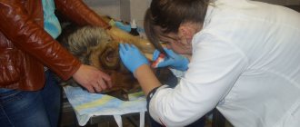
To make a correct diagnosis, the dog owner must inform the veterinarian about recent injuries, fights, surgeries, and long-term illnesses of the pet. The decisive diagnostic value is puncture of the inflamed area with the release of pus.
To determine the cause of the pathology and the general condition of the four-legged patient, laboratory tests of urine and blood tests are prescribed.
Treatment of pathology is aimed at destroying pathogenic microflora and restoring the animal’s body after severe intoxication.
The initial stages of the disease are treated conservatively with the use of warm compresses, novocaine blockades, antibiotic therapy, alcohol dressings with ichthyol and camphor oil.
Necrotic lesions must be surgically removed in a veterinary clinic with complete excision of dead tissue and postoperative wound treatment. To relieve intoxication, cardiovascular drugs, drip infusions of sterile physiological solutions, antibacterial, immunostimulating drugs and vitamins are used.
Prevention
The formation of phlegmon in a dog can be prevented by following preventive measures:
- Open injuries should be immediately washed with a solution of hydrogen peroxide; large injuries should preferably be treated in a veterinary clinic;
- It is recommended to feed your pet a balanced diet with vitamin and mineral supplements;
- It is advisable to immediately treat all infectious and non-infectious diseases of the pet;
- Once a year, it is necessary to carry out preventive examinations of the dog in a veterinary clinic with laboratory testing of urine and blood tests;
- At the first symptoms of the disease, you should immediately consult a specialist.
Cellulitis in dogs is a very serious and deadly pathology; the prognosis of the disease directly depends on the timely contact of the dog owner to specialists.
Symptoms and diagnosis
What are the symptoms of phlegmon in a dog, if observed, you should urgently take your pet to the veterinarian? Here is their main list:
- Hot and reddened swelling in the subcutaneous tissue.
- Decreased appetite.
- Apathy, indifference.
- Reluctance to move, jump, play. Any movement can cause pain, and the animal squeals and whines.
- Upon palpation, a pronounced pain reaction is noted.
- Fever accompanied by a strong increase in overall body temperature.
Be sure to tell the veterinarian about everything that contributed to the development of phlegmon: fights with other animals, surgical interventions, mention that the dog has any other diseases. This data will help the specialist make the correct diagnosis faster and more accurately. Upon examination, the veterinarian should determine whether the inflammation has a clearly defined border. A complete blood count is indicated, which reveals the total number of leukocytes. If there is a suspicion of the appearance of some kind of aggressive neoplasm (sarcoma, for example), a biopsy is necessary with subsequent study of the pathological material.
Cellulitis in dogs
Cellulitis is a serious purulent inflammation, aggravated by necrosis (death) of adipose tissue (including skin). Unlike diseases of this type, in the case of phlegmon, necrotic processes prevail over inflammatory ones. Unlike other similar diseases, such as an abscess, it does not have clear outlines. It usually originates due to the penetration of putrefactive or purulent microorganisms into the tissue. In rare cases, certain chemical exposures can trigger the disease.
Cellulitis can be primary and secondary. In the first case, the causes of the disease are wounds or closed injuries. Carbuncles, abscesses and boils, purulent arthritis, pulpitis and osteomyelitis become provoking factors that contribute to the occurrence of secondary phlegmon.
Depending on the location of the spread of the disease, phlegmon occurs:
- parachondral;
- subfascial;
- subcutaneous;
- mucous;
- intermuscular.
Based on the type of fluid released from the tissue or cavity, it can be classified as:
- purulent or purulent-hemorrhagic;
- gas;
- putrefactive;
- serous.
The course of the disease can be divided into six stages:
- at the very beginning of the disease, inflammatory swelling appears on the dog’s body;
- a cell barrier is formed;
- necrotic formations form, the process of abscess formation begins;
- the final maturation of phlegmon occurs, all necrotic processes are completed;
- granulation occurs;
- the scarring process begins.
Abscesses and cellulitis in animals
Tracheal collapse is characterized by C-shaped flattened cartilaginous tracheal rings and the development of a loose, redundant dorsal tracheal membrane, followed by tracheal narrowing and obstruction.A chronic progressive disease that occurs predominantly in small breed dogs (Yorkshire terrier, dwarf Spitz, toy terrier, chihuahua, etc.). The congenital form can appear in young dogs. It is believed that congenital tracheal collapse is associated with a decrease in glycoproteins and glycosaminoglycans in the cartilage of the tracheal rings. These and other changes lead
to a decrease in the rigidity of the tracheal rings - during breathing they collapse or one of the walls of the trachea sags into its lumen. In addition to the fact that the trachea loses its shape, changes also occur inside it. The collapse of the trachea leads to its chronic inflammation, because of this, changes occur in the epithelium - the special protective membrane lining the trachea; the number of ciliated cells decreases (they are responsible for cleansing the trachea), and the production of thick mucus increases. These changes contribute to worsening breathing and increased coughing.
Compression may be present for years without symptoms and discovered incidentally on an x-ray or endoscopy. Thus, the symptoms of this disease can appear at any time. Various contributing factors, such as tracheobronchitis, increased respiratory resistance in the upper respiratory tract, obesity and mitral valve regurgitation, favor the manifestation of symptoms.
Symptoms
Paroxysmal, dry cough, which is easily caused by excitement, pulling on a leash or palpation of the trachea. In severe cases, stridor (wheezing) is observed between coughing attacks, as well as shortness of breath, cyanosis, and sometimes attacks of suffocation and collapse. With cervical collapse of the trachea, predominantly inspiratory dyspnea occurs; when there is collapse inside the chest due to increased transtracheobronchial pressure during exhalation, predominantly expiratory shortness of breath occurs; sometimes an additional pop is heard from the collision of the tracheal walls with each other.
Diagnosis
diagnosed on the basis of x-rays and tracheoscopy. X-rays are taken in the lateral projection in the desired phase, in case of cervical collapse - in the expiratory phase, in case of intrathoracic collapse - in the inhalation phase.
Tracheoscopy is an endoscopic examination, which involves inserting a special device into the trachea of an animal under anesthesia - an endoscope. It allows you to examine the trachea from the inside along its entire length, assess the condition of its mucous membrane, and evaluate the movement of the trachea during the respiratory cycle. Tracheoscopy is considered the “gold standard” and is used to confirm the diagnosis. Tracheoscopy reveals a flattened tracheal lumen and sagging of the dorsal tracheal membrane.
As the lumen narrows, 4 degrees of tracheal collapse are distinguished:
Membrane offset 25%
| Membrane offset 50% |
| Membrane offset 75% |
| Complete closure of the tracheal lumen |
In the video, the tracheoscopy process shows the collapse of the animal’s trachea
Tracheal collapse is treated with therapeutic methods, surgical correction, and also with the help of intra- and extra-intracavitary prostheses (depending on the degree of collapse). Therapeutic treatment in conjunction with a weight loss program is generally used to treat mild cases of collapse. Antitussives, bronchodilators, broad-spectrum antibiotics are used (if tracheobronchitis is suspected), secretolytic and expectorant drugs are sometimes indicated.
Surgical treatments described include folding of the dorsal tracheal membrane, dissection of the tracheal annular cartilage, and intraluminal/extraluminal stabilization with polypropylene rings or nickel-titanium mesh. All surgical techniques are used in cases where therapeutic treatment is ineffective or in the presence of a large degree of collapse.
Despite the known success of external luminal stabilization, this method requires open surgery. Although an external luminal prosthesis can be applied to cervical tracheal collapse, it cannot be applied to intrathoracic collapse, which typically occurs in dogs. Recent studies have found that intraluminal stents have advantages over external luminal stents.
Intraluminal tracheal restoration using a self-expanding nitinol stent (an alloy of titanium and nickel) has recently begun to be used. This prosthesis was originally designed to protect the tracheal cavity from tumors compressing the trachea or to dilate damaged arteries and bile ducts in humans. Because nitinol is flexible and resilient and has physical properties similar to tracheal cartilage, most self-expanding stents are made from this alloy. After deployment of the stent in the tracheal cavity, it gradually adapts to the size of the tracheal cavity. The advantages of this procedure over surgical stabilization are that it is atraumatic, does not require intensive care after implantation and takes only 5 to 10 minutes, depending on practical skill.
X-ray before stent placement
X-ray after stent placement
Stent in the trachea after placement
Clinical signs of cellulitis in dogs
- At the initial stage of the disease, dense and warm swelling forms. Touching this swelling causes pain.
- The dermis at the site of the edema is tense, and you can also notice that the temperature around it is much higher than in other parts of the body.
- During illness, an animal becomes lethargic, apathetic and much less active.
- The pet's breathing and heart rate increase greatly.
- There is a complete or partial loss of appetite.
- In later stages of the disease, necrotic foci appear around the edema.
During the illness, general intoxication of the dog’s body occurs; the number of neutrophils in the blood increases, which indicates increasing leukocytosis.
It is impossible to accurately diagnose phlegmon at home, since differentiation of this disease requires a puncture, which is only possible in a veterinary clinic.
Diagnosis of bronchial asthma
The disease is difficult to diagnose because attacks can occur at any unforeseen time, so the pet may appear completely healthy upon arrival at the veterinary clinic.
To determine as accurately as possible the cause of the pet’s deterioration in well-being, the doctor conducts a number of tests:
- The initial collection of the necessary data helps to determine the severity of the pet’s condition and identify the prerequisites for the disease. Includes a general and chemical blood test, as well as urine and stool tests.
- Heartworm detection test.
- Chest X-ray – allows you to see changes in the lungs.
- Cytology, tracheal wash and respiratory lavage - identifies harmful microorganisms, fungal infections or helminths.
- Tracheoscopy and bronchoscopy - helps to study the condition from the inside by introducing a microscopic camera.
- Auscultation – helps to identify the location of the disease, allows you to listen to the work of the lungs.
- Ultrasound of the heart - diagnoses the presence of heart problems that can lead to asthma.
If bronchial asthma is suspected, the veterinarian will conduct a full examination of the dog.
Important. A complete diagnosis does not always help to identify the root cause of asthma, but the presence of properly selected medical care allows the dog to lead a full and long life.
Treatment of cellulitis in dogs
Treatment of phlegmon largely depends on what stage of the disease the animal is at.
If the disease has not had time to develop significantly (stages 1-3), warming compresses, mainly alcohol, are used. Tissue cleansing is carried out at the site of swelling. After this, the site of inflammation is lubricated with ichthyol. Antibiotics such as penicillin and streptomycin are prescribed intramuscularly to the dog. If the disease area is severely swollen, small incisions 1-1.5 cm long are made to remove interstitial fluid and reduce interstitial pressure, which are staggered at a distance of 2-3 cm from each other. The incised area is covered with a bandage containing a hypertonic solution of medium salts, ethacridine and furatsilin.
It is important to remember that one of the most important conditions for a pet’s recovery is to provide it with complete rest.
If the disease is at a late stage, the phlegmon must be opened through surgery. It allows you to open all necrotic formations and allows the infiltrate to come out. In this case, the operation is performed under complete anesthesia or immobilization of the dog. In this case, large incisions are made to ensure free flow of fluid from the tissue. The wound is powdered with various antiseptic powders; an emulsion of synthomycin and applications of Vishnevsky's liniment can be applied. In addition to opening the phlegmon in the later stages, calcium chloride and a course of antibiotics are prescribed.
To eliminate intoxication, the animal is usually prescribed intravenous glucose, and pyrogenal is used to quickly heal wounds, resolve scar tissue and form elastic scars.
No matter what stage of the disease the dog is at, if you suspect phlegmon, you should not self-medicate, but take it to the veterinarian as soon as possible.
Asthma in dogs
1 Thursday, April 5, 2012 19:16:30
- Dear Administrator:)
- From: Russia
- Registered: Sunday, February 13, 2011
- Invitations. 0
- Posts: 16193
- Respect. [+387/-0]
- Positive. [+720/-0]
- Female gender
- Awards: activnost,god,pozitiv,yvajenie,pobeditel,vip,smex,pyt,sto,dvesti,trista,pyat,sem,vosem,vipp,vipp,viip
- Cat breed: Domestic
- Spent on the forum: 1 month 27 days
- Last visit: Tuesday, March 29, 2021 21:02:49
The cause of asthma is
an allergic reaction of the body to any irritant substance (allergen). Symptoms of this disease include coughing and wheezing. Often the allergen is plant pollen.
Asthma
is a very serious disease in dogs, also known as chronic bronchitis. This chronic condition is characterized by inflammation of the small bronchi. When a pet inhales air, its airways become very narrow, making the dog's breathing difficult. In severe cases of asthma, these pathways become blocked by mucus and the smooth muscles that surround them, restricting breathing (bronchial spasm).
Dogs of all ages can suffer from asthma, but it is more common in young and middle-aged pets. Poodles are especially prone to asthma. Veterinarians are confident that commercial pet food (often filled with dyes and extracts), together with excessive vaccination, harms the immune system of animals and thereby causes allergies. Most asthma attacks are triggered by natural allergens.
Bronchial asthma in dogs is characterized by prolonged attacks of suffocation. The most common cause of occurrence is. The disease can occur in dogs regardless of age, but most often occurs in young pets and in older dogs, between the ages of two and seven years. The Maltese are the most susceptible to asthma.
Seizures often occur as a result of exposure to natural allergens or chemicals that are included in many specialized dog foods. The main distinguishing signs are the presence of cough and strained breathing, in some cases there is vomiting, sneezing and pronounced wheezing.
With bronchial asthma, a dog has difficulty breathing and severe wheezing.
The main danger of the disease in question is that the inability to independently clear the bronchi of accumulated mucus makes the pet vulnerable to the development of secondary infections.
The most common causes of asthma, in addition to chronic bronchitis, are:
- a sharp change in usual conditions - humidity, increased or decreased room temperature;
- excessive physical activity disrupts the regular breathing rhythm and causes an attack of suffocation;
- severe stressful situation - leads to rapid heartbeat, which can cause shortness of breath;
- damage to the mucous membrane of the respiratory tract as a result of inhalation of smoke, dust or chemicals;
- allergens of non-infectious and infectious nature.
In rare cases, the disease in question is transmitted hereditarily. In this case, the animal experiences swelling of the respiratory tract and uncontrolled mucus formation, which leads to the appearance of general symptoms.
Bronchial asthma can develop due to excessive exercise or allergies.