Breathing disorders are one of the most dangerous complications. A prolonged lack of oxygen can be fatal, so it is very important not to miss the first signs of illness. Similar pathologies include collapse of the trachea in dogs, accompanied by deformation of the rings of the respiratory tube.
What is tracheal collapse
To understand the real danger, it is necessary to understand the structure of the trachea. It is located at the intersection of the larynx and bronchi and performs 2 functions: conductive and protective.
Anatomical features
The breathing tube is hollow inside, and outside it is surrounded by rigid half rings that form its frame. Their open side faces the spine, where it is covered by a special membrane. It is very mobile and consists of muscle fibers and connective tissue. It makes the rings look complete.
Such a well-thought-out structure does not restrict the mobility of the neck and facilitates the passage of food and air currents. The flexible organ constantly contracts and expands, regulating inhalation and exhalation during the breathing process.
No less interesting is what is inside. The walls of the hollow tube are covered with ciliated epithelium, containing a huge number of special cilia. It is responsible for the production of mucus, which traps harmful microorganisms. Bacteria stuck in it are coughed up along with sputum or spat out with saliva.
Collapse and what it can be confused with
As a result of tissue softening, the tracheal half-rings are flattened and deformed. The membrane covering them sags into the tube, narrowing the existing lumen. The walls of the deformed organ begin to come into contact with each other, irritating the nerve endings.
In response to these changes, the body launches an inflammatory reaction, accompanied by a debilitating cough. In this way, he tries to clear the clogged passage, as the epithelial cells cease to cope with their work. Gradually they are replaced by fibrous tissue.
Due to lack of oxygen, internal pressure also changes. It increases in the pulmonary artery, causing right ventricular failure. Without timely medical care, the animal may die not only due to hypoxia, but also due to cardiac arrest.
The symptoms of the pathology are similar to most respiratory diseases. For this reason, it is necessary to be confident in the diagnosis and not self-medicate before contacting a veterinarian.
Main causes of attacks
The wear and tear of cartilage tissue that causes tracheal collapse in dogs occurs due to a genetic predisposition or as a complication of a number of diseases. In the first case, the disorder is often diagnosed 6 months after birth.
Adverse factors and health problems
Tissue deformation develops against the background of acute glucosamine deficiency. This substance is part of the fluid that lubricates joints. Its deficiency results in faster wear of the articular surfaces and loss of shock-absorbing function.
Unfavorable factors that accelerate the process of compression of the tracheal tube include:
- excess weight;
- frequent colds;
- allergic reactions and regular inhalation of aggressive substances that irritate the nasopharyngeal mucosa (household chemicals, tobacco smoke, perfume);
- indoor air is too dry;
- prolonged stress or excessive emotionality;
- mechanical damage to the neck;
- reverse sneezing syndrome;
- an incorrectly selected collar that compresses the trachea.
The secondary type of pathology is diagnosed in the presence of cardiovascular diseases, extensive neoplasms, respiratory infections and inflammation of the respiratory system. In these cases, the main treatment is aimed at eliminating the underlying cause.
Breed predisposition
The risk group includes decorative dog breeds that suffer from a lack of physical activity. Miniature Poodles, Chihuahuas, Pomeranians, Pekingese and Yorkies spend most of their time on the couch. If you don’t monitor their weight, it will go up very quickly.
But the mentioned factor is not fundamental. It only exacerbates a problem that has been present since birth. The fact is that all of the listed breeds have much softer tracheal rings than those of shepherd dogs, Labradors and other large dogs. Signs of a congenital anomaly worsen with age, reaching their peak by 6-7 years.
What is this
This is a chronic disease, as a result of which the respiratory passage is deformed, the lumen of the tracheal tube narrows due to flattening of the C-shaped cartilaginous rings.
Wear of cartilage tissue is caused by various reasons. The prerequisites for the occurrence of the pathology have not yet been precisely established, but experts note that tracheal collapse occurs both independently and against the background of other pathologies occurring in the body.
The primary form is usually caused by genetics and can be diagnosed at an early age. Manifests itself in the form of initially soft cartilage. The secondary form is observed in dogs that suffer from diseases of the respiratory or cardiovascular systems.
Take the Attention Test! Find 10 differences! (click right here!)
Find the answer Are you bothered by some problem or question? Enter “Breed” or “Name of the problem” into the form, press Enter and you will find out everything about the issue that interests you.
There are other causes of pathology. According to doctors, allergic reactions, regular presence of the pet in a room with polluted air, and excess weight can lead to it.
This leads to a deficiency of glycosamines and glycoproteins, due to which the cartilaginous rings are shortened and deformed, as a result of which the respiratory lumen narrows and the tracheal membrane becomes mobile.
Symptoms and manifestations
When the trachea collapses, the dog develops a chronic cough. This is the main symptom, the severity of which can be used to judge the advanced stage of the disease.
Other symptoms include the following:
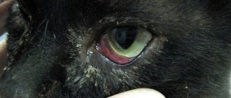
- cyanosis of the mucous membranes, paw pads and ears on the inside;
- gurgling wheezing and shortness of breath;
- fatigue and lethargy;
- attacks of suffocation and short-term loss of consciousness.
Breathing problems can occur when you inhale and exhale simultaneously, or when you inhale air exclusively. The first option is noted when the thoracic vertebrae are affected, and the second option when the cervical vertebrae are affected.
The latent period of the disease, accompanied by rare coughing, often drags on, preventing timely diagnosis. Despite this, distinguishing pathology from a common cold is not so difficult. When the tracheal rings are damaged, most attacks occur during physical exertion, accidental pulling on the leash, and excessive expression of emotions at the sight of their beloved owner.
Diagnostics :
Palpation assessment of the shape of the dog's trachea in the cervical region. This method allows you to detect slight thickening or the formation of a depression in the trachea at the border of the transition to the thoracic region. Even the absence of clinical manifestations indicates the risk of developing tracheal collapse.- X-ray examination. Radiographs are obtained in standard projections. Sometimes the veterinarian will order a ventrodorsal radiograph during inhalation and exhalation. This radiograph is more informative for collapse in the cervical trachea. With collapse, the thoracic trachea may be dilated and the cervical trachea may be distended. X-ray examination is advisable for collapse of the main bronchi, thoracic trachea, and combined collapse. A coughing fit during the examination can improve accuracy. However, this diagnostic method does not always allow the diagnosis of respiratory diseases to be correctly interpreted; according to statistics, only about 60% of collapses can be determined using x-rays.
- Ultrasonography. Using ultrasound, the lumen of the cervical trachea is established, and its work is monitored during the respiratory act. Such a study is prescribed when fluoroscopy is not possible. However, ultrasound does not detect inflammatory processes in the trachea and is only applicable for cervical spine collapse.
- Bronchoscopy. The examinations are carried out under general anesthesia. This method is used to study fluid samples from the lower respiratory tract. The results will allow us to evaluate the contribution of inflammatory processes to the overall symptoms and select the correct treatment.
- Ultrasound of the heart. Animals that suffer from tracheal collapse have increased pressure in the pulmonary circle, which is a consequence of the disease. In connection with this, it is necessary to establish the cause of changes in the heart, whether they are primary or secondary.
- Endoscopy. This method is considered the best for assessing the condition of the trachea and bronchi. Its disadvantage is that it is performed only under general anesthesia. Allows you to trace the trachea along its entire length, conduct a full assessment of the condition of its mucous membranes, record the work of the trachea throughout the full respiratory cycle, and assess the degree of collapse.
- Laboratory research. Complete blood count, biochemical parameters, urinalysis - all these studies are necessary to diagnose possible concomitant diseases.
Severity of the problem
The method of treatment depends not only on the cause, but also on the severity of the disease. Depending on the nature and scale of the deformation, there are 4 degrees of severity:
- The tracheal lumen is narrowed within 25%
. The sagging of the membrane at this stage is insignificant, so the rings operate as usual without losing their functionality.
- The lumen narrows by half
. The membrane membrane sags heavily, and the cartilage becomes denser. There is a partial loss of function.
- The percentage of narrowing increases to 75%
. The sagging membrane almost touches the rings, which take the shape of an ellipse.
- The lumen narrows to 95-100%
completely blocking breathing. The membrane shell fits closely to the rings. At this stage, suffocation occurs even with complete rest and can result in the death of the animal.
It is important to note that the recommended rate of full tube dilatation is not typical for Yorkies. The tracheal cartilages of these dogs have an ellipsoidal shape from birth.
How is diagnostics carried out?
It is possible to understand how to treat tracheal collapse in a dog only after a complete diagnosis. To do this, you will have to make an appointment at the veterinary clinic.
The condition of the affected organ is examined using:
- palpation, revealing noticeable deformation of the walls of the tracheal tube;
- X-ray, which determines the size of the lumen and the presence of foreign bodies;
- fluoroscopy, which allows you to observe a specific area in dynamics;
- endoscopy, necessary to take mucus samples and assess the actual extent of the lesion;
- Ultrasound to check for tumors and associated diseases;
- clinical and biochemical blood test confirming pathological deviations in basic parameters.
X-rays and endoscopy are performed under sedation, since the dog’s movement may interfere with taking a picture or taking a piece of biomaterial. For other studies, the administration of anesthetic is not practiced.
Fluoroscopy is considered the most effective diagnostic method. Despite the high radiation dose to everyone present, the data obtained turns out to be 100% correct. This avoids the false positive and false negative results that are common with x-rays.
How to treat tracheal collapse in small dogs?
We have found out what tracheal collapse in dogs is, now we need to figure out how to treat it. How to help your four-legged friend during a paroxysmal cough, how to relieve it.
Removing respiratory irritants from the air
A simple treatment for mild tracheal collapse is to remove all respiratory irritants from your dog's environment. Smoke or tobacco smoke of all types must be eliminated. Some irritants can be carried into your pet's environment on clothing and hair, such as pollen and dust.
Weight loss
Obesity can cause your dog's body to work too hard to perform simple tasks. Reducing his weight will help him breathe easier; however, strenuous exercise may cause coughing fits. If your dog has a collapsed trachea, keeping him at an ideal weight or even a low weight can be beneficial for his health.
Drug therapy
Mild tracheal collapse can be managed with cough suppressants or medications to reduce the occurrence of cramps. They can also be treated with medications that widen the airways. Glucocorticosteroids, such as prednisolone, are used to stimulate breathing, bronchodilators, such as terbutaline, are used to relieve cough, and expectorants, such as mucaltin, are also used.
However, some of these drugs are controversial because they dilate the airways rather than the trachea itself. Antibiotics may also be used if tracheal collapse is caused by or associated with infection. Finally, some dogs may require a sedative to reduce the anxiety associated with coughing.
Surgery
According to veterinary surgeons, up to 70 percent of all dogs with tracheal collapse can be treated without surgery. Surgery can be performed in either the neck or chest area. When surgery is performed in the chest, treatment usually involves placing a spring device inside the trachea. This device, called a stent, holds the windpipe open. Surgery performed in the neck area usually involves replacing decaying cartilage with C-shaped plastic rings. Even when stents are placed, many dogs still have surgery to replace the ring in their neck.
How to help your pet
Emergency assistance from the owner is permissible only in case of a severe attack. In all other cases, the animal must be treated by a veterinarian.
Conservative therapy is effective for disease of 1st and 2nd severity. Four-legged patients with a more severe diagnosis are required to undergo mandatory surgery.
First aid for an attack
First aid is aimed only at temporarily improving the condition, so the victim will still have to be shown to a veterinarian. If you experience an attack of suffocation, follow these steps:
- Calm your pet with an approving voice and strokes. Try to remain calm, as your excitement will certainly be transmitted to the patient.
- Try to eliminate the provoking factor. To do this, you need to remove the collar and ventilate the room if you are not outside.
- Wet your dog's head with cool water and apply a cold compress wrapped in a thick cloth to his chest.
- Contact your veterinary clinic by phone to receive personalized advice from your veterinarian.
An oxygen cylinder, which can be purchased at a regular pharmacy, will help normalize breathing. Codeine, which blocks the cough center, also copes well with the problem. If you don’t have any of these remedies on hand, don’t waste precious time looking for them and go to the veterinary clinic.
Drug therapy
To stimulate respiratory function, glucocorticosteroids (Prednisolone) are used, and to suppress spasm and cough - bronchodilators (Albuterol, Terbutaline, Theophylline), antitussives (Butamirate) and expectorants (Mukaltin, Bronchipret) drugs. The list of other medications depends on the cause of the disease. It may include:
- cardiotonics and vasopressors that lower blood pressure and normalize the force of heart contraction;
- antibiotics (Baytril, Amoxicillin, Tsiprovet) and antiviral (Anandin, Neotime, Fosprenil), eliminating the causative agent of infection;
- anti-inflammatory, dulling the body's response to irritation;
- immunomodulators that restore defenses;
- sedatives that have a calming effect.
Thanks to drug therapy, stabilization of the condition is observed in 71-93% of four-legged patients, depending on the severity of their disease. In other cases, surgical intervention is resorted to.
If a large amount of mucus is detected, the respiratory tract is sanitized. The frequency of procedures depends on the rate of sputum formation and the characteristics of the cough, since when it is dry, the natural discharge of pathological secretions is difficult.
Surgical intervention
The operation carries many risks, so it is used only when the threat to life outweighs the possible complications. Its main task is to correct the narrowed lumen using prosthetics. To do this, use one of two methods:
- Installation of external implants
, replacing damaged areas. Suitable exclusively for pathologies of the cervical spine and is often accompanied by rejection of the material.
- Installation of a special metal stent
, performing a frame function, from the inside. It is used for dysfunction of both the cervical and thoracic vertebrae, but does not solve the problem of constant coughing and copious sputum discharge.
In addition to possible rejection, the disadvantages of implants include the high likelihood of damage to the laryngeal nerves during surgery, which can lead to paralysis of the larynx. For this reason, many owners agree to stenting, which is attractive not only due to lower risks, but also due to more gentle anesthesia.
The persistence of chronic cough during such an operation is explained by the atrophy of the cilia on the mucous membrane, which are forced to constantly contact the tube inserted into the lumen. Despite this, stenting does make breathing easier, so with stable use of symptomatic medications, the patient’s condition is successfully stabilized.
Treatment scheme for tracheitis in the clinic
Severe disease of the respiratory system in dogs is carried out in a hospital setting. In the absence of the ability to breathe independently, the animal is intubated - insertion of a tube, or a tracheostomy - an opening in the trachea. To relieve intoxication, drip infusion of electrolytic solutions is used.
Treatment regimen for tracheitis in dogs in a veterinary clinic:
- eliminating the cause of the disease with the help of antibacterial, antiviral, antifungal agents if the disease is infectious in nature;
- symptomatic therapy: use of expectorants, antitussives, antipyretics, antiallergic drugs;
- stimulating the body's defenses by prescribing vitamins and immunostimulants.
Important! The prescription of medications is carried out after a bacteriological examination of sputum to determine the sensitivity of the pathogen to the drugs.
Are there any complications?
As a result of deformation of the tracheal tube, not only the organs of the respiratory system, but also the heart and brain are affected. Without timely treatment, the disease provokes the development of the following disorders:
- pulmonary hypertension, characterized by an increase in blood pressure due to narrowing of blood vessels;
- right ventricular heart failure, which can lead to death due to gradual heart failure;
- death of brain neurons suffering from prolonged lack of oxygen.
Long-term use of antibiotics can also cause complications. These drugs destroy not only pathogenic microorganisms, but also their own immune cells. As a result of such therapy, the existing disease is often complicated by a secondary infection.