Anatomy of the prostate gland
The prostate gland (prostate) is an accessory sex gland in males, performing a secretory function; it is the only accessory sex gland in males.
The prostate surrounds the proximal urethra at the bladder neck, and its ducts flow into the urethra in a circumferential manner. On the dorsal surface it is divided into two lobes with a septum in the middle. The prostate gland does not contain microorganisms. The function of the prostate gland is to produce secretion, which is a supporting and transporting medium for sperm during ejaculation.
Also, seminal fluid mechanically dilutes the sperm produced by the testes, increases the volume of ejaculate, and promotes the movement of viable sperm outside the male body. Basal secretion of small amounts of secretion leads to its constant entry into the excretory ducts and the prostatic part of the urethra.
The hormone testosterone is required to maintain the size and growth of the prostate gland. When a male dog is castrated before puberty, normal prostate growth is suppressed. When a male dog is castrated as an adult, the gland involutes up to 20% of its normal size in an adult animal.
Prevention of prostatitis in dogs
Effective methods for preventing prostatitis in dogs are:
- castration of males not participating in breeding;
- timely treatment of infectious diseases of the genitourinary system;
- Regularly walking your pet to empty its bladder and bowels.
It is also necessary to provide the dog with a complete diet, balanced in the content of vitamins and minerals, which will provide a high level of immunity and minimize the risk of developing hormonal pathologies and obesity.
Over the last 60 days, 2 issues (1-2 times every 2 months)
Statistics
Diagnosis of prostate diseases
- Anamnesis collection.
- Examination with rectal palpation.
- Cytological examination of any discharge from the urethra.
- Analysis of urine.
- Biochemical and clinical blood test.
- X-ray examination.
- Cytoloic and microbiological study of prostate secretion.
- Ultrasonography.
- Aspiration or percutaneous biopsy.
History taking
It is necessary to collect a complete history, take into account the main complaint and the general condition of the patient. Pay attention to the state of urination and defecation.
Physical examination (prostate palpation)
It is advisable to use the two-handed method. The prostate gland is palpated with a finger through the rectum, in the ventral part of the pelvic canal. During palpation, it is necessary to evaluate the size, consistency, symmetry, contours, whether it is fixed or removable. Physical examination of the prostate gland through the rectum can be facilitated by simultaneous palpation of the caudal part of the abdominal cavity, pressing on which can displace the gland more caudally along the pelvic canal. Normally, a healthy gland is smooth, symmetrical, and painless.
Urinalysis and bacteriological culture
Detection of hematuria, bacteriuria/pyuria in the urine analysis of an uncastrated male dog can always indicate the possibility of prostate diseases.
If an infectious process in the prostate is suspected, it is recommended to take sperm or urine for bacteriological examination. In this case, urine can be taken for bacteriological culture in several ways:
1) By cystocentesis (the gold standard for culture testing for urinary tract infections), but it should be taken into account that urine culture may be falsely negative if the prostatic secretion has not contaminated the contents of the bladder.
2) By taking a mid-portion of urine (in this case, possible contamination of pathogenic microflora from the distal part of the urethra should be taken into account).
Blood tests
Blood tests are necessary to exclude systemic diseases and screen for hidden diseases in aging animals.
Serological tests designed specifically for the diagnosis of prostate diseases are currently not available.
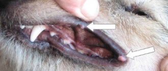
Cytological examination of any urethral discharge
Discharge from the urethra
If prostate disease is suspected, it is necessary to examine the secretion of the gland in a male dog in order to determine the cause of the pathological process.
Any discharge from the urethra should be examined microscopically.
Bacteriological culture of the urethra should not be performed due to the presence of bacterial flora in the distal part of the urethra.
Sperogram
In males, sperm consists of 3 fractions
- Urethral
- Spermatic
- Prostatic.
For diagnostic purposes, 2-3 ml of the third fraction is collected. For the accuracy of the result, cytological and cultural examination is required, since bacterial flora is normally present in the distal part of the urethra.
With diseases of the testes and appendages, the appearance and color of the ejaculate may change.
The prostatic secretion of a healthy dog contains a small amount of leukocytes, epithelial cells, and bacteria. Sperm pH 6.0-6.7.
Deviations during the study:
- a large number of leukocytes.
- a large number of red blood cells
- bacteria in large numbers located inside leukocytes and macrophages.
-macrophages containing hemosiderin.
When culturing the ejaculate, a large number of gram-negative microorganisms in combination with a large number of leukocytes indicates an infectious process, unless the sample was contaminated with preputial contents during collection.
In cases of suspected prostate neoplasm involving the prostatic urethra, the likelihood of the presence of atypical cells in samples obtained from prostate massage is greater compared with the likelihood of their presence in the ejaculate.
Radiography
X-rays provide information about the size and location of the prostate gland. The photographs can reveal its increase. However, radiography is of limited benefit. In many cases, palpation of the prostate can provide more accurate results.
For dysuria in dogs, the test of choice for prostate disease is remote retrograde urethrocystography.
With asymmetry of the prostate in relation to the urethra, narrowing of the prostatic part of the urethra, the following are most likely: abscess formation, parenchymal cysts, neoplasms, hyperplasia. If the X-ray shows marked enlargement of the prostate, presumably due to a neoplasm, radiographs of the chest and abdomen should be examined for signs of metastasis.
Ultrasonography
Ultrasound diagnostics is the best method for assessing the prostate gland. In this case, it is possible to determine the size and homogeneity of the prostate tissue.
The prostate parenchyma is homogeneous, of medium echogenicity with fine or medium granularity and smooth edges. In the sagittal projection, the shape of the organ is round or oval; in the transverse projection, it is estimated that both lobes are symmetrical.
The prostatic part of the urethra with its surrounding muscles and vertical suture is visualized as a hypoechoic structure located between the two lobes. Ultrasound diagnostics can determine the presence of inflammation of the prostate, abscess, prostate cysts, and prostate tumors.
It is necessary to examine the sublumbar lymph nodes. During infectious processes or neoplasms, their increase or change in echogenicity may be observed.
Prostate biopsy
Under ultrasound control, a needle aspiration biopsy (14-18 G biopsy needles) is performed from intraprostatic cavity lesions to collect cellular material for cytological examination. When receiving an aspiration biopsy, the prostatic fluid should be examined microscopically, as well as bacteriological culture to detect pathogenic microflora.
The most common complication of prostate biopsy is mild hematuria, but significant bleeding is also possible. To avoid complications, before performing a biopsy, it is recommended to take a test to study the patient’s blood clotting - a coagulogram.
1. Percutaneous biopsy. Conducted via transabdominal access under ultrasound guidance, the animal is under sedation.
2. Surgical biopsy. X surgical biopsy is performed using a biopsy needle by wedge-shaped resection of the parenchyma. Before performing a biopsy, cysts or areas of potential abscess should be aspirated. A sample of sublumbar lymph nodes should also be taken.
Urethroscopy
The prostatic urethra can be visualized using urethroscopy. In this case, it is possible to visualize the flow of exudate or bleeding into the prostatic part of the urethra and exclude non-prostate urethral lesions as the cause of urethral discharge.
Therapy
The following drugs are used in the treatment of prostatitis:
- To relieve the inflammatory process, antibiotics are prescribed, but in order for such therapy to be successful, it is necessary to conduct a sensitivity study to different types of antibiotics. To do this, swabs are taken from the urethra and sent to the laboratory.
- The specialist can prescribe broad-spectrum drugs that are typical for the prostate microflora. But it is worth considering this fact: not every antibiotic is able to penetrate the epithelium of the gland; it must have a high acidity constant and be slightly alkaline.
- Macrolides are considered the least toxic drugs. They remain in the tissues of the gland for a long time and maintain antibacterial conditions. An example of such a medicine would be Azithromycin.
- Experienced veterinarians often resort to fluoroquinolones. These are modern drugs that have a pronounced antibacterial effect. The drug leaves the body unchanged along with urine, and due to its high concentration it gives a positive effect. One such remedy is Profloxacin. For an animal up to ten kilograms, 1/4 tablet twice a day is enough.
- Such drugs as Marbofloxacin, Enrofloxacin, Trimetaprim and Clindamycin have proven themselves to be effective. The course of treatment with these drugs lasts from four to five weeks, depending on the form of the disease.
- For prostatitis against the background of hyperplasia, complex therapy using antiandrogen drugs is used.
- For older dogs, experts prescribe a chidectomy.
- Often, with a complex course of the disease, surgical treatment of prostatitis in dogs is prescribed. This therapy means castration of the animal. If the dog is young and this method is unacceptable, you can use progestogen agents. These include: medroxyprogesterone acetate, megestrol acetate and chlormadinone acetate.
Diseases of the prostate gland in males
Prostatitis, prostate abscess
Prostatitis is a nonspecific inflammation of the prostate gland. There are acute and chronic prostatitis. The most common causative agents of infection are Escherichia coli, staphylococci, and streptococci. The development of a chronic process is facilitated by the accumulation and stagnation of prostate secretions. Pathogenic microflora enters the prostate tissue ascendingly from the urethra. Most often, a prostate abscess occurs after incomplete cure of acute prostatitis.
Abscesses develop when the infection is severe and pus is encapsulated.
Abscess formation is the result of chronic infection.
The source of infection of the prostate gland is usually the urethra. The inflammatory process can spread to the overlying parts of the urinary tract, which causes damage to the bladder, ureters and kidneys.
Symptoms: Difficulty urinating (stranguria), bloody or purulent discharge from the urethra, tenesmus, fever, anorexia, difficulty defecating.
Diagnosis: To make a diagnosis, an animal's medical history, a general clinical blood test, a biochemical blood test, a urinalysis and urine culture results, and an assessment of prostate secretions are collected.
Treatment: The antibiotic selected based on culture results should be used for at least 28 days.
Castration is beneficial and may be a necessary condition for resolving a chronic infectious process in the prostate gland.
Prostate abscesses require surgical drainage. Drainage is carried out under an ultrasonic king.
Benign hyperplasia, cystic hyperplasia
Benign prostatic hyperplasia is an increase in the number and size of epithelial cells. Prostatic hyperplasia is the most common pathology of the prostate gland. Almost 100% of uncastrated males, starting from 2.5 years of age, develop signs of prostatic hyperplasia as they age. This is a hormone-dependent process associated with a violation of the ratio of androgens and estrogens, and occurs exclusively in the presence of testes. Due to hyperplasia, intraparenchymal fluid cysts may develop. Most often, this pathology manifests itself over the age of 4 years.
Symptoms: In most male dogs, the lesion may be asymptomatic. Sometimes there may be bloody discharge from the urethra, hematuria, hematospermia.
Diagnosis: Ultrasound diagnosis, prostate biopsy. The diagnosis is confirmed by a positive response to castration.
Treatment: There are several treatments for prostatic hyperplasia.
- Surgical castration results in a 75% reduction in prostate size within 8-10 weeks.
- Chemical castration involves the use of estrogens or antiandrogens.
Hormone therapy in the form of estrogen is rarely used due to a number of side effects, toxic effects on the body, bone marrow suppression, and the development of diabetes.
Antiandrogens do not cause a number of side effects, unlike estrogens. There are drugs licensed for veterinary use.
At the moment, antiandrogens are the therapeutic choice of the veterinarian.
Prostate neoplasms
In older animals, the prostate gland is susceptible to neoplastic transformation.
Most often these are malignant neoplasms. The prostate can be the site of metastasis or the site of primary formation. There are such neoplasms as: carcinoma, transitional cell carcinoma, adenocacyoma, lymphosarcoma, squamous cell carcinoma and hemangiosarcoma. Benign prostate tumors, such as leiomyoma, are rare.
Symptoms: Enlarged prostate on rectal examination, asymmetrical prostate gland. Difficulty urinating and defecating, urethral obstruction.
The tumor can grow into the bladder neck and can cause ureteral obstruction.
Pathological changes in urine and hematuria predominate.
Diagnosis: If a male dog was neutered at a young age and has obvious enlargement of the prostate gland, it is most likely that the enlargement may be due to a neoplasm.
To check for the presence of metastases in the lungs, it is necessary to conduct an x-ray of the organs of the ore cavity.
The bodies of the lumbar vertebrae and pelvic bones should be examined to identify foci of proliferative changes that suggest the presence of metastases.
It is necessary to conduct ultrasound diagnostics. In non-castrated males with a noticeable enlargement of the prostate gland, the neoplasm should be differentiated from prostate abscessation and paraprostatic cysts. To establish a diagnosis, a prostate biopsy is necessary. The final diagnosis is made based on cytological or histopathological examination of prostate tissue samples.
Treatment: There is no effective treatment for prostate cancer.
Sometimes, in the absence of metastases, surgery can be performed to completely remove the prostate. But owners should be warned about the likely development of urinary incontinence after surgery. The goal is usually temporary tumor control and relief of clinical signs. Castration leads to a slight positive effect.
What is prostate adenoma in dogs?
Prostate adenoma is a benign tumor that occurs as a result of the proliferation of glandular tissue of the organ.
Normally, in male dogs, whose average weight is up to 10 kg, the size of the prostate does not exceed the diameter of a hazelnut. With adenoma, it can increase tens or even hundreds of times, taking on the size of a chicken egg or a small apple.
As the gland grows, it begins to compress the urethra, ureters and rectum , leading to the formation of diverticula (pockets) in it, the removal of which subsequently requires surgical intervention.
The secretion of the prostate gland of a healthy male contains bactericidal substances that suppress the development of pathogenic microflora in the organs of the genitourinary system. When prostate function is impaired due to the proliferation of glandular tissue, this function is lost, and bacteria penetrate into the organ tissue, provoking the development of prostatitis. We can say that these two pathologies are inextricably linked, which should be taken into account when developing a course of therapeutic measures.
Knowing what prostate adenoma is in dogs and what serious consequences it can lead to, it is necessary to contact a veterinarian for help in a timely manner.