Description of the pathology
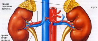
Cryptorchidism is a common hereditary condition in which one or two testicles do not descend into the scrotum. The testes may be located in the inguinal canal, perineum, or abdominal cavity. Sometimes they cannot be detected even with ultrasound diagnostics. With cryptorchidism, the puppy does not have any complaints, the pet feels great and nothing bothers him.
Typically, the descent of the testes into the scrotum occurs on days 5-10 of the puppy’s life. Depending on the breed, this phenomenon can last up to 2-5 months.
If the testicles are not fixed in the scrotum by six months of age, doctors make a preliminary diagnosis - cryptorchidism. However, veterinarians often advise waiting up to 10 months and re-examining.
Small breeds are more susceptible to the anomaly than large ones. For example, cryptorchidism is often found in Spitz, Yorkshire terrier, pug, Chihuahua, toy terrier, Maltese, poodle, and Pekingese. The probability of pathology is 7-13% of all diseases in male dogs. With this phenomenon, the dog retains its sexual desire and hormone production.
Reasons for appearance
Despite the fact that the pathology is purely “male”, from which only males suffer, it is genetically transmitted from the female. This is an autosomal recessive inherited abnormality. But this is not always the reason. Among the factors that influence cryptorchidism are:
- Anatomical reasons: narrow inguinal canal, large testes, underdeveloped scrotum, short spermatic cord, formation of connecting folds at the base of the scrotum.
- Mechanical - injuries or bruises of the scrotum and groin area, which slow down the process of descent of the testis.
- Hormonal factors. Insufficient production of testosterone and gonadotropin, which affects the connection of the fundus of the scrotum and testicle.
- Inflammatory processes that lead to reduction or complete obstruction of the inguinal canal and acquired cryptorchidism in dogs.
Most pathologies occur at the intrauterine stage of fetal development, which is associated with improper care of the pregnant bitch by the breeder. Viral diseases or malnutrition can have a bad effect on her offspring.
Prevention
The main preventive measures to prevent cryptorchidism are the following.
Caring for a pregnant pet
- careful and balanced nutrition with high-quality food is necessary;
- We need protection from possible diseases, in particular infectious ones. It is advisable to avoid contact with untested animals;
- avoidance of taking medications that can affect fetal development.
Proper care for puppies
- isolation from untested animals;
- balanced nutrition according to age;
- compliance with safety conditions to avoid injury to young animals.
In the article I talked about cryptorchidism in dogs. She explained what it is. She described the types of disease, causes and methods of treatment. She gave preventive measures for the disease.
Kinds
The following types of anomaly are distinguished:
- bilateral or monolateral cryptorchidism;
- abdominal or inguinal;
- false or true.
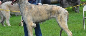
Monolateral or unilateral cryptorchidism is a pathology in which one testicle does not descend. This is a common phenomenon in which a male dog retains reproductive function.
Bilateral - when both testicles are fixed outside the scrotum. This form of anomaly is less common. Males with bilateral cryptorchidism are sterile. This is explained by the fact that high temperature in the abdominal cavity disrupts normal spermatogenesis.
The abdominal form is also called the abdominal form. With this phenomenon, the testes can become fixed under the skin of the peritoneum. Often they are detected only during laparoscopy, since ultrasound does not give an accurate result. Inguinal cryptorchidism - when the testicles are located in the groin area. They are easy to detect visually or by palpation.
The true form of the anomaly is when the reduction of the testicle into the scrotum is only permissible through surgery. The testis may be located in the inguinal canal, in the abdominal cavity, in the upper part of the scrotum, or at the inguinal ring.
False cryptorchidism or wandering testicle syndrome. With this phenomenon, the testicles can periodically be retracted into the abdominal cavity and back. This usually happens when there is stress or a sudden change in weather. This anomaly is not considered serious, but male dogs with “wandering” testes are not allowed for breeding.
Types of cryptorchidism in dogs
- Congenital.
- Acquired.
- Bilateral (both testes have not descended).
- Monolateral (right or left testis has not descended)
- Intra-abdominal (abdominal).
- Inguinal.
True - the testis can be located in the upper or middle part of the scrotum, inguinal canal, at the inguinal ring or in the abdominal cavity. It is impossible to descend the testis into the scrotum in a non-surgical way.
False—reduction of the testis into the scrotum is possible regardless of its original location.
An elevated testis is a phenomenon that occurs when the growth of the spermatic cord slows down after the testicle descends into the scrotum, which, as the puppy grows, leads to a reverse rise of the testis.
Classification of cryptorchidism by palpability.
1) Retractile - are located in the upper part of the scrotum and are pulled up due to excessive activity of the muscle that lifts the testis.
2) Ectopic - located in an atypical location (inner thigh, etc.).
3) Undescended - any testes that are not located in the scrotum (excluding retractile and ectopic ones)
1) Canalicular - located in the inguinal canal, not palpable.
2) Intra-abdominal – located in the abdominal cavity.
3) Developing - not reaching the determined size in time.
4) Absent - testes that have undergone atrophy or agenesis (complete congenital absence).
Classification by palpability
Testicles can be palpable or non-palpable. The first group includes:
- Ectopic testes located in an atypical place for them. For example, the inner thighs.
- Retractile testicles are located in the upper zone of the scrotum and can be retracted into the abdominal cavity due to the high activity of the muscles of the spermatic cord. This is the cause of false cryptorchidism.
- Undescended testicles are any testicles located outside the scrotum (except the first two types).
Non-palpable testicles are:
- Intra-abdominal (located in the abdominal cavity).
- Canalicular (in the inguinal canal).
- Absent (testicles that did not form in the womb or were atrophied).
- Developing (testes that have not reached the required size).
What types of disease are there?
The following types of cryptorchidism are distinguished in puppies:
- a congenital species that developed during the embryonic period;
- an acquired appearance that appeared due to some external factors (for example, illness or physical injury);
- bilateral view, when not descended, both testes;
- monolateral type, when one of the testes has not descended (this is unilateral cryptorchidism in dogs);
- intra-abdominal – this is the abdominal type of the disease;
- true - this is when the testis is located in the upper or lower part of the scrotum and cannot descend to the end;
- raised testis - this symptom occurs when the growth of the spermatic cord is simply inhibited, when the testicle has already descended into the scrotum and the testis begins to rise again;
- Retractile. The testes are located at the top of the scrotum and are constantly pulled up due to the fact that the muscle that elevates the testis is overactive.
- Ectopic. They can be found in places that are not typical for the testis.
- Undescended. These include absolutely all testes that are not located in the scrotum (exceptions are retractile and ectopic).
We invite you to read: Wet food for dogs: tips for choosing, veterinarian review
Classification of testes by type of non-palpable species:
- Canalicular. They may be located in the inguinal canal and cannot be palpated.
- Intra-abdominal. Found in the abdominal cavity.
- Developing. These are those types of testes that have not developed and because of this cannot be identified physically.
- Absent. Atrophied or agenesis testes (i.e. their complete absence).
Consequences
The unnatural location of the testes can pose a risk to your pet's health. There are several reasons why you need to get rid of cryptorchidism in time:
- Decreased reproductive function and risk of tumor formation. For normal spermatogenesis in a male dog, the temperature of the testes should be 2–4°C lower than the dog’s body temperature. Overheating of the testicles leads to the extinction of the process of sperm formation and the risk of tumor degeneration. Semenomas, sertoliomas or leydigomas can often develop at a fairly early age. The dog is in danger of death.
- The occurrence of tumors leads to hormonal imbalance, the production of estrogen. This provokes hyperplasia of the anal glands, cystic prostatitis and decreased bone marrow activity in the dog.
- Cryptorchid males also need to remove the stuck testicle because spermatic cord torsion may develop. This pathology is accompanied by the symptom of “acute abdomen” and requires separate treatment.
Answer
A pug's testicles, like other breeds, should be approximately the same size. A slight difference is acceptable. How long ago did you notice the difference, did the differences appear recently or is there a congenital defect? Ask the breeder from whom you bought the puppy if there are any similar cases in the pedigree? Gently touch the enlarged testicle; it should not be hard or painful. Carefully check whether both testicles are present in the scrotum? Probably the second one did not have time to descend.
The puppy should be examined by a veterinarian to rule out a tumor, abscess, orchitis or brucellosis. The doctor will conduct a thorough medical examination, taking into account a history of symptoms and incidents that may play a role in the onset of the disease.
Possible causes of the above symptoms include scrotal hernia, scrotal dermatitis, twisted spermatic cord, sperm-filled mass of inflamed tissue (granuloma), fluid-filled sacs on the spermatic cord (hydrocele), prostatitis, cystitis, and abnormal cell growth (neoplasia). Before starting treatment, these diseases must be excluded.
Testicular swelling occurs in male dogs without preference for a specific breed. Epididymitis occurs more often in older dogs, but can occur at any age. The tumor is easy to diagnose at home; it is important to determine changes in the size of the dog’s testicles. A common cause of testicular swelling is scrotal trauma. Male dogs suffer from orchitis, an inflammation of the testicles when one or both become hard and swollen due to inflammation of the tube containing sperm or the epididymis.
Diagnostics
A diagnosis of cryptorchidism can only be made after collecting a detailed history and a series of studies, including:
- inspection;
- Ultrasound diagnostics;
- hormonal test.
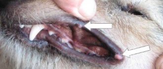
First, the veterinarian examines the animal and palpates it. If the testicle is fixed in the area of the inguinal canals or the upper part of the scrotum, it is easy to detect. With this study, false or true cryptorchidism can be determined. To do this, the doctor tries to bring the testis into the scrotum. If this can be done, it is false. Otherwise, true.
Ultrasound diagnosis is appropriate for palpable types of anomaly, when scrotal fat or inguinal lymph nodes can mimic testicles. However, in practice, this method is not very effective, since its result can be influenced by the quality of the equipment, the accumulation of gases in the intestines, and the experience of the diagnostician.
Doctors may use a hormonal test to confirm the diagnosis. Its principle of action is as follows: the dog's testosterone level is measured before and 60 minutes after the gonadotropin injection. With cryptorchidism, an increased level of testosterone in the blood will be noticed.
Treatment
There are several ways to treat cryptorchidism. Depending on the type of anomaly and the desire of the dog owner, this may be:
- drug therapy;
- orchiopexy (surgical removal of the testicle into the scrotum);
- orchiectomy (castration).
Conservative
Drug treatment includes hormonal medications and therapeutic massages. The prognosis depends on how quickly you start therapy. The greatest effectiveness was observed in pets under 6 months of age. When the inguinal canal closes, it will be more difficult to achieve results.
As a rule, the animal is prescribed medications to increase the activity of luteotropic hormone, which promotes prolapse of the testicles. For example, Gonadotropin-releasing or Choriogonadotropin. It is advisable to do massage when the stuck testicle is close to the scrotum and can be palpated.
Hormonal therapy can only be indicated if the causes of cryptorchidism are in the endocrine system. This treatment is carried out under the supervision of a veterinarian.
Orchiopexy
Surgical placement of a testicle into the scrotum is a controversial operation. Considering the heredity of this anomaly, orchiopexy in the Russian Federation and a number of other countries is regarded as fraud and is prohibited by law. It is customary to reject males with cryptorchidism from breeding.
Technically, such an operation is quite difficult and is not always possible, even if the testicle is located in the inguinal canal. The intervention is performed using laparoscopy. Most often, the operation lasts 20 minutes under general anesthesia and is completed in one step. Less commonly used is a two-stage technique, when the testis is first fixed on the thigh and then moved to the scrotum.
Most veterinarians advise not to break the law and carry out castration.
Orchiectomy
Castration is an effective way to get rid of cryptorchidism, as well as limit its spread in the population. Orchiectomy is also performed under general anesthesia and can be performed in two ways:
- For inguinal cryptorchidism, abdominal surgery is not performed. It lasts about 15-25 minutes and is minimally traumatic.
- To treat abdominal cryptorchidism, abdominal surgery is performed, lasting about 40-60 minutes. The complexity of this method is determined by the location of the testis in the cavity.
The price of an orchiectomy is in the range of 7-20 thousand rubles, which depends on the method of surgery, anesthesia drugs, the experience of the doctor and the reputation of the clinic.
Treatment of cryptorchidism in dogs
1. Conservative (therapeutic)
In the conservative treatment of cryptorchidism in males, massage courses and hormone therapy are used. The earlier treatment is started, the better the prognosis. The greatest result (about 20%) can be achieved in patients under the age of 6 months; later, when the inguinal canal closes, the effectiveness of the procedures decreases significantly. The use of massage is rational if the testis can be palpated and it is in close proximity to the scrotum. During the procedure, the testis is gradually pulled up in the caudal direction, lowered into the scrotum and held there for some time. The more often the sessions are performed, the more effective the treatment.
Hormone therapy uses drugs such as choriogonadotropin or gonadotropin-releasing hormone. The drugs increase the production of endogenous luteotropic hormone, which promotes testicular descent. It should be noted that hormone therapy is used only when the disease is caused by a disruption of the endocrine system. If cryptorchidism is caused by other reasons, then the use of drugs may cause an acceleration of neoplastic changes in the undescended testis. Such treatment should be carried out under the strict supervision of a veterinarian.
Orchidopexy is an operation whose purpose is to bring down the testis and secure it in the scrotum. There are various methods for carrying out this procedure. In terms of invasiveness, trauma and duration, this surgical intervention is practically no different from castration. There is evidence of fruitful matings and a gradual improvement in the quality of spermatogenesis in 20-30% of operated male dogs. Considering the hereditary nature of the disease, the use of orchiopexy in most countries, including the Russian Federation, is recognized as fraud and is prohibited by law. It is recommended to cull the male dog from breeding and castrate it. This operation is not performed in our clinic.
An orchiectomy is an operation to remove the testes. It is performed under general anesthesia using sedatives and painkillers. There are various techniques for performing orchiectomy and their choice depends on the type of cryptorchidism. With the inguinal type, the operation does not require opening the abdominal wall (the operation is not abdominal), the invasiveness and trauma are low, the duration is about 15-25 minutes, that is, the same as during castration of a normal male dog. In the treatment of intra-abdominal cryptorchidism, abdominal surgery is performed. Its duration (30-60 minutes) and complexity vary depending on the location of the testis in the abdominal cavity.
Carrying out castration
To undergo an orchiectomy, the dog must be completely healthy. The general picture of the operation looks like this:
- First, the pet is given general anesthesia. When the animal loses activity, it can be placed on the operating table with its belly up and immobilized.
- The abdominal area is sterilized with an antiseptic drug.
- The doctor then performs an opening of the abdominal cavity. The skin over the location of the testicle is cut layer by layer.
- The cryptorchoid testis is removed.
- Then the spermatic cord, testicular vein and artery are excised.
- The testicle is removed and the wound is sutured in layers.
- An incision is then made between the penis and the scrotum, from which a normal testicle is removed.
- The spermatic cord and vessels coagulate. The wound is stitched up.
Progress of the operation
Let us consider the progress of orchiectomy in a specific case. The patient is a male, 7 years old, mixed breed. The left testis is cryptoroid - located intra-abdominally in the hypogastrium. A diagnosis was made of true unilateral cryptorchidism with intra-abdominal localization of the left testis. First, before the operation, premedication is performed and then the animal is put under anesthesia.
We recommend reading: Feces and oncology in dogs
The first stage - preparation of the surgical field is carried out taking into account generally accepted rules of asepsis.
Rice. 1. Preparation of the surgical field.
The next stage is surgical access. Surgical access is carried out layer by layer, above the location of the testis. The dissected vessels are coagulated.
Rice. 2. Surgical access.
Rice. 3. Surgical access, coagulation of blood vessels.
Rice. 4. Opening of the abdominal cavity.
The next step is to remove the left (cryptorchoid) testis. As you can see, it is significantly enlarged, which indicates its neoplastic degeneration.
Rice. 5. Extraction of the cryptorchoid testis.
Then the testicular vein, artery and spermatic cord are ligated and dissected. The testis is removed.
Next, the wound is sutured in layers.
Rice. 6. Layer-by-layer suturing of the wound.
The last step is to remove the normal testis. This happens as during normal castration of a male dog.
Rice. 7. Surgical access to the right (healthy) testis.
Rice. 8. View of the right testis.
Rice. 9. The vessels and spermatic cord are ligated/coagulated and dissected.
Rice. 10. The testis is removed. The wound is sutured.
Rice. 11. View of the patient immediately after surgery.
Let us consider the progress of orchiectomy in a specific case. The patient is a male, 7 years old, mixed breed. The left testis is cryptoroid - located intra-abdominally in the hypogastrium. A diagnosis was made of true unilateral cryptorchidism with intra-abdominal localization of the left testis. First, before the operation, premedication is performed and then the animal is put under anesthesia.
Sometimes puppies have congenital defects that can only be discovered over time. Cryptorchidism in dogs is one of these anomalies that owners of male dogs often encounter. Pathology puts an end to the pet's exhibition career and can cause the development of cancer.
- Description of the pathology
- Reasons for appearance
- Kinds
- Classification by palpability
- Consequences
- Diagnostics
- Treatment
- Conservative
- Orchiopexy
- Orchiectomy
- Preparing for surgery
- Carrying out castration
- Caring for your dog after surgery
- What to pay attention to after anesthesia
- Reviews
- Irina, 37 years old, Krasnodar
- Olga, 29 years old, Moscow
- Arthur, 41 years old, Omsk
Caring for your dog after surgery
Typically, the pet regains consciousness after anesthesia 30-90 minutes after surgery. The time it takes for the condition to normalize depends on many factors: the dog’s age and weight, anesthesia drug, physiological state, etc. At this time, it is important to provide him with the necessary care.
Immediately after you pick up your dog from the clinic, wrap it in a thin blanket. Lay out a disposable diaper, since the pet is not yet fully in control of consciousness and may wet itself. When you arrive home, put the dog in a pre-prepared place. It should be a soft, clean bed in a quiet corner, without drafts, away from heating appliances. Turn your pet from side to side every half hour. At the same time, check if the diaper is wet. Replace with a clean one as needed.
When recovering from anesthesia, the dog may be disoriented in space. This is normal, no need to panic. Make sure that the animal is not injured. You can feed or water your dog only after all symptoms of lethargy have passed.
Rehabilitation will also require wound care. For 10-14 days, treat the seams with brilliant green 1-2 times a day. The doctor may prescribe another antiseptic. To avoid licking the stitches and complications, you can put a post-operative blanket on your pet. Usually 2 weeks after surgery, the sutures are safely removed.
What to pay attention to after anesthesia
Normally, the dog recovers from anesthesia calmly. Breathing is smooth, mucous membranes may be pale pink, but without unnecessary discharge or anxiety. Defecation occurs 1–2 days after surgery, thirst remains at the usual level. However, you should immediately consult a doctor if you notice one or more of the following symptoms:
- Body temperature exceeds 39.0 degrees Celsius.
- Weak intermittent breathing, wheezing in the sternum.
- Vomiting more than 2 times a day.
- Weak pulse.
- No signs of normalization more than 6 hours after anesthesia (the dog does not move, does not open its eyes).
The prognosis for surgical treatment of cryptorchidism is positive. After 2-3 months, the dog can swim in ponds and live a normal life.
Reviews
Irina, 37 years old, Krasnodar
“I have a Yorkie, unilateral cryptorchid. We went to the doctor when it was still 4 months, the doctor said to wait until 6. If this happens, he will refer me for surgery and we will suture the testicle to the scrotum. As agreed, we showed our Caesar at 6 months, and the veterinarian advised us to wait another two months or, even better, up to a year. We waited and hoped that it would go down on its own. Alas. As a result, we were prescribed to inject Gonadotropin and repeat it in the future (after 6 weeks). The canals can still be stimulated and the testicle will come out. We are waiting for the results.”
Olga, 29 years old, Moscow
“My pug has been suffering from cryptorchidism for 3 years now and his testicle can’t even be felt. The veterinarian advises castration, but so far they have refused. The dog feels good, no complaints. Once every six months we undergo an ultrasound and examine him to catch the tumor. I think we’ll take you to surgery when you’re 5 years old.”