At-risk groups
Neoplasms and growths on the eyelids are a common problem. The growths usually appear in older dogs, but can develop at any age. Growths on the eyelids harm the health of the eyes and reduce the quality of life of the pet. Fortunately, most eyelid growths are benign.
The growths increase over time, leading to structural and functional changes in the eyelids and irritation of the surface of the eye (cornea). Malignant growths are rare; if diagnosed and excised in a timely manner, they do not pose a threat to the life of the animal.
If neoplasms are ignored and they increase in size, there is a risk of:
- Deterioration of vision.
- Damage to the mucous membrane of the eye.
- Ulceration or clouding of the cornea.
- Inflammation of adjacent tissues.
Eye diseases in dogs: main symptoms and treatment methods
Canine diseases affecting the organs of vision do not always occur clearly. Owners may not notice the pathology in the early stages, so such ailments often develop into serious symptoms. Only an attentive dog breeder is able to distinguish the deviation at the very beginning of its appearance.
Redness and unusual discharge from the eyes are the first signs that something is wrong with your pet’s vision. If they are detected, you must immediately contact a veterinarian. Timely diagnosis will allow the dog to be treated before irreparable complications arise.
What eye diseases occur in dogs?
Eye diseases in dogs can be of 3 types:
- Infectious – occurs when infected with viruses or bacteria as a primary disease or complication of infection of another organ.
- Non-infectious - mechanical damage, neoplasms, inflammation due to improper growth of eyelashes, eversion of the eyelids.
- Congenital - characteristic of some breeds, for example, Shar Peis. Includes eversion, entropion of the eyelids, deformations of the eyes and lenses. They are usually corrected surgically.
Each disease has a characteristic description, diagnostic methods and treatment.
Keratitis
Keratitis is inflammation of the eye cornea.
The disease causes significant discomfort to the dog and threatens serious complications on the visual system. Keratitis occurs due to mechanical injuries, burns, acute vitamin deficiencies, infectious diseases (hepatitis, enteritis, plague), and allergic reactions.
They can also affect dogs with diabetes mellitus and weakened immune and endocrine systems. In rare cases, it develops at the genetic level.
Keratitis can be identified by swelling of the eye and conjunctiva, cloudy cornea and lens, purulent discharge of white, yellow and gray shades. There is profuse lacrimation and photophobia. The white becomes red, the eye membrane becomes rough. There may be frequent blinking in an attempt to clear vision. Symptoms appear within 2-4 hours of infection or injury.
Keratitis threatens the development of glaucoma, cataracts, and loss of vision. Treatment depends on the cause of the appearance. General procedures for all forms of pathology are washing with a solution of furatsilin or another antiseptic, drops of levomecithin, application of erythromycin ointment, a course of oral antibiotic. Pain relief injections and a hypoallergenic diet may be prescribed. On average, treatment gives effect in 1-2 months.
Check out the full article on the eye disease in dogs - keratitis.
Cataract
Cataract is expressed in severe clouding of the lens of the eye, its swelling and increased intraocular pressure.
The disease may appear due to exposure to toxic substances, tissue wear and tear with age, or be congenital. Cataracts are dangerous due to complete or partial loss of vision as a result of rupture of the tissues of the eyeball. Drug treatment is practiced (drops of viceine, catachorm, vitamin compounds), but is considered ineffective. The result is achieved only by surgical intervention followed by maintenance therapy.
Learn more about the symptoms and treatment of cataracts in dogs.
Ectropion and entropion
Ectropion and entropion are the medical names for eversion and entropion of the eyelids.
Both diseases usually develop in parallel, so they are practically not separated. They are expressed in a change in the shape of the eyelids (turning outwards or inwards), loss of their natural functions. When eversion occurs, the eyeball is left without moisture and protection from germs. When curled, the eyelashes irritate the pupil and can grow into the protein tissue.
In both cases, the dog begins to blink constantly, profuse lacrimation occurs, and the animal experiences pain.
Treatment is surgical when the dog is an adult, when its growth has stopped. In the early stages and during recovery after surgery, medical support is provided with hormones, antiseptics, and moisturizing drops.
Shar-Peis, Staffordshire Terriers, Dachshunds, Great Danes, Newfoundlands, St. Bernards, Basset Hounds, Great Danes, Spaniels and Ridgebacks are particularly susceptible to the disease.
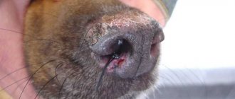
Corneal ulcer
One of the most common eye diseases in dogs.
Also called ulcerative keratitis. It is expressed in inflammation of the upper layer of the epithelium of the eyes. The tissue becomes thinner, wears out, becomes covered with ulcers, gradually growing into one large wound. All layers of the epithelium are affected. Watery eyes appear, pus is released, the outline of the pupil blurs, and the white of the pupil turns red. The color of the cornea may change. The dog begins to squint, close its paws and lower its eyes. It may develop into a chronic form.
Occurs due to physical damage to the skin, infection, burns with chemicals, as a complication of acute conjunctivitis, volvulus and neoplasms. It is rarely hereditary.
Treatment ranges from surgical excision of the affected tissue to conservative treatment. Medication methods take up to six months, drugs are selected strictly individually.
Lens luxation
Lens luxation (or luxation) involves displacement of the corresponding part of the eye from the hyaloid fossa.
Dislocation can be partial or complete. It occurs for genetic reasons, due to glaucoma, cataracts or as a complication of severe injuries and infections. It can lead to loss of visual function and is therefore considered a serious disease.
Different types of terriers are most susceptible.
Dislocation occurs after the ligaments of the lens and ciliary line are torn. The pupil becomes deformed, moves away from the center and swells, and the shape of the apple may change. The movement of fluid in the ocular body is disrupted.
Treatment is carried out by surgical correction. The earlier the stage of the disease, the more positive the prognosis for saving the eye. After removal of the lens, an intraocular lens implant is placed. In rare cases, implantation of the entire eyeball is indicated.
Eyeball luxation
It consists in the exit of the eyeball from the orbit behind the eyelid completely or partially.
Caused by mechanical damage to the bones of the head and temples, large muscle tension as a result of the shallow depth of the bone orbit. The apple extends far beyond its natural boundaries, the conjunctiva swells, dries out, and takes the form of a hanging cushion. Such a dislocation threatens with blindness, ulcers, and necrosis of eye tissue. Often found in Pekingese, Japanese Chins and similar breeds.
As first aid for loss, it is suggested to carefully irrigate the apple with a solution of furatsilin or novocaine to reduce pain and prevent drying out. It is corrected surgically under anesthesia after excision of the eyelid adhesion. A fixing suture is applied, which is removed after a few days. Then corticosteroid therapy is used to relieve inflammation.
Treatment of eye diseases in dogs
How to treat eye diseases in dogs is a matter of an individual approach. Only a thorough diagnosis gives a complete understanding of the causes, forms of the disease, and possible methods of correcting the problem.
Ointments and drops are used to treat various eye diseases.
Treatment of eye diseases in dogs is carried out with different drugs, but has common principles:
- bandages are used to apply astringents and antiviral ointments;
- before instilling drops, the eyes are washed to remove pus and other secretions (tea brewing or saline solution works well);
- antibacterial drugs are most often administered intramuscularly after testing for sensitivity to medications of the selected group;
- in acute forms of inflammation, antibiotics and anti-inflammatory drugs are combined.
Important. Dogs' eyes are very sensitive to drops, so dosages and precautions must be observed. In case of a moderate course of the disease, it is enough to drip 2-6 times a day, in case of acute disease – up to 10 times. The number of drops is determined only by the doctor.
It is also important to wear a collar or socks on the paws to prevent the dog from scratching its sore eyes. With purulent discharge, a diet with a minimum of fermentation products will not harm. Clean pieces of bandage, cotton pads or lint-free swabs are suitable for washing the eyes.
Daily careful monitoring of your pet’s condition and regular hygiene are the key to the health of his eyes.
Detection of alarming signs at the first stages of their appearance will allow you to treat your dog as quickly as possible, preserving his full vision.
Genetic predisposition must be found out when purchasing a puppy by asking the breeder or previous owner about the medical history of the dog’s parents and relatives.
We offer you to watch a video in which a veterinarian talks about eye diseases in animals. We wish you pleasant viewing!
Source: https://sobaki-pesiki.ru/bolezni-glaz-u-sobak-siptomy-lechenie-foto.html
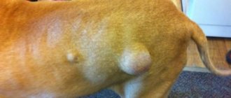
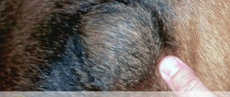
Types of growths and neoplasms
The growths can affect any layer of eyelid tissue. Statistically, the most common neoplasms on the eyelids in dogs are:
- Adenomas are benign tumors.
- Papillomas are benign growths that can become malignant.
- Adenocarcinomas are rare but are the most common malignant tumor affecting the eyelids.
As you age, the risk of developing:
- Meibomian gland adenocarcinomas.
- Melanomas.
All of the above types of growths are malignant and require timely diagnosis. If cancer is diagnosed at an early stage, the affected cells are removed surgically and healing is monitored.
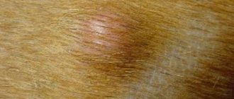
Less commonly, growths appear on the eyelids of dogs, which are:
- Histiocytomas are benign tumors of the skin.
- Mastocytomas are a benign tumor of the skin and/or subcutaneous tissue.
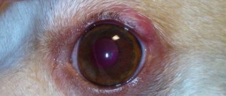
Meibomian gland adenocarcinoma and adenocarcinoma
They are modified cells of the meibomian glands (lining the upper and lower eyelids). The meibomian glands secrete an oily lubricant that helps and protects the tear glands and the eye itself.
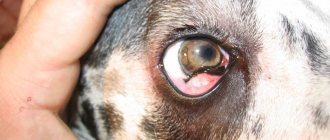
Growths can appear on both the outer and inner sides of the eyelid. In the second case, the neoplasms are visible - they protrude or protrude from the eyelid. These growths have a rich pink color and a lobed structure. If the situation is ignored, the tumors begin to bleed and/or self-opening.
Melanoma
There are two types of eyelid melanoma in dogs. The first type occurs due to degeneration of the skin cells of the eyelids. The growth is a solid, protruding, smooth, pigmented layer of tissue that can be easily removed surgically.
The second type of melanoma occurs when the cells of the pigmented edge of the eyelid degenerate. The growth is noticeable even at an early stage; it is flat, wide, and grows quickly. This type of melanoma is more dangerous because the tumor quickly takes root and may require removal of a large area of the eyelid to completely excise it.
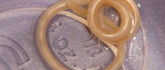
Papilloma
The growth is of viral origin! Papillomas vary in color (from white to pink and red) and have an uneven surface.
Note! Papillomas are more common in young dogs.
Neoplasms and infections in diseases of the eyes of dogs
Very often, owners do not notice eye disease in dogs until all the symptoms have appeared and the appearance of the eye has changed. This is precisely the pathology that does not manifest itself for a long time and cannot be diagnosed. There are a large number of eye diseases in dogs and they occur in puppies or appear in adult dogs. The appearance is also influenced by genetics and breed characteristics.
Symptoms of eye diseases in dogs
In some dogs, eye disease is considered genetic, possibly due to breed or genetic predisposition. When adopting a dog, you should be familiar with this. In other cases, you need to pay attention to the type of animal as well as signs of disease.
The main symptoms are:
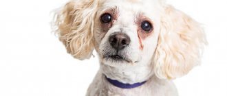
- The appearance of discharge in the eyes that is not natural in appearance and consistency;
- Excessive lacrimation (more common in puppies);
- Changes in the appearance of the eye (tissue inflammation, slight swelling, clouding, spots and bulges);
- New growths may appear, as well as unnatural trembling of the iris;
- Possible photophobia, sometimes loss of vision.
These symptoms occur in dogs whose facial skin hangs loosely. The eyelid everts and infection or bacteria can enter the eye.
At the first symptoms, contact your veterinarian; this could be an infection or small objects getting into the visual system. Due to the fact that the dog has vision problems, the general condition also worsens.
Very often the animal feels discomfort and becomes less active.
Primary and secondary (usually infectious) eye diseases in dogs are classified. And there are many diseases.
Conjunctivitis
Inflammation of the outer layer of the eye, which is called conjunctivitis. The disease can be infectious or non-infectious. Most often it occurs due to a virus, bacteria or fungus. And this is just a symptom that may indicate a disease.
The non-infectious variety may indicate a possible allergic reaction or foreign body ingestion. It could even be a small grain of sand or dust.
An animal may also have conjunctivitis from various chemicals, drafts, and even minor hypothermia.
There are a large number of types of this disease and each of them requires separate treatment, which is why it is so important to see a doctor, even if you have already had problems with eye diseases.
Conjunctivitis is more common in certain dog breeds, namely pugs and chihuahuas. This occurs due to the structure of the skull and eyes. Symptoms are: dullness of the eyes, green discharge of a rough consistency. The dog rubs its eyes all the time, which can cause wounds and ulcers. Partial loss of vision up to and including loss of vision is also typical. Very often, the feeling of malaise does not go away for a long period.
Only a doctor can diagnose and prescribe treatment after conducting the necessary tests. The doctor needs to find out what caused the disease. If you have chronic diseases, you need to warn your doctor about them.
Blepharospasm
A disease in which the animal's eyelid contracts quickly, unconsciously. Visually, this disease is noticeable, since the dog blinks frequently and unnaturally. During the daytime, photophobia is observed, to the point that the dog constantly keeps his eyes closed. Very often, you can observe a small discharge of fluid from the eyes of the animal.
More often, the disease indicates the presence of more serious diseases in your pet’s body. Sometimes this is an indication of inflammation of the trigeminal nerve.
The disease can cause various injuries or inflammation of the eyes. The pet's eye system often swells, and upon palpation the dog feels unpleasant painful sensations.
Treatment is to eliminate the cause of the disease.
And to eliminate the symptoms, use lidocaine drops or other pain medications. Animals suspected of having blepharospasm should be placed under hospital treatment, as the drugs used are highly toxic and use at home is not recommended.
Third eyelid prolapse
A severe disease during which the third eyelid moves to the corner of the eye; the disease often manifests itself in only one eye. And the name “cherry eye” arose because of the specific type of eye, which becomes very red and resembles a cherry.
There are reasons why the disease can occur, but most often, the reason for this is the thinning or weakening of the tissues that support the eyelid.
In dog breeds: bulldog, spaniel, such prolapse occurs much more often than in others. These dog breeds have genetically weak eye tissues and are prone to third eyelid prolapse.
Also, veterinarians say that the disease can be transmitted hereditarily.
And although the disease is not dangerous, it can significantly complicate the work of the lacrimal glands, as well as provoke conjunctivitis and keratitis.
It is worth treating as soon as you notice symptoms. And the treatment itself is to perform a surgical operation. Afterwards, the animal will need to receive special drops for the rest of its life.
Eversion and inversion of the eyelid
This disease is more common in dogs. And more often for specific breeds:
- Dachshund;
- Saint Bernard;
- Basset;
- Great Dane;
- Newfoundland.
Eversion and entropion should be treated together, as these diseases can develop in parallel. During inversion of the eyelids, the animal experiences pain due to improperly grown eyelashes. This disease can develop over many years. And its symptoms are as follows: severe tearing of the eyes, pus, pain on palpation.
Eversion of the eyelids is usually treated surgically. But the operation is best performed on an adult dog that will tolerate the use of anesthesia well. If the first signs appear, you can use various ointments and drops to slow down the disease process. On a case-by-case basis, your doctor will decide which treatment is appropriate for your dog.
Disease of the lacrimal apparatus
A disease in which there is a small amount of tear fluid. It is also called keratoconjunctivitis sicca. This disease can occur in various terriers, as well as in Bichon Frize dogs. This disease can be inherited and will be noticeable even in a puppy.
This causes disorders of sex hormones, with various injuries of the skull, as well as with the use of medications. Upon examination, the dog has pronounced blinking, as well as dry edges of the eyes, very often with a crust. There is also unevenness of the cornea; in addition, the disease affects the nasal area, and often provokes diseases of the facial nerve.
The main thing in treatment is to eliminate the main cause of the disease. The affected areas should be washed with saline solution every two hours, then apply the medicine. The disease is considered quite serious, since the animal requires constant care and visits to the veterinarian.
Dermatitis of the century
It occurs more often in animals with long hair and drooping ears. A pathology that often causes eye disease.
And its signs are:
- Inflammation of the skin around the eye;
- Redness of the eyelid;
- Sometimes the appearance of purulent formations;
- A specific odor is often felt from a dog;
- The animal's eyes become very sour;
- Exudate release.
Dermatitis most often spreads to the conjunctiva and is diagnosed in long-haired animals. And because the animal involuntarily scratches its wounds. For treatment, general antibiotics are used, and it is advisable to cut off the hair on the affected areas and lubricate them daily with antiseptic ointment for animals.
Antimicrobial eye drops are placed into the eyes. It is necessary to promptly rinse the eyes with saline solution or an antibacterial agent. To prevent the dog from injuring the eye and scratching the wound, it is necessary to wear a collar.
Blepharitis
Infectious eye diseases such as blepharitis can occur due to injury or infection. The disease can also be affected by food allergies, staphylococcus, and mycosis.
Only a doctor can make a diagnosis after tests. But externally, the animal can observe severe redness of the eyelids, slight swelling, scales and erosion may form.
If the cause of blepharitis is an infection, it is worth identifying the pathogen and reducing the animal’s contact with it. The animal must also undergo treatment with antihistamines. If it is an infection, treatment is carried out comprehensively, using strong antibiotics. Monitoring by a doctor during treatment is mandatory.
Diagnosis and treatment of eye disease in dogs requires a lot of effort and time. This is often due to the fact that diseases may have the same symptoms, but differ in treatment. To do this, you need to contact your veterinarian when the first symptoms appear.
Do not self-medicate the animal under any circumstances. It is also worth warning your doctor about possible chronic diseases of your pet and possible allergies. Most eye diseases in dogs are treatable, but can seriously affect the state of vision, often the animal completely loses the ability to see.
Source: https://RealPet.ru/zdorovie/bolezni-glaz-u-sobaki.html
Treatment and prognosis
Surgical removal is the only type of treatment for most types of eyelid growths.
Even if the growth is benign (this is confirmed by biopsy), removal is recommended, since the growth of the tumor affects the functioning of the eye and quality of life. In addition, growths always tend to grow larger. In advanced cases, complete removal of the growth can cause deformation of the eyelid.
Due to possible irritation of the mucous membranes of the eye, growth of the growth and potential discomfort, there is no definite prognosis. Surgical removal is considered the optimal method of solving the problem, since with complete excision of degenerated cells, the risk of relapse is less than 15%. There are cases where papillomas in young dogs went away without surgical intervention.
Small growths can be removed under local anesthesia - the risks are minimal. If the growth is several years old or there is a risk of injury to the eye, general anesthesia is needed - this is always a risk.
Diagnostics
Papilloma in dogs on the eye is clearly visible to the veterinarian. In most cases, to select a treatment regimen and method of removing the growth, the doctor only performs an examination.
PCR diagnostics allows you to determine the type of papilloma, the concentration of the virus in the blood, and the time of infection of the animal.
This analysis is extremely effective, but very expensive. Therefore, it is rarely carried out. If a malignant form of fibrosarcoma is suspected, the veterinarian sends the removed wart for cytological examination.