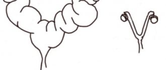
System structure
The drip device consists of several parts. Before you start using it, you need to know what all these parts are for.
Main part
This is a flexible tube. Along it, the drug gradually moves from the container to the needle and then enters the body.
Fence needles
There are only two of them in the set. Inserted into the lid of the container with the drug. One of them is connected to the tube, the second is equipped with an expander and provides air flow into the container with the infusion solution.
Dispenser
This is a box made of plastic, equipped with a slider or wheel. The dispenser is designed to regulate the speed of liquid movement.
Container chamber
The device is made of transparent soft plastic. A filter is placed inside the part to prevent air from entering the tube and blood.
Cannula
The main purpose of this part of the structure is to introduce additional drugs into the solution.
The part is a container made of plastic or a rubber pad.
Dropper assembly
The procedure for assembling the system is as follows:
- Wear gloves and treat your hands with antiseptic.
- Prepare a bottle of medicine and hang it on a tripod.
- Check your doctor's prescription: often the dropper is a saline solution to which other components are added.
- If necessary, open the ampoules with medications, draw the contents with a syringe and inject them into the sodium chloride solution.
- Print out the system for installing the dropper.
- Find the plastic tube and the regulator on it - it needs to be moved to the closed position. This is not difficult to do - the tube will seem to be pinched.
- Insert the needle into the cap of the bottle or a special hole in the bag with the injected solution.
- Remove the extra needle from the package and insert it next to the main one to prevent a vacuum from forming.
- Find a cylinder-shaped extension on the plastic tube. Press it several times - it is necessary that it is filled with the medicine to about half. Using this cylinder you will determine the rate of introduction of the solution.
Types of infusion needles
When placing an IV, different games are used. They have a number of features. If you lack the skills, not all types can be used at home. This issue needs to be examined in detail.
Infusion disposable
Attached to the system.
They differ in thickness and length. For subcutaneous injection into the withers, a medium-thick long needle is used. For intravenous infusion, which is considered more traumatic, a needle of medium length and minimal thickness is used.
Butterfly needle
A short, fairly thin needle. In people it is injected into small veins. Suitable for small pets. It has a special design: “wings” made of plastic are placed in front of the expander. With their help, the needle is conveniently fixed.
Intravenous catheter
This is a plastic tube designed to be inserted into a vein. The drug enters through the catheter, and then the gateway is closed with a stopper.
It is worth knowing how many days such a needle can be used: it can remain in the vein for up to six days, but only with proper care of the puncture site.
Can all needles be used independently?
Not all needles are allowed to be installed on your own by a puppy or an adult dog.
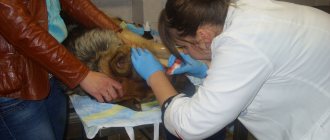
The catheter must be inserted exclusively by a veterinarian . In the absence of experience, there is a risk of damage to nerve endings and veins.
How is infusion therapy performed?
Typically, infusion therapy is carried out in the presence of intravenous access - for this, a plastic intravenous catheter is installed in the saphenous vein on the paw or the jugular vein on the neck of the animal for continuous wear for several days. Sometimes, more often in newborn puppies and kittens (due to the extremely small diameter of the saphenous veins available for manipulation by a doctor), the infusion is carried out using intraosseous or subcutaneous infusion.
Owners often worry that a metal needle remaining in the catheter may damage the vein and soft tissue of their pet's skin and muscles when the animal moves. But this is impossible, since the metal needle (mandrel) is in the catheter only at the moment of puncture of the skin and vein wall. After the catheter enters the vein, the mandrel is immediately removed by the doctor, and only a small-diameter plastic tube remains in the lumen of the vein.
Ivanova Nadezhda Viktorovna Veterinarian. Specialization: emergency care, therapy, reproduction, ultrasound.
Typically, inserting a catheter takes only a few seconds, so the owner does not always have time to notice the order of the doctor’s actions. But we can assure you that wearing an intravenous catheter taped to the paw with an adhesive bandage for several days cannot cause the animal severe pain. If for some reason it is necessary to deliver a liquid drug into the patient’s vascular bed once, and in a short time, the doctor can use not a plastic catheter, but a “butterfly” needle, but it is never left to be worn continuously.
Often animals, especially excitable cats, show anxiety when fixing and wearing a catheter. If the animal tightens and refuses to step on the paw in which the intravenous catheter is located, it is necessary to check whether the paw is tied too tightly with a plaster or fixation bandage.
Nowadays, almost all veterinary clinics use infusion pumps, or syringe pumps, for infusion therapy. Unlike a standard dropper, where a system is attached to the solution bottle, and the rate of supply of the solution is calculated by the doctor approximately, “by eye,” tracking the speed of the falling drops, the electronic infusion pump allows you to accurately calculate the volume and speed of the injected solution. For intensive care unit patients who frequently require low-volume infusion, this is, of course, the preferred method.
Algorithm for preparing a dropper
Before performing basic manipulations, it is important to properly prepare the IV. This procedure is performed in several stages.
Preparation
Hands are treated with an antiseptic. After this, gloves are put on.
They should also be degreased.
Opening the bottle
The next step is to open the bottle with the solution and the system itself. The entire metal cap should not be removed: only its central part is removed.
Inserting needles into the bottle and fixing the container
Both needles are inserted into the rubber stopper.
After this, you need to fix the bottle so that it is located upside down. At home, you can use improvised materials for this purpose, for example, a mop handle and tape.
It is necessary that the container is located at a height of 45-50 cm from the dog.
Preparing the dispenser
The dispenser lever should be moved all the way and thus clamp the tube.
After this, you need to press the reservoir with the filter several times so that it draws in the drug. All that remains is to shake the chamber to remove excess air and slightly lower the lever on the dispenser, thus adjusting the speed of movement of the liquid.
When should a dog be given an IV?
There are many cases when an IV may be needed. This:
- acute poisoning,
- prolonged diarrhea or vomiting
- recovery process after surgery,
- kidney failure in a dog, in which it is urgent to stop the loss of water from the body, and in many other cases, both simple and serious.
Preparing the animal
When the dropper is ready for use, you need to calm the dog and prepare it for the procedure.
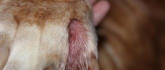
Optimal places for infusion
It is recommended to insert the needle into the wide veins located on the pet's front legs.
Removing hair at the injection site
The next step is to remove the hair between the elbow and wrist. It is shaved with a machine or cut with scissors.
Bandaging the leg with a tourniquet
The limb is tightened before the puncture. The tourniquet is applied slightly above the place where the infusion will be performed.
Inserting a needle into a vein and fixing it with an adhesive plaster
The needle is disconnected from the IV and carefully inserted into the vein parallel to the limb.
Actions are performed slowly, very carefully. The pressure should be moderate. If everything was done without errors, a small amount of venous dark blood will be released from the dilator.
The needle is then secured with adhesive tape. Only after this is the tourniquet removed.
Connecting the system
At the final stage, the needle is connected to the system. All that remains is to monitor the condition of the animal and observe the puncture site.
Diagnosis of animal diseases
Unfortunately, our pets cannot always make their owners understand that something is bothering them in the early stages of the disease. Owners usually notice that something is wrong with their animal when it is already showing severe symptoms of the disease. Usually this is loss of appetite and thirst for several days, diarrhea, repeated vomiting, and difficulty urinating.
When such a patient sees a veterinarian, as a rule, dehydration of one degree or another is always diagnosed. This explains the need to prescribe infusion therapy. Therefore, in addition to standard prescriptions, including tablets and injections of drugs, the patient’s chart contains a note about the appointment of IVs.
Optimal replenishment of fluid deficiency in a dehydrated body is carried out continuously and gradually. To do this, the animal must be left in the inpatient department of the veterinary clinic, where the cat or dog is constantly receiving intravenous correction solutions, and the hospital doctor monitors the dynamics of fluid replenishment 24 hours a day. If for some reason the animal cannot be left in the hospital, or its condition does not require round-the-clock infusion under the supervision of a specialist, the veterinarian may recommend that the owners come in for a drip 1-2 times a day - usually such an infusion lasts about an hour.
Sometimes the doctor diagnoses a decrease in the volume of fluid in the animal’s vessels. Typically, these are animals in a state of blood loss, loss of protein-rich fluid, or in a state of shock. In this case, replenishment of fluid volume in the vascular bed should be carried out as soon as possible. Depending on the pathological condition of the animal, either donor blood or colloidal solutions that normalize oncotic pressure in the vessels are used as administered solutions in these situations.
Unforeseen circumstances and their consequences
Even with strict adherence to all rules, there is a risk of unforeseen situations. It is worth considering the most common ones in order to take the necessary measures in time.
The drug was administered incorrectly
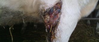
A lump forms at the puncture site, the dog becomes restless and experiences pain in the affected area. Such problems often arise even with correct insertion: the animal periodically relaxes and tenses the veins, and the needle can come out.
For such problems, the lump can be injected with novocaine, but it is better to show your pet to a veterinarian to avoid negative consequences.
Allergy upon administration of the drug
The animal may develop an allergy to the administered drug. In this case, difficulty breathing occurs, lips and throat become swollen.
If such changes are observed during the procedure, you need to perform the following actions:
Close the dispenser.- Tighten the limb with a tourniquet. The main thing is not to clamp the paw for more than a quarter of an hour, monitor the pulse and temperature of the limb.
- Inject the leg with a solution of adrenaline (0.1%) or administer it intramuscularly. The dosage is calculated taking into account the weight of the animal.
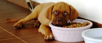
Refusal to eat and drink after IV drip
With such changes, you need to constantly monitor your pet's condition. It is recommended to measure the animal’s body temperature, monitor its breathing rate and heart rate.
If all indicators are normal and the condition is improving, then you should wait a little. After this, the animal is given a warm drink.
If the condition worsens, you should contact a veterinary clinic.
What you need to know when placing an IV
When carrying out the procedure, many points must be taken into account, otherwise irreparable harm will be caused to the animal.
Solution temperature
The drug should not be hot or cold. The best option is a drug at room temperature.
What to do if your dog's temperature is low
If your pet’s temperature is below normal, part of the plastic pipe intended for transporting the solution should be placed in a container with water heated to +70...+80 °C. Due to this, the drug passing through the system will warm up slightly. This helps normalize the animal's body temperature.
Solution infusion rate
The speed at which the solution is administered deserves special attention. It is unacceptable for the drug to enter the body too quickly. No more than two drops should be poured per second.
As the speed increases, the dog’s condition can deteriorate sharply, resulting in:
- gagging;
- increased blood pressure;
- shiver.
Consequences of vein congestion
It is unacceptable for air to enter the vein, otherwise a gas embolism will develop.
Constant monitoring of the pet during the procedure
During the procedure, the pet must be under constant supervision. The owner’s main task is to prevent the dog from running away and to promptly identify negative changes.
Useful tips
Be prepared for the fact that not everything will go smoothly the first time, especially since you are performing the procedure not on an adult, from whom you can learn about pain and well-being, but on an animal.
Whenever possible, contact your veterinarian for fluid therapy . Do it at home only if absolutely necessary.
Remember that all stages of this procedure must be carried out using sterile gloves and sterile instruments. Otherwise, you can introduce an infection into the blood.
Be sure to talk to the animal during the procedure; a confident and calm tone will help him in this unpleasant situation. The dog experiences stress from manipulations, even if they are painless.
In a sick animal, these quantities and ratios may change depending on the causes of the disease, which leads to very specific problems. A loss of more than 5% of fluid (all fluid - that is, about 700 ml of water for a young dog weighing 20 kg) will be visible to the doctor at the appointment in the form of reduced skin turgor, slower straightening of the skin fold, with a loss of 10% (1400 ml) this will become noticeable and to the owner - the animal will be lethargic, apathetic, the skin fold will straighten out very slowly, the mucous membranes will become dry, and the first signs of shock may appear. If the degree of dehydration is more than 12-15% (1800-2100 ml for our invented dog), the patient will be in a state of shock (pale mucous membranes, non-retractable skin fold, decreased body temperature, tachycardia, shortness of breath) and will die in the coming hours.
Where can this amount of liquid go? How does the patient lose it? Every day, warm-blooded animals should consume about 30-50 mlkg of water weight. This is both the liquid you drink and the water that is part of the food (meat, porridge, wet or dry food). Situations in which a deficiency of water and electrolytes in the body develops: 1. For some reason, the patient may be deprived of access to water. This is especially true for cats. Some of them get used to drinking under certain conditions - for example, from a bucket in the bathroom, or only from the tap, and then the bucket was moved to the toilet, and the tap was no longer opened often, or an adult cat could be transferred from natural or wet food to dry, and out of habit, she continues to drink small amounts of water. All of these cats will begin to experience fluid deficiency. 2. The animal became ill with something (infection, poisoning, exacerbation of chronic diseases of internal organs, for example, chronic renal failure) and stopped eating and began to drink little. 3. The underlying disease occurs with symptoms of fever or shortness of breath (fluid loss occurs with breathing), vomiting and/or diarrhea (loose stools) - the patient begins to lose not only water, but also electrolytes (microelements). These are infectious diseases, poisoning, diseases of the gastrointestinal tract, chronic diseases of internal organs in the acute stage, etc. 4. The underlying disease occurs with signs of polyuria (increased urine output). This is common in chronic kidney failure, diabetes, and some rarer conditions. Of course, following this, thirst also increases, but sooner or later there comes a time when the patient is not able to compensate for the losses by drinking. 5. Acute blood loss. 6. Shock caused by any reason (painful, traumatic, due to blood loss, neurogenic, toxic, septic) leads to a redistribution of fluid and the development of a condition called hypovolemia, which ultimately comes down to a decrease in the volume of circulating blood - hereinafter referred to as BCC. This can happen with pyometra and acute urinary retention. 7. Fluid retention in the third space. With peritonitis, intestinal obstruction, gastric volvulus, internal bleeding, pancreatitis, with intestinal atony, up to several liters of fluid that does not participate in circulation can accumulate in its lumen. 8. Surgical interventions always require infusion support. On the day of surgery, the patient, as a rule, does not eat and is deprived of water, almost any surgical intervention is accompanied by blood loss, add here evaporation from the surface of the surgical wound (this applies to a greater extent to operations on the abdominal and thoracic cavity), the need to remove drugs used for general anesthesia and some other points, and it becomes obvious that the patient needs additional volumes of solutions. It is known that during thoracic operations the patient loses 3-5 mlkg per hour, during abdominal operations - 6-8 mlkgh, 2 mlkgh evaporates from the surface of the wound.